Carotid Intraplaque Hemorrhage is Associated with Acute Cerebral Ischemic Events and Progression of Stenosis on Magnetic Resonance Imaging
INTRODUCTION
Extracranial carotid artery stenosis is a critical cause of stroke that accounts for approximately 20% of all ischemic strokes (1). At present, symptoms and severity of
This is an Open Access article distributed under the terms of the Creative Commons Attribution Non-Commercial License (http://creativecommons.org/licenses/
by-nc/3.0/) which permits unrestricted non-commercial use, distribution, and reproduction in any medium, provided the original work is properly cited.
Received: September 17, 2017 Revised: September 29, 2017 Accepted: October 12, 2017 Correspondence to:
Se Jeong Jeon, M.D.
Department of Radiology, Wonkwang University Hospital, 895 Muwang-ro, Iksan 54538, Korea.
Tel. +82-63-859-1920 Fax. +82-63-859-4749 E-mail: medicalq@hanmail.net
Copyright © 2017 Korean Society of Magnetic Resonance in Medicine (KSMRM)
Original Article
Purpose: To investigate the association of carotid intraplaque hemorrhage (IPH) with acute cerebral ischemic events and progression of stenosis using magnetic resonance (MR) imaging.Materials and Methods: From April 2014 to December 2016, 53 patients underwent carotid plaque MR imaging, including magnetization-prepared rapid acquisition with gradient-echo (MPRAGE) sequence. A total of 66 carotid arteries in 53 patients had carotid stenosis, and they were included in this study. Carotid arteries were classified according to the presence of IPH, the age of hemorrhage, and degree of stenosis. We assessed ipsilateral cerebrovascular event rates and progression of stenosis between the IPH and no-IPH groups.
Results: Of the 61 carotid arteries assessed, 34 (56%) had IPH, and 27 (44%) had no IPH. Acute cerebral ischemic events were more frequent in the IPH group (47% vs.
22%, P = 0.045), especially in the < 30%-stenosis group (100% vs. 0%, P = 0.028).
However, there was no significant difference in the incidence of ischemic events according to the age of hemorrhage (50% vs. 44%, P = 0.492). Among the 61 carotid arteries, 20 carotid arteries had previously undergone carotid artery imaging and were evaluated for plaque progression. The trend for progression of stenosis favored the IPH group versus the no-IPH group, with a marginal P-value (20% ± 12.7 vs. 9.6%
± 5.7, P = 0.063).
Conclusion: IPH was associated with an increased incidence of acute ischemic events, especially in the mild-stenosis group and it was also associated with progression of stenosis. Evaluation of the carotid IPH by carotid plaque MR could improve discrimination of carotid plaques that cause ischemic events and progression of stenosis.
Keywords: Intraplaque hemorrhage; Stroke; Plaque progression; Magnetic resonance imaging
Hye Ji Ryu, Se Jeong Jeon, See Sung Choi
Department of Radiology, Wonkwang University Hospital, Iksan, Korea Magnetic resonance imaging
stenosis are used to determine the treatment in patients with carotid artery stenosis. However, ischemic strokes in patients with carotid artery disease are not restricted to arteries with severe stenosis, and there is growing evidence that other factor such as plaque vulnerability may help to improve the existing risk stratification criteria for the best therapeutic approach (2, 3).
While intraplaque hemorrhage (IPH) was considered as a minor criterion of plaque vulnerability in the coronary artery (4), many studies have nevertheless focused on the vulnerable features of IPH. Lusby et al. (5) first reported in 1982 that IPH was more frequent in symptomatic patients based on carotid endarterectomy specimens. After the development of magnetic resonance (MR) imaging techniques for identifying plaque components, many studies investigated the association of MR-depicted IPH with cerebral ischemic events and plaque progression. A recent meta-analysis found that IPH was associated with an approximately 6-fold higher risk of cerebrovascular events (6). But they also emphasized the necessity of studying homogeneous populations under standardized conditions in the future due to the substantial heterogeneity in study populations and definition of IPH among published studies.
Although the pre-existing literature has assessed the effects of IPH on acute cerebrovascular events, these effects have not been fully evaluated in terms of correlations to the degree of carotid stenosis and hemorrhage age of the plaque.
The aims of our study were 1) to investigate the association between IPH and ipsilateral acute cerebral ischemic events by further stratifying carotid plaques according to the degree of stenosis and age of hemorrhage, and 2) to identify the association of IPH with stenosis progression.
MATERIALS AND METHODS
Study PopulationThis was a retrospective study that received the Institutional Review Board approval and the requirement for obtaining informed consent was waived. From April 2014 to December 2016, 53 consecutive patients underwent carotid plaque MR imaging for further evaluation of carotid plaques detected by stroke MR or routine brain MR (n
= 44) imaging evaluations that included time-of-flight magnetic resonance angiography (TOF-MRA), or computed tomography angiography (CTA) (n = 9). A total of 66 carotid
arteries in 53 patients had stenosis, and they were included in our study. From these 66 carotid arteries, 2 arteries were excluded due to intracarotid sources of ischemia such as cardioembolism (n = 1, atrial fibrillation) and non- atherosclerotic vasculopathy (n = 1, Moyamoya disease), and 3 arteries were excluded due to total carotid occlusion.
All patients with acute ischemic stroke underwent echocardiography for evaluations of cardioembolic sources of stroke. Also, although there were 3 carotid arteries with ipsilateral intracranial artery stenosis on TOF-MRA or CTA, the stenosis measured < 50% and the arteries were therefore not excluded from this study. Consequentially, 61 carotid arteries in 48 patients were assessed for data analysis. Presenting symptoms of these patients were as follows; 26 patients with dysarthria and/or unilateral motor weakness, 8 patients with dizziness, 2 patients with headaches, 1 patient with vision loss, 1 patient with blurred vision, and 10 patients with no symptom (follow-up of the known vascular lesion).
Imaging Protocol (Carotid Plaque MR Imaging and Stroke/Routine Brain MR Imaging)
All MR studies were performed on a 3T MR scanner (Achieva; Philips Healthcare, Best, The Netherlands).
For carotid plaque analysis, carotid plaque MR imaging including TOF-MRA, T1-weighted, T2-weighted, post- contrast T1-weighted and magnetization-prepared rapid acquisition with gradient-echo (MPRAGE) sequences was acquired with a 16-channel head and neck coil by placing carotid bifurcation at the center. T1-weighted, T2-weighted, and post-contrast T1-weighted sequences were acquired with a 2.5 mm-section thickness, and TOF, MPRAGE sequences were obtained with 1.2 and 1.0 mm-section thicknesses, respectively. All sequences had no intersection spacing and were scanned with a field of view (FOV) of 140
× 140 mm and a matrix size of 216 × 192. More details of scan parameters used in our study were as follows: 1) TOF- MRA; repetition time (TR)/echo time (TE)/flip angle, 16 ms/3 ms/20°, 2) T1-weighted and post-contrast T1-weighted sequence; TR/TE, 800/10 ms, 3) T2-weighted sequence; TR/
TE, 3000/80 ms, 4) MPRAGE sequence; TR/TE/flip angle, 9 ms/5 ms/15°.
Our protocol for stroke/routine brain MR imaging studies included diffusion-weighted imaging (DWI) with an apparent diffusion coefficient (ADC) map and TOF- MRA of carotid and intracranial arteries. DWI was obtained with 3 b-values of 0, 1000, and 3000 s/mm2 and with the following scan parameters: TR/TE, 7500/36 ms for b-value
3000 and 3478/77 ms for b-value 1000; flip angle, 90°;
FOV, 210 × 210 mm; matrix, 124 × 122; section thickness/
gap, 4 mm/0.4 mm.
Total scan time was usually 30 minutes each for plaque MR imaging and stroke/routine brain MR imaging.
Evaluation of Carotid IPH and Acute Ischemic Events Carotid plaque MR imaging and stroke/routine brain MR imaging results were interpreted by two neuroradiologists with 10 and 25 years of experience, respectively, and results were summarized through consensus interpretations.
First, the presence of IPH was evaluated on the MPRAGE sequence. IPH was defined when there was an increased signal intensity of the plaque, which was more than twice the signal intensity of the sternocleidomastoid (SCM) muscle, as previously described (7). Signal intensities were measured on the MR imaging console by placing a circular region of interest (4-9 mm in diameter). After identifying IPH, the age of hemorrhage was categorized according to the signal intensity on T1- and T2-weighted images by visual assessment, based on previously published criteria (8). SCM muscle was used as a reference. A previous histopathological study by Lusby et al. (5) was the first to report 3 different stages of IPH; fresh (age < 1 week), recent (2-6 weeks old), and old (> 6 weeks old). A later study using multi-contrast MRI reported that MRI could accurately detect the age of IPH with high sensitivity of 90% and moderate specificity of 74% (8). An IPH with a high signal intensity on T1-weighted images and iso- or hypo-signal intensity on T2-weighted images was classified as fresh IPH (type I), and IPH with a high signal intensity on both T1 and T2-weighted images were classified as a recent IPH (type II).
Luminal stenosis was measured on TOF-MRA according to the North American Symptomatic Carotid Endarterectomy Trial (NASCET) criteria and was further categorized into the following 3 groups: mild stenosis (< 30%), moderate stenosis (30-69%), and severe stenosis (70-99%).
Acute ischemic events were defined as ipsilateral ICA territorial lesions with increased signal intensities on DWI and correspondingly decreased signal intensities on the ADC map. Although 9 carotid arteries in 9 patients only underwent CTA without stroke or routine brain MR imaging evaluations, they were considered to have no ipsilateral acute ischemic lesions because these patients did not have any acute neurologic symptoms, suggesting anterior circulation stroke or transient ischemic attack (TIA). They underwent CTA for following reasons; 6 follow up for the known vascular lesion, 1 vision loss, 1 chronic dizziness, 1
a chronic headache. Mean time interval between stroke/
routine brain MR and plaque MR was 5.9 days (range, 0-30 days).
Evaluation of Stenosis Progression
Among the 61 included carotid arteries in 48 patients, 28 carotid arteries in 27 patients had previously undergone MRA (TOF or contrast-enhanced [CE]-MRA) or CTA. In 8 carotid arteries of 7 patients, there were no corresponding carotid plaques on previous carotid artery imaging studies.
Therefore, a total of 20 carotid arteries in 20 patients were evaluated for plaque progression. Initial carotid stenosis was measured on previous TOF-MRA with the exception of 6 carotid arteries. Two carotid arteries were assessed with CE-MRA, and 4 carotid arteries were evaluated with CTA.
Statistical Analysis
Carotid arteries with plaques were divided into the IPH, and the no-IPH groups, and they were further classified according to the grade of stenosis and age of hemorrhage.
A two-sample t-test was used to determine the differences in continuous variables between subgroups. Chi-square and Fisher’s exact test was used to evaluate significant differences in categorical variables. The tendency of plaque- type according to the grade of stenosis was analyzed with the linear-by-linear association. Odds ratio (OR) was calculated to evaluate the risk of acute ischemic events caused by IPH. P < 0.05 was considered to indicate statistical significance. All statistical analyses were performed using SPSS, version 18.0 (SPSS Inc., Chicago, IL, USA).
RESULTS
Baseline Clinical and Imaging Characteristics
Of the total 61 carotid arteries, 34 arteries (56%) had MPRAGE-positive signal and were classified into the IPH group. The remaining 27 arteries (44%) were classified into the no-IPH group. In the 34 carotid arteries that had IPH, type I IPH was found in 18 carotid arteries (52.9%) and type II IPH was found in 16 carotid arteries (47.1%).
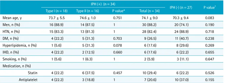
Baseline clinical characteristics of patients with IPH (type I and type II) and without IPH were shown in Table 1. There were no significant differences between the two groups in terms of age, sex, past history, smoking status, and medications.
Baseline characteristics of carotid plaque MR imaging in
the two groups were shown in Table 2. Mean stenosis and distribution according to the degree of stenosis also were not significantly different between the two groups.
Incidence of Acute Ischemic Events
Association between IPH and acute ischemic events according to the degree of stenosis and age of hemorrhage was evaluated. The incidence of acute ischemic events was 36.1%, and it was increased with an increase in the degree of stenosis (mild stenosis vs. moderate stenosis vs. severe stenosis; 22.2% vs. 28.6% vs. 50%). The incidence of IPH in the mild stenosis, moderate stenosis, and severe stenosis groups was 22.2%, 67.9%, and 54.2%, respectively. Acute cerebral ischemic events were more frequently found in the IPH group (47% vs. 22%, P = 0.045), resulting in an odds ratio of 3.111 (95% confidence interval [CI] = 1.005-9.630) (Figs. 1, 2). When these results were evaluated according
to the degree of stenosis, there was a significant difference only in the mild stenosis group with a P-value of 0.028 (Fig.
3). But the odds ratio was not calculated due to the small sample size in the mild stenosis group. In the moderate and severe stenosis groups, the odds ratio decreased with an increase in the degree of stenosis (4.667 in the moderate stenosis group, 1.4 in the severe stenosis group), although it was not statistically significant. With respect to age of hemorrhage, there was no significant difference in the incidence of acute cerebral ischemic events between type I and type II hemorrhages in the IPH group (50% vs. 43.8%, P
= 0.492) (Fig. 4).
Progression of Carotid Stenosis
Among the 20 carotid arteries with previous carotid imaging, 9 were in the IPH group, and 11 were in the no-IPH group. Baseline clinical characteristics including
Table 1. Baseline Clinical Characteristics of Patients with and without IPH IPH (+) (n = 34)
IPH (-) (n = 27) P value† Type I (n = 18) Type II (n = 16) P value* Total (n = 34)
Mean age, y 73.7 ± 5.5 74.6 ± 1.0 0.751 74.1 ± 9.0 70.3 ± 9.4 0.083
Men, n (%) 16 (88.9) 14 (87.5) 1 30 (88.2) 20 (74.1) 0.190
HTN, n (%) 15 (83.3) 13 (81.3) 1 28 (82.4) 24 (88.9) 0.718
DM, n (%) 4 (22.2) 5 (31.3) 0.703 9 (26.5) 11 (40.7) 0.238
Hyperlipidemia, n (%) 1 (5.6) 5 (31.3) 0.078 6 (17.6) 8 (29.6) 0.269
IHD, n (%) 4 (22.2) 2 (12.5) 0.660 6 (17.6) 6 (22.2) 0.655
Smoking, n (%) 1 (5.6) 1 (6.3) 1 2 (5.9) 3 (11.1) 0.647
Medication, n (%)
Statin 4 (22.2) 6 (37.5) 0.457 10 (29.4) 6 (22.2) 0.526
Antiplatelet 4 (22.2) 3 (18.8) 1 7 (20.6) 10 (37.0) 0.155
Data are presented as mean ± standard deviation for continuous variables and number (percentage) for categorical variables. *P values between type I and type II IPH group, †P values between IPH and no IPH group. DM = diabetes mellitus; HTN = hypertension; IHD = ischemic heart disease; IPH = intraplaque hemorrhage
Table 2. Baseline Characteristics of Carotid Plaque MR Imaging in Carotid Arteries with and without IPH IPH (+) (n = 34)
IPH (-) (n = 27) P value† Type I (n = 18) Type II (n = 16) P value* Total (n = 34)
Mean stenosis, % 62.9 ± 22.8 55.9 ± 20.0 0.351 59.6 ± 21.5 50.9 ± 27.6 0.167
Grade of stenosis
< 30%, n (%) 2 (11.1) 0 (0) 0.492 2 (5.9) 7 (25.9) 0.330
30-69%, n (%) 7 (38.9) 12 (75) 19 (55.9) 9 (33.3)
70-99%, n (%) 9 (50) 4 (25) 13 (38.2) 11 (40.7)
Data are presented as mean ± standard deviation for continuous variables and number (percentage) for categorical variables. *P values between type I and type II IPH group, †P values between IPH and no IPH group. IPH = intraplaque hemorrhage
medication were not significantly different between the two groups. Mean time intervals between preceding carotid imaging and plaque MR imaging in the IPH and no-IPH groups were 29.8 months (range, 0.3-48 months) and 20.2 months (range, 0.3-48.4 months), respectively, without any significant difference. Also, the degree of initial stenosis was not significantly different between the two groups (51.3% ± 18 vs. 56.8% ± 14.4, P = 0.458). In the IPH group, 8 out of the 9 carotid arteries showed stenosis progression, and one carotid artery showed stenosis regression. In the no-IPH group, 7 out of the 11 carotid arteries showed stenosis progression. Among the remaining 4 arteries in the no-IPH group, 2 arteries showed plaque regression, and 2
arteries showed no change. When arteries that showed the progression of stenosis were evaluated separately, the IPH group showed a trend favoring higher stenosis progression compared to the no-IPH group, with a marginal P-value (20% ± 12.7 vs. 9.6% ± 5.7, P = 0.063) (Table 3, Fig. 5).
DISCUSSION
In this study, we found that carotid plaques with IPH on the MPRAGE sequence were associated with ipsilateral acute cerebral ischemic events on DWI. When carotid plaques were classified according to the degree of stenosis,
a b c
d e
Fig. 1. An example of a 67-year-old man with type I IPH and ipsilateral cerebral infarction. (a) 3D TOF angiogram shows the high signal intensity of the plaque causing mild stenosis. (b) MPRAGE image also shows the high signal intensity of the plaque, which suggests IPH. (c, d) High signal intensity on the T1-weighted image (c) and iso-signal intensity on the T2- weighted image (d) indicate type I IPH (arrows). (e) DWI shows diffusion restriction in the left ICA territory. The asterisk indicates the ICA lumen. 3D = three-dimensional; DWI = diffusion-weighted imaging; ICA = internal carotid artery; IPH = intraplaque hemorrhage; TOF = time of flight
this association was statistically significant in the mild stenosis group. Age of hemorrhage did not have any influence on the incidence of ischemic events. Also, the IPH group showed more substantial stenosis progression than the no-IPH group.
IPH was mainly thought to result from neovessel leakage because of leaky endothelial junctions and insufficient support by smooth muscle cells (9, 10). Correlation of IPH with acute ischemic events and plaque progression observed in our study was supported by previous pathophysiologic studies which demonstrated that IPH may contribute to
several biologic processes involved in local inflammation, leading to deposition of free cholesterol and release of proteolytic enzymes, which may result in plaque growth and plaque destabilization (11, 12). As the importance of IPH has been emphasized by many authors, there have been new developments in MR techniques for detection of IPH. T1-weighted sequences were commonly used for IPH detection because degradation of the hemorrhage into methemoglobin caused T1-shortening and a resultant high signal intensity on T1-weighted images (13). Our study used MPRAGE sequence for IPH detection, which demonstrated a
d e
a b c
Fig. 2. An example of a 62-year-old man with type II IPH and territorial infarction. (a) 3D TOF angiogram shows moderate focal stenosis in the left proximal ICA. (b) MPRAGE image shows the high signal intensity of the plaque, which suggests IPH. (c, d) A high signal intensity lesion on T1- and T2-weighted images (arrows) corresponds to a high signal intensity lesion on MPRAGE sequence. Also, note the iso-signal intensity lesion within the plaque (arrowheads), which shows no contrast enhancement on the contrast-enhanced T1-weighted image (not shown). This finding indicates type II IPH with a lipid-rich necrotic core. (e) DWI demonstrates territorial ischemic events. The asterisk indicates the ICA lumen. 3D = three- dimensional; DWI = diffusion-weighted imaging; ICA = internal carotid artery; IPH = intraplaque hemorrhage; TOF = time of flight
significantly higher diagnostic capability with a sensitivity of 80% and a specificity of 97% in detecting IPH compared with T1-weighted fast spin-echo and TOF (14). MPRAGE sequence facilitates suppression of background tissue signals by a non-selective inversion pulse and either spectrally selective water excitation or fat suppression (13, 15). Also, plaque components with relatively long T1, such as a lipid-rich necrotic core and fibrous tissue, were suppressed by the inversion-recovery preparation. So uniform hypointensity of background tissue and suppressed blood signal and other plaque component signal result in the higher diagnostic performance of MPRAGE sequence for detection and quantification of IPH (14).
Although previous longitudinal studies have shown the association of IPH with ischemic stroke, this relationship is still a controversial topic, possibly due to substantial heterogeneity among published studies. Sitzer et al. (16) reported that plaque ulceration and lumen thrombus were related to cerebral microemboli in high-grade ICA stenosis (≥
70%). Another recent study by de Rotte et al. (17) reported that IPH and a thin/ruptured fibrous cap were not related
to the old and current cortical and subcortical infarctions in patients with 30-69% carotid artery stenosis. In our study, IPH on MPRAGE was associated with an increased incidence of acute ischemic events on DWI and this impact was more significant in mild stenosis group than in moderate and severe stenosis groups. Considering the results of two previous studies by Sitzer et al. (16) and de Rotte et al.
(17), which could not identify any association between IPH and ischemic events in moderate to high carotid artery stenosis, it was suggested that the impact of IPH on plaque vulnerability and embolization may be much higher in low- degree stenotic lesions that were overlooked as a cause of acute ischemic events. Because the number of lesions in the mild-stenosis group in our study was small, future studies with a larger number of lesions are needed to confirm our result.
We also classified IPH according to the age of hemorrhage using carotid plaque MR imaging studies. Saam et al. (18) revealed that the age of hemorrhage could be important in determining the patient’s symptoms. They found that type I IPH occurred more often in symptomatic plaques than in
Table 3. Progression of Stenosis According to the Presence or Absence of IPH
IPH (+) (n = 8/9) IPH (-) (n = 7/11) P value
Initial carotid stenosis, % 52.4 ± 18.9 56.9 ± 23.1 0.686
Stenosis progression, % 20 ± 12.7 9.6 ± 5.7 0.063
Interval between preceding carotid imaging and plaque MR imaging, months 27.6 ± 16.1 22.7 ± 18.2 0.593 Data are presented as mean ± standard deviation. IPH = intraplaque hemorrhage; MR = magnetic resonance
Fig. 4. Association between IPH and acute ischemic events by further stratifying IPH according to the age of hemorrhage. IPH = intraplaque hemorrhage
P P
Fig. 3. Association between IPH and acute ischemic events by further stratifying carotid plaques according to the grade of stenosis. IPH = intraplaque hemorrhage
P
asymptomatic plaques, while this did not occur in patients with type II IPH. There was no significant difference in the incidence of IPH between symptomatic and asymptomatic
plaques in their study. In our study, we obtained an opposite result. Although there was an association between IPH and ischemic events, the incidence of acute ischemic events Fig. 5. A 78-year-old man with IPH and progression of carotid stenosis. (a, b) TOF-MRA image (a) and axial 3D TOF source image (b) examined 45 months ago show moderate stenosis (arrows) in the right proximal ICA with the high signal intensity of the plaque. (c-e) Plaque MR images examined due to dysarthria show progression of luminal stenosis (arrows) and high signal intensity of the plaque on the TOF source image (d) and MPRAGE (e). The asterisk indicates the ICA lumen. ICA = internal carotid artery; IPH = intraplaque hemorrhage; MR = magnetic resonance; TOF-MRA = time-of-flight magnetic resonance angiography
a b c
d e
was not significantly different between type I and typed II hemorrhages in the IPH group. Our findings suggest that the presence of IPH itself was more critical to plaque vulnerability, regardless of the age of hemorrhage, and type II hemorrhage had just as many vulnerable features as type I hemorrhage. Two pathways of plaque destabilization and plaque stabilization after hemorrhage were reported.
IPH could contribute to plaque instability via induction of an inflammatory response, enlargement of the lipid-rich necrotic core, and development of new vessels. Also, it could ultimately lead to plaque stabilization via induction of the healing process with formation of calcified and fibrous tissue (11, 19). Type II IPH encompasses not an only early organization, but also increased interstitial fluid associated with inflammation (8). Therefore, type II IPH is still involved in the process of plaque destabilization with inflammation before complete stabilization, and IPH itself could be an essential feature of plaque vulnerability, regardless of the age of hemorrhage.
In previous serial MR imaging studies that evaluated plaque progression, IPH was associated with an increase in the necrotic core and plaque progression (20-22). In accordance with these studies, IPH group tended to show the higher progression of luminal stenosis compared to the no-IPH group in our study. Among the 8 arteries with plaque progression in the IPH group, 6 carotid arteries had undergone previous TOF-MRA, and 5/6 carotid arteries showed high signal intensities in the plaque, and 1/6 carotid artery showed an iso-signal intensity in the plaque.
Among the 7 arteries with plaque progression in the no- IPH group, 6 carotid arteries had undergone previous TOF- MRA, and none of them showed high signal intensity in the plaque. Therefore, the potent atherogenic stimulus of IPH and acceleration of plaque growth with new IPH could be the possible factors for plaque progression in the IPH group of our study.
Our study has several limitations. First, we only focused on the IPH as a vulnerable feature of a plaque due to its increasing importance. When considering the greater impact of IPH on acute ischemic events in mild stenosis group, which was observed in our study, in moderate and high-grade stenosis, other plaque features such as ruptured or a thin fibrous cap, large lipid-rich necrotic core, and inflammation could have more impact on plaque vulnerability. The impact of these features on ischemic events according to the degree of stenosis needs to be investigated further. Second, we evaluated carotid stenosis on TOF-MRA images, and for evaluating plaque progression,
in few cases, carotid stenosis was assessed with CE- MRA and CTA. Although DSA was the standard method for evaluating carotid stenosis to date, TOF-MRA was a frequently used imaging technique in a clinical setting because of its non-invasiveness and absence of need for contrast injection. Although TOF-MRA was known to be sensitive to artifacts, there was also increasing evidence that TOF-MRA has similar accuracy or it could even be more accurate than CE-MRA, especially in severe grade stenosis (23-25). Third, we evaluated the percentage of luminal stenosis as an indicator of plaque progression. Plaque volume could also be increased without significant luminal narrowing due to positive remodeling. In the coronary artery, remodeling type was considered to be essential for the assessment of plaque vulnerability. Therefore, a further study evaluating arterial remodeling in the carotid artery would help us in understanding a carotid plaque in many aspects. Lastly, this was a retrospective study with a relatively small sample size performed at a single institution.
In conclusion, IPH was associated with increased incidence of acute ischemic events on DWI, especially in the mild stenosis group and it was also associated with progression of stenosis. Our results suggest that identification of IPH by carotid plaque MR, particularly in mild stenosis group, could improve discrimination of carotid plaques that caused ischemic events and stenosis progression, and ultimately, it could be important in guiding treatment in patients with carotid artery disease.
REFERENCES
1. Go AS, Mozaffarian D, Roger VL, et al. Heart disease and stroke statistics--2014 update: a report from the American Heart Association. Circulation 2014;129:e28-e292
2. Kwee RM, van Oostenbrugge RJ, Mess WH, et al. MRI of carotid atherosclerosis to identify TIA and stroke patients who are at risk of a recurrence. J Magn Reson Imaging 2013;37:1189-1194
3. Naylor AR, Schroeder TV, Sillesen H. Clinical and imaging features associated with an increased risk of late stroke in patients with asymptomatic carotid disease. Eur J Vasc Endovasc Surg 2014;48:633-640
4. Naghavi M, Libby P, Falk E, et al. From vulnerable plaque to vulnerable patient: a call for new definitions and risk assessment strategies: Part I. Circulation 2003;108:1664- 1672
5. Lusby RJ, Ferrell LD, Ehrenfeld WK, Stoney RJ, Wylie EJ.
Carotid plaque hemorrhage. Its role in production of cerebral ischemia. Arch Surg 1982;117:1479-1488
6. Saam T, Hetterich H, Hoffmann V, et al. Meta-analysis and systematic review of the predictive value of carotid plaque hemorrhage on cerebrovascular events by magnetic resonance imaging. J Am Coll Cardiol 2013;62:1081-1091 7. Yamada N, Higashi M, Otsubo R, et al. Association between
signal hyperintensity on T1-weighted MR imaging of carotid plaques and ipsilateral ischemic events. AJNR Am J Neuroradiol 2007;28:287-292
8. Chu B, Kampschulte A, Ferguson MS, et al. Hemorrhage in the atherosclerotic carotid plaque: a high-resolution MRI study. Stroke 2004;35:1079-1084
9. Virmani R, Kolodgie FD, Burke AP, et al. Atherosclerotic plaque progression and vulnerability to rupture: angio- genesis as a source of intraplaque hemorrhage. Arterioscler Thromb Vasc Biol 2005;25:2054-2061
10. Virmani R, Narula J, Farb A. When neoangiogenesis ricochets. Am Heart J 1998;136:937-939
11. Michel JB, Delbosc S, Ho-Tin-Noe B, et al. From intraplaque haemorrhages to plaque vulnerability: biological consequences of intraplaque haemorrhages. J Cardiovasc Med (Hagerstown) 2012;13:628-634
12. Michel JB, Virmani R, Arbustini E, Pasterkamp G. Intra- plaque haemorrhages as the trigger of plaque vulnerability.
Eur Heart J 2011;32:1977-1985, 1985a, 1985b, 1985c 13. Moody AR, Murphy RE, Morgan PS, et al. Characterization
of complicated carotid plaque with magnetic resonance direct thrombus imaging in patients with cerebral ischemia.
Circulation 2003;107:3047-3052
14. Ota H, Yarnykh VL, Ferguson MS, et al. Carotid intraplaque hemorrhage imaging at 3.0-T MR imaging: comparison of the diagnostic performance of three T1-weighted sequences. Radiology 2010;254:551-563
15. Zhu DC, Ferguson MS, DeMarco JK. An optimized 3D inversion recovery prepared fast spoiled gradient recalled sequence for carotid plaque hemorrhage imaging at 3.0 T.
Magn Reson Imaging 2008;26:1360-1366
16. Sitzer M, Muller W, Siebler M, et al. Plaque ulceration and lumen thrombus are the main sources of cerebral microemboli in high-grade internal carotid artery stenosis.
Stroke 1995;26:1231-1233
17. de Rotte AA, Truijman MT, van Dijk AC, et al. Plaque components in symptomatic moderately stenosed carotid arteries related to cerebral infarcts: the plaque at RISK study. Stroke 2015;46:568-571
18. Saam T, Cai J, Ma L, et al. Comparison of symptomatic and asymptomatic atherosclerotic carotid plaque features with in vivo MR imaging. Radiology 2006;240:464-472
19. Moreno PR, Purushothaman M, Purushothaman KR. Plaque neovascularization: defense mechanisms, betrayal, or a war in progress. Ann N Y Acad Sci 2012;1254:7-17
20. Takaya N, Yuan C, Chu B, et al. Presence of intraplaque hemorrhage stimulates progression of carotid ather- osclerotic plaques: a high-resolution magnetic resonance imaging study. Circulation 2005;111:2768-2775
21. Sun J, Underhill HR, Hippe DS, Xue Y, Yuan C, Hatsukami TS. Sustained acceleration in carotid atherosclerotic plaque progression with intraplaque hemorrhage: a long-term time course study. JACC Cardiovasc Imaging 2012;5:798- 804
22. Underhill HR, Yuan C, Yarnykh VL, et al. Arterial remodeling in [corrected] subclinical carotid artery disease. JACC Cardiovasc Imaging 2009;2:1381-1389
23. DeMarco JK, Huston J 3rd, Bernstein MA. Evaluation of classic 2D time-of-flight MR angiography in the depiction of severe carotid stenosis. AJR Am J Roentgenol 2004;183:787-793
24. Babiarz LS, Romero JM, Murphy EK, et al. Contrast- enhanced MR angiography is not more accurate than unenhanced 2D time-of-flight MR angiography for determining > or = 70% internal carotid artery stenosis.
AJNR Am J Neuroradiol 2009;30:761-768
25. Townsend TC, Saloner D, Pan XM, Rapp JH. Contrast material-enhanced MRA overestimates severity of carotid stenosis, compared with 3D time-of-flight MRA. J Vasc Surg 2003;38:36-40