The skull is a very complex skeletal framework made up of numerous bones. The temporal bone is one of the most complex bones in the skull, and may present with several developmental anomalies. Anatomical variations in the human skull can be puzzling and pose diagnostic challen
ges on conebeam computed tomography(CBCT) evalu
ation. In the past, anatomical variants were studied using cadavers or dried skulls. With the development of improv
ed imaging technology, especially threedimensional(3D) imaging, anatomical variants can be readily assessed dur
ing routine examinations. Despite advances in technology, certain structures of the skull remain relatively unexplored, such as the mastoid emissary vein.1,2 The mastoid canal transmits the mastoid emissary vein and a branch of the
occipital artery.3,4 Reis et al.5 referred to the first descrip
tion of the mastoid canal by Gruber in 1875. Reis and col
leagues5 further elaborated that the mastoid canal follows a winding course and if present, is located near the occip
itomastoid suture or the posterior border of the mastoid portion of the temporal bone.
Emissary veins are residual embryonic venous tracts that connect the intracranial sinuses with the extracranial ve
nous system through minute bony apertures in the skull.6 Emissary veins are named depending on their anatomical location(e.g., frontal, occipital, parietal, and mastoid).3 These emissary veins are valveless veins, and hence blood can flow in both directions.3,7 The number of mastoid emissary foramina may range from zero to four.7 Mastoid emissary veins with a diameter >3.5mm are considered large.8 The prevalence of mastoid emissary veins is great
er in males than in females.9 The etiology of the enlarge
ment of these vessels is unknown, but is associated with conditions such as vascular malformations, severe cranial
Incidental occurrence of an unusually large mastoid foramen on cone-beam computed tomography and review of the literature
Ali Z. Syed1, Cleo Sin1, Raquel Rios1, Mel Mupparapu2,*
1Department of Oral and Maxillofacial Medicine and Diagnostic Sciences, School of Dental Medicine, Case Western Reserve University, Cleveland, OH, USA
2Division of Radiology, University of Pennsylvania School of Dental Medicine, Philadelphia, PA, USA
AbstrAct
The incidental finding of an enlarged mastoid foramen on the right posterior mastoid region of temporal bone is reported, together with a discussion of its clinical significance. A 67-year-old female underwent the pre-implant assessment of a maxillary left edentulous region. A conebeam computed tomographic(CBCT) image was acquired and referred for consultation. Axial CBCT slices revealed a unilateral, well-defined, noncorticated, low-attenuation, transosseous defect posterior to the mastoid air cells in the right temporal bone. The borders of the osseous defect were smooth and continuous. No other radiographic signs suggestive of erosion or sclerosis were noted in the vicinity. The density within the defect was homogenous and consistent with a foramen and/or soft tissue.
The patient’s history and physical examination revealed no significant medical issues, and she was referred to a neuroradiologist for a second opinion. The diagnosis of an enlarged mastoid foramen was made and the patient was reassured.(Imaging Sci Dent 2016; 46: 39-45)
Key words: ConeBeam Computed Tomography; Mastoid; Temporal Bone; Cortical Defect
Copyright ⓒ 2016 by Korean Academy of Oral and Maxillofacial Radiology
This is an Open Access article distributed under the terms of the Creative Commons Attribution NonCommercial License(http://creativecommons.org/licenses/bync/3.0) which permits unrestricted noncommercial use, distribution, and reproduction in any medium, provided the original work is properly cited.
Imaging Science in Dentistry·pISSN 22337822 eISSN 22337830 Received August 20, 2015; Revised September 10, 2015; Accepted September 20, 2015
*Correspondence to : Prof. Mel Mupparapu
Division of Radiology, University of Pennsylvania School of Dental Medicine, 240 S 40th Street, Philadelphia, PA 19104, USA
Tel) 12157468869, Fax) 12155737853, Email) [email protected]
hypoplasia, and craniosynostosis.10 Generally, the emis
sary veins are relatively small, asymptomatic, and cannot be visualized in imaging studies.6,10
case report
The volumetric CBCT dataset of a 67year old female was referred to our department for the preimplant eval
uation of an edentulous left maxilla in the region corre
sponding to the first molar(Figs. 1 and 2). A mediumvol
ume CBCT was prescribed by the implantologist and was acquired in the office of the periodontist who referred the case to us. A Carestream CBCT CS 9300(Carestream Inc., Atlanta, GA, USA) was used with a voxel resolution of 200μm. A limited field of view was used to capture the maxilla. No significant medical or dental history was re
ported by the periodontist. The patient had not been hos
pitalized in the recent past, and she was stable and alert at the time of presentation. The dental examination revealed multiple dental restorations, including root canal treat
ments, that had been done over a period of time. The left maxillary first molar was missing in the arch and the pa
tient expressed interest in having an implant placed in that region. The office staff proceeded with the radiographic examination after the clinical examination was completed.
The limitedvolume CBCT was referred for radiographic consultation and reporting to the Case Western University School of Dental Medicine.
Multiplanar CBCT reconstructions and 3D reformations demonstrated the area in question and appropriate mea
surements were made as part of the preimplant evaluation of the left maxillary first molar region(Figs. 1 and 2). In addition, an incidental finding was noted, unrelated to the clinical question. In the axial slices, a large lowattenua
tion area within the occipital region on the right side in close proximity to the mastoid region was observed(Figs.
35). It was a welldefined, noncorticated, transosseous
defect with a homogenous density similar to that of soft tissue. Volume rendering helped isolate the mastoid fora
men for better visualization(Figs. 68). Based on the ra
diographic examination, and since no other clinical signs and symptoms indicated intracranial abnormalities, the diagnosis of a mastoid emissary vein was made. Interest
ingly, a small mastoid foramen was also noted on the left side corresponding to the area on the right. In order to rule out other intracranial pathology and to confirm the diag
nosis, a second opinion was sought from a neuroradiolo
gist at the Case Western Reserve University Hospital who concurred with our diagnosis based on the history, physi
cal data, and radiographic findings. No further advanced imaging was ordered once the diagnosis of this anatomi
cal variation within the skull was confirmed.
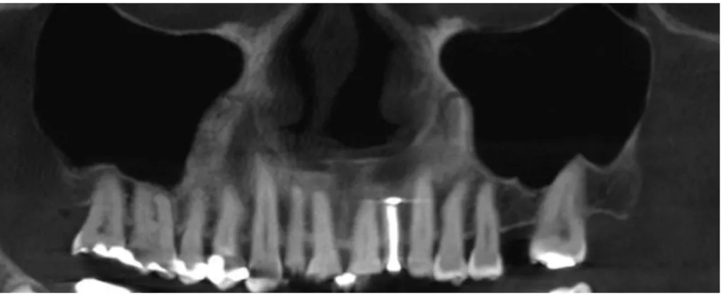
Fig. 1. Cone beam CT panoramic re
construction of the maxilla shows a missing maxillary left first molar as well as missing third molars bilaterally.
The maxillary sinuses are well pneuma
tized bilaterally.
Fig. 2. An orthogonal section of the edentulous area in the region of the left maxillary first molar showing the residual bone in the region, the vicinity of both maxillary sinuses, and the nasal fossa.
discussion
CBCT is interpreted by oral and maxillofacial radiolo
gists for a variety of reasons, including preimplant im
aging, and therefore, the usefulness of this imaging tech
nology has come to be appreciated by the dental profes
sion.11 The immense popularity of CBCT and its wide use are not just restricted to dentistry; ear, nose, and throat (ENT) specialists have found it to be useful in evaluating
the temporal bone and paranasal sinuses due to its spa
tial resolution, which is superior to that of multidetector computed tomography. CBCT machines come in different designs, with variations in detector design and multiple fields of view. The medium and larger fields of view are
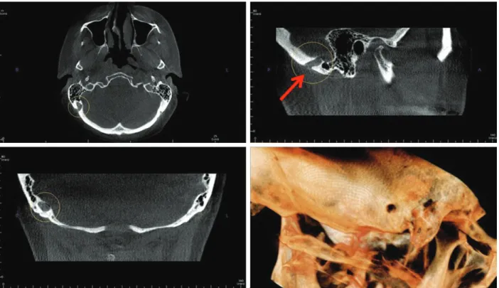
Fig. 3. Cone beam CT multiplanar reconstruction images show an enlarged right mastoid emissary foramen. Axial(top left), sagittal(top right with arrow pointing to the foramen), coronal(bottom left) and threedimensional volume rendering views(bottom right) demonstrate this anatomical variation.
Fig. 4. Threedimensional volume rendering shows the large mas
toid foramen on the right side(arrow).
Fig. 5. Axial cone beam CT image demonstrates the large right mastoid foramen and a smaller left mastoid foramen(arrowhead).
used extensively in a range of diagnostic procedures, such as the assessment of craniofacial growth, orthognathic surgery, and so on.11,12 Small fields of view are used in the preimplant evaluation of selected maxillary or man
dibular edentulous sites. Since the medium and large field of views capture not only the dentoalveolar region, but also extragnathic regions, such as parts of the calvarium, vertebrae, and the base of the skull, it is imperative to rec
ognize normal anatomical structures and their anatomical variants, in contrast to pathological findings, in order to avoid unnecessary additional investigative procedures.
Recognizing emissary veins radiographically is very im
portant because they are clinically significant, especially for oral and maxillofacial surgeons.6 Preoperative knowl
edge of these veins could assist surgeons in preventing potential complications such as profuse bleeding, tinnitus, thrombosis, and infections.1315 Any potential surgery in the mastoid region requires a thorough evaluation of the relevant skeletal structures. A review of the Englishlan
guage literature revealed a dearth of studies on this ana
tomical variant.7 An emissary vein of this magnitude has never before been studied using CBCT imaging.
The temporal bone is one of the most complex bones of the calvarium and may present with anomalies. In the
past, craniofacial anatomical variants were studied using cadavers. With the increase in the use of 3D imaging mo
dalities such as CBCT in dental settings, these anatomi
cal variants can be more readily observed and studied. In order to differentiate healthy anatomical variation from tumors or pathologies, it is necessary to carefully analyze the borders, contents, and surrounding structures.15
Emissary veins are valveless veins, comprising residual embryonic venous paths that pass through the cranium via minute apertures.6,16 The major emissary veins that have been noted include the mastoid emissary vein, posterior condylar vein, petrosquamosal sinus, and occipital emis
sary vein.16 The mastoid canal follows a winding course, traversing the occipitomastoid suture before terminating in the occipital bone.17 The mastoid emissary vein follows a winding route connecting the sigmoid sinus and the pos
terior auricular or occipital vein.6 The emissary veins are valveless veins that permit blood to flow in either direc
tion.6 Cabanac and Brinnel18 reported that emissary veins helped the brain to cool. Emissary veins can measure any
where between 1mm and 4mm.13,14 If they are more than 3.5mm in diameter, then they are considered prominent or enlarged mastoid emissary veins.8,13 In our case, the
Fig. 7. Intracranial side of the right mastoid foramen(arrow). Note
the small mastoid foramen on the left side(arrowhead). Fig. 8. Extracranial side of the right mastoid foramen(arrow). The smaller left mastoid foramen is still noticeable in this section(ar
rowhead).
Fig. 6. Sagittal image of the right mas
toid region shows the mastoid foramen measurements(11.2×7.2mm).
maximum diameter of the vein was over 5mm.
The importance of recognizing emissary veins is increa
singly appreciated due to their clinical significance and because doing so helps surgeons in the pretreatment plan
ning of procedures related to the mastoid region. Syndro
mic conditions such as Apert syndrome frequently show abnormal venous anatomy, which is information that should be conveyed to the operating surgeon.1 With recent ad
vances in neuroradiology for endovascular procedures, enlarged emissary veins may be used to obtain access to the sigmoid sinus.9,10,19
Lang and Samii9 reported that mastoid emissary foram
ina were more prevalent in males than in females. Boyd20 noted that the right foramen was larger than the foramina on the left side. Keskil et al.21 reported similar findings, adding that larger foramina tended to be more prevalent on the right side. The study of Reis et al.5 using anatomi
cal specimens found the average diameter of the mastoid foramen to be 2.15mm. Table 1 provides an overview of the anatomical and radiographic studies included in this review. Hadeishi et al.17 observed in their study that the diameter of the mastoid canal varied from 1mm to 6mm.
Louis et al.3 reported diameters ranging from 3.8mm to 11.8mm. Koesling et al.15 reported that the prevalence of
Table 1. Summary of the anatomical studies included in this review.
Author Sample size Study type Prevalence Mean diameter Largest diameter Side
Kim et al.1 Reis et al.5 Murlimanju et al.7 Boyd20
Keskil et al.21 Hadeishi et al.17
10614 150096
20016
Dry skulls Cadavers Dry skulls Dry skulls Dry skulls Dry skulls
Unknown Unknown 91.7%
34.4%
88.5%
87.5%
1.64mm 2.15mm
- 2 - 16mm
7mm 4mm - 45mm
- 6
Unknown Unknown Unknown UnknownLeft Unknown
Table 2. Summary of the radiological studies included in this review.
Author Imaging
modality Sample size Gender Age Additional findings Side
MarsotDupuch et al.23 CT, MRI 6 M1
F2 6, 25, and
47 years Craniosynostosis and
hydrocephalus Jugular vein Hypoplasia
Koesling et al.15 CT 223 M117
F106 13 days to
88 years Hearing loss Inflammation Unknown
Hadeishi et al.17 CT 161 - - Hemifacial spasm,
trigeminal neuralgia, cerebellar tumor
Bone wax migrated into sigmoid sinus
Pekcevik et al.16 CT 182 M90
F96 Mean age,
52.7 years - Unknown
CT, computed tomography. MRI, magnetic resonance imaging.
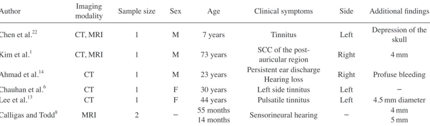
Table 3. Summary of the case studies included in this review.
Author Imaging
modality Sample size Sex Age Clinical symptoms Side Additional findings
Chen et al.22 CT, MRI 1 M 7 years Tinnitus Left Depression of the
skull
Kim et al.1 CT, MRI 1 M 73 years SCC of the post
auricular region Right 4mm
Ahmad et al.14 CT 1 M 23 years Persistent ear discharge
Hearing loss Right Profuse bleeding
Chauhan et al.6 CT 1 F 30 years Left side tinnitus Left -
Lee et al.13 CT 1 F 44 years Pulsatile tinnitus Left 4.5mm diameter
Calligas and Todd8 MRI 2 - 55 months
14 months Sensorineural hearing - 4mm
5mm CT, computed tomography; MRI, magnetic resonance imaging; SCC, squamous cell carcinoma.
a prominent mastoid emissary vein was approximately 6%
in their study, which used highresolution CT scans. Kim et al.1 found the prevalence to be 4.3% based on cadaver dry skull specimens. Hadeishi et al.17 observed that seven out of 161 patient scans reviewed retrospectively revealed a large mastoid foramen and reported that postoperatively, bone wax had migrated into the sigmoid sinus. Chauhan et al.6 reported that an enlarged mastoid vein may lead to tinnitus. A review by Pekcevik et al.16 noted that only five patients showed a large mastoid measuring more than 5 mm, with two on the right side and three on the left side.
Chen et al.22 described a large mastoid emissary vein in a sevenyear old boy in their study that used CT imaging.
A review of the literature showed that some case series and studies described the presence of large, prominent mastoid emissary veins.6 Table 2 provides an overview of the radiographic studies included in this review. Table 3 provides a summary of all case studies included in this re
port. In the present case study, the mastoid emissary vein measured 11.2mm×7.2mm, which was consistent with the measurements from the study of Louis et al.3
A minimal amount of information has been published about enlarged mastoid emissary veins in the literature,1 although these variations in vascular structures are im
portant to recognize, especially during pretreatment plan
ning, in order to avoid potential complications if surgery is planned around the mastoid region.3,7,14 On many occa
sions, tinnitus(pulsatile or not) may be the only symptom of an enlarged mastoid emissary vein.23
Failure to recognize this variant can potentially lead to an incorrect diagnosis or cause iatrogenic lifethreatening bleeding during attempted surgery in the region of the mastoid.7,14,15 The mastoid emissary vein is considered by anatomists to be the remnant of a primitive jugular vein.2 In the large majority of cases, the vein is unilateral and small. It originates on the outer edge of the lateral sinus groove and displays a short intracranial course with an upward and backward direction. It opens on the surface just behind the upper posterior edge of the base of the mastoid process to enter into the occipital or posterior au
ricular vein, eventually forming part of the external jugu
lar vein.
Unilateral, enlarged mastoid foramina are extremely rare anatomical variants that transmit the mastoid emissary vein, and should be noted during the presurgical planning of surgical procedures involving the mastoid region due to their potential complications, such as profuse bleed
ing and thrombosis, which may be fatal if unrecognized.
This vein may be infected from the lateral sinus, where
swelling due to local edema or an abscess may form. This has been referred to as Griesinger’s sign of lateral sinus thrombosis.2 Overall, the early recognition of dilated or enlarged emissary veins within the skull is helpful to sur
geons, especially if they are a potential source of tinnitus, pain in and around the temporomandibular joint, or unex
pected bleeding. All incidental findings must be thorou
ghly investigated. Both sides of the mastoid region must be compared with one another to identify anatomic vari
ations, and reassuring the patient and documenting such entities in the electronic health record are mandatory parts of the treatment process.
Acknowledgements
We would like to thank Dr. Charles Lanzieri of Case University Hospitals for his expert second opinion.
references
1. Kim LK, Ahn CS, Fernandes AE. Mastoid emissary vein:
anatomy and clinical relevance in plastic & reconstructive surgery. J Plast Reconstr Aesthet Surg 2014; 67: 77580.
2. Cheatle A. The mastoid emissary vein and its surgical impor
tance. Proc R Soc Med 1925; 18: 2934.
3. Louis RG Jr, Loukas M, Wartmann CT, Tubbs RS, Apaydin N, Gupta AA, et al. Clinical anatomy of the mastoid and occipital emissary veins in a large series. Surg Radiol Anat 2009; 31:
13944.
4. Freire AR, Rossi AC, de Oliveira VC, Prado FB, Caria PH, Botacin PR. Emissary foramens of the human skull: anatomi
cal characteristics and its relations with clinical neurosurgery.
Int J Morphol 2013; 31: 28792.
5. Reis CV, Deshmukh V, Zabramski JM, Crusius M, Desmukh P, Spetzler RF, et al. Anatomy of the mastoid emissary vein and venous system of the posterior neck region: neurosurgical implications. Neurosurgery 2007; 61(Suppl 2): 193201.
6. Chauhan NS, Sharma YP, Bhagra T, Sud B. Persistence of multiple emissary veins of posterior fossa with unusual origin of left petrosquamosal sinus from mastoid emissary. Surg Ra
diol Anat 2011; 33: 82731.
7. Murlimanju BV, Chettiar GK, Prameela MD, Tonse M, Kumar N, Saralaya VV, et al. Mastoid emissary foramina: an anatom
ical morphological study with discussion on their evolutionary and clinical implications. Anat Cell Biol 2014; 47: 2026.
8. Calligas JP, Todd NW Jr. Hemorrhage from large mastoid em
issary vein: pedicled, rotated, indented, periosteal-galeal flap.
Laryngoscope 2014; 124: 5513.
9. Lang J Jr, Samii A. Retrosigmoidal approach to the posterior cranial fossa. An anatomical study. Acta Neurochir(Wien) 1991; 111: 14753.
10. Pekçevik Y, Pekçevik R. Why should we report posterior fossa emissary veins? Diagn Interv Radiol 2014; 20: 7881.
11. Dawood A, Patel S, Brown J. Cone beam CT in dental prac
tice. Br Dent J 2009; 207: 238.
12. Quereshy FA, Savell TA, Palomo JM. Applications of cone beam computed tomography in the practice of oral and maxil
lofacial surgery. J Oral Maxillofac Surg 2008; 66: 7916.
13. Lee SH, Kim SS, Sung KY, Nam EC. Pulsatile tinnitus caused by a dilated mastoid emissary vein. J Korean Med Sci 2013;
28: 62830.
14. Ahmad R, Ali I, Naikoo GM, Choo NA, Jan F. Giant mastoid emissary vein: source of profuse bleeding during mastoid sur
gery. Indian J Otolaryngol Head Neck Surg 2011; 63(Suppl 1):
1023.
15. Koesling S, Kunkel P, Schul T. Vascular anomalies, sutures and small canals of the temporal bone on axial CT. Eur J Ra
diol 2005; 54: 33543.
16. Pekcevik Y, Sahin H, Pekcevik R. Prevalence of clinically important posterior fossa emissary veins on CT angiography.
J Neurosci Rural Pract 2014; 5: 1358.
17. Hadeishi H, Yasui N, Suzuki A. Mastoid canal and migrated bone wax in the sigmoid sinus: technical report. Neurosurgery 1995; 36: 12204.
18. Cabanac M, Brinnel H. Blood flow in the emissary veins of the human head during hyperthermia. Eur J Appl Physiol Oc
cup Physiol 1985; 54: 1726.
19. Rivet DJ, Goddard JK 3rd, Rich KM, Derdeyn CP. Percutane
ous transvenous embolization of a dural arteriovenous fistula through a mastoid emissary vein. Technical note. J Neurosurg 2006; 105: 6369.
20. Boyd GI. The emissary foramina of the cranium in man and the anthropoids. J Anat 1930; 65: 10821.
21. Keskil S, Gözil R, Çalgüner E. Common surgical pitfalls in the skull. Surg Neurol 2003; 59: 22831.
22. Chen Z, Feng H, Zhu G, Wu N, Lin J. Anomalous intracranial venous drainage associated with basal ganglia calcification.
AJNR Am J Neuroradiol 2007; 28: 224.
23. MarsotDupuch K, GayetDelacroix M, ElmalehBerges M, Bonneville F, Lasjaunias P. The petrosquamosal sinus: CT and MR findings of a rare emisssary vein. AJNR Am J Neuroradi
ol 2001; 22: 118693.