Total hip arthroplasty (THA) has shown good clinical outcomes and high rates of long-term survival in patients with end-stage hip diseases.1) However, it remains difficult to treat a patient complicated with an ankylosed hip joint2) because the preceding lesion results in changes in bony structure, contracture of soft tissue, atrophy of the external rotator, and acetabular subchondral bone defects.3)
Comparison of Outcomes of Total Hip
Arthroplasty between Patients with Ankylosing Spondylitis and Avascular Necrosis
of the Femoral Head
Sun-Ho Lee, MD, Gun-Woo Lee, MD, Young-Jun Seol, MD, Kyung-Soon Park, MD, Taek-Rim Yoon, MD
Department of Orthopedic Surgery, Center for Joint Disease, Chonnam National University Hwasun Hospital, Hwasun, Korea
Background: The objective of this study was to compare clinical and radiological outcomes of total hip arthroplasty (THA) be- tween ankylosing spondylitis (AS) of the hip joint and avascular necrosis (AVN) of the femoral head.
Methods: Thirty patients (30 hips) underwent cementless THA for AS between 2003 and 2012. They were compared to 30 patients (30 hips) who underwent the same procedure for AVN of the femoral head. Each group was matched for age and gender, and both groups had similar preoperative demographic characteristics. All cases were followed for minimum 4 postoperative years. Clinical evaluation was based on operation time, intraoperative blood loss, quantity of postoperative drainage, Harris Hip Score (HHS), and range of motion (ROM). Radiological results were evaluated by acetabular cup anteversion and inclination, femoral stem orienta- tion, pre- and postoperative leg length discrepancy, and postoperative complications.
Results: The operation time was significantly longer in the AS group (120.2 ± 26.2 min) than in the AVN group (79.5 ± 11.1 min).
The volume of postoperative drainage was significantly greater in the AS group (764.5 ± 355.4 mL vs. 510.5 ± 195.6 mL). Preopera- tive HHS was lower in the AS group (55.6 ± 13.8 vs. 59.2 ± 2.8). Similarly, postoperative HHS was significantly lower in the AS group (92.8 ± 2.7 vs. 97.4 ± 2.6). The arc of ROM was improved from 146.5° ± 13.2° preoperatively to 254.7° ± 17.2° postoperative- ly in the AS group and from 182.6° ± 15.5° to 260.4° ± 13.7° in the AVN group. Implant position and postoperative leg length dis- crepancy were not different between the groups. However, three cases of heterotopic ossification was observed in the AS group, whereas only 1 case was found in the AVN group. One deep infection and one aseptic stem loosening were found in the AS group, whereas none was observed in the AVN group.
Conclusions: Cementless THA showed satisfactory clinical and radiological results in both groups, despite the longer operation time, larger blood loss volume, and lower HHS score of the AS group. Our findings suggest that cementless THA is an effective and reliable treatment for both AS and AVN.
Keywords: Ankylosing spondylitis, Total hip arthroplasty, Avascular necrosis
Copyright © 2017 by The Korean Orthopaedic Association
This is an Open Access article distributed under the terms of the Creative Commons Attribution Non-Commercial License (http://creativecommons.org/licenses/by-nc/4.0) which permits unrestricted non-commercial use, distribution, and reproduction in any medium, provided the original work is properly cited.
Clinics in Orthopedic Surgery • pISSN 2005-291X eISSN 2005-4408 Received November 13, 2016; Accepted May 22, 2017
Correspondence to: Taek-Rim Yoon, MD
Department of Orthopedic Surgery, Center for Joint Disease, Chonnam National University Hwasun Hospital, 322 Seoyang-ro, Hwasun 58128, Korea
Tel: +82-61-379-7676, Fax: +82-61-379-7681 E-mail: [email protected]
Ankylosing spondylitis (AS) results in severe limi- tation of motion and functional disability, which affects posture and gait. AS tends to manifest at a younger age and the extent of skeletal involvement can vary widely.
The accompanying pain and impaired gait severely disturb daily life activities. The incidence of hip involvement in AS is between 30% and 50%, and 47% to 90% of patients have it bilaterally. Several reports have focused on the treatment of AS with THA showing excellent outcomes and suggest THA as the most well-established treatment of choice for
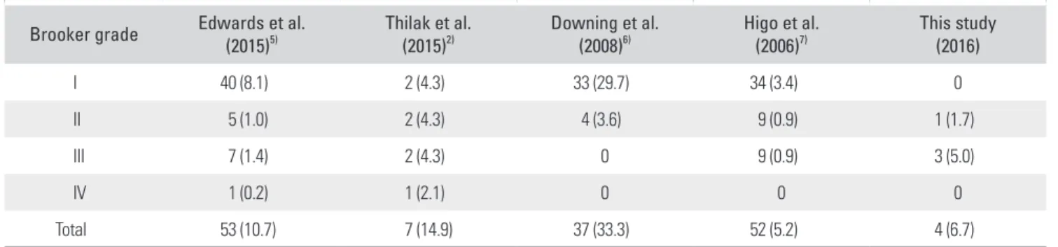
AS with severe hip involvement.2-4) However, the exact lo- cation for the implant is difficult to determine during the operation due to changes in bony structure of the lesion.4) In addition, the balance among the vertebral column, pel- vis, and lower extremities should also be considered when performing THA in AS patients. A number of studies have reported postoperative loosening of the acetabular compo- nent and heterotopic ossification (Table 1).2,5-7) However, few reports have evaluated subsequent surgical outcomes and compared with the results of other diseases involving
Table 1. Comparison of Heterotopic Ossification with Other Studies Brooker grade Edwards et al.
(2015)5) Thilak et al.
(2015)2) Downing et al.
(2008)6) Higo et al.
(2006)7) This study
(2016)
I 40 (8.1) 2 (4.3) 33 (29.7) 34 (3.4) 0
II 5 (1.0) 2 (4.3) 4 (3.6) 9 (0.9) 1 (1.7)
III 7 (1.4) 2 (4.3) 0 9 (0.9) 3 (5.0)
IV 1 (0.2) 1 (2.1) 0 0 0
Total 53 (10.7) 7 (14.9) 37 (33.3) 52 (5.2) 4 (6.7)
Values are presented as frequency (%).
Table 2. Demographic Characteristics of the Patients
AS AVN p-value
Hip (patient) 30 (30) 30 (30) -
Age (yr) 39.6 (22–55) 42.6 (33–58) 0.334
Sex (male/female) 26/4 26/4 -
BMI (kg/m2) 25.7 (19.0–34.1) 24.9 (19.2–32.2) 0.485
Follow-up (mo) 69.0 (50–118) 71.5 (50–110) 0.334
Acetabular cup -
Securefit (Stryker, USA) 13 0
Duraloc (Depuy, USA) 1 0
Pinacle (Depuy, USA) 0 1
Delta PF (Lima Corp., Italy) 16 29
Femoral stem -
Wagner cone (Zimmer, USA) 7 2
Accolade (Stryker, USA) 6 0
CLS (Zimmer, USA) 1 0
M/L taper (Zimmer, USA) 16 28
Values are presented as mean (range).
AS: ankylosing spondylitis, AVN: avascular necrosis, BMI: body mass index.
the hip joint such as osteonecrosis of the femoral head or of primary osteoarthritis (OA).4) Therefore, the objective of this study was to analyze outcomes of cementless THA in patients with AS and also clinically and radiologically compare patients with AVN of the femoral head who un- derwent the same procedure.
METHODS
Thirty patients (30 hips) with AS underwent THA be- tween 2003 and 2012. These patients were followed for >
4 years. Their mean patient age was 39.6 years (range, 22 to 55 years) at the time of the operation. Twenty-six pa- tients were male, and 4 were female. All hips underwent unilateral procedure. The mean follow-up period was 69.0 months (range, 50 to 118 months). The control group was selected from patients with avascular necrosis (AVN) of the femoral head, who underwent the same cementless hip arthroplasty procedure during the same period. The mean age of the patients in the control group (30 patients, 30 hips)
were 42.6 years (range, 33 to 58 years). Twenty-six were male, and 4 were female, and the mean follow-up period was 71.5 months (range, 50 to 110 months) (Table 2).
Surgical Technique
All surgeries were performed by the same surgeon (TRY).
Cementless implants were used as the fixation method for both the acetabular cup and femoral stem. Minimally in- vasive two-incision technique was used in all hips.8) Plac- ing the patient in a lateral decubitus position, we utilized the Watson-Jones anterolateral approach to insert the ace- tabular component and made a posterior incision through the intermuscular interval between the gluteus medius and piriformis for femoral component insertion.
Ceramic-on-ceramic hips were implanted in all pa- tients and data on all acetabular cups and femoral stems are described in Table 2. An adductor tenotomy was per- formed after implantation in all hips except for one in the AVN group. Drains were removed when drain volume de- creased to < 100 mL daily. Tolerable weight bearing ambu-
A B C
Fig. 1. Radiographs of a patient with ankylosing spondylitis who underwent primary cementless total hip arthroplasty. (A) Preoperative radiograph. (B) One-year postoperative radiograph. (C) Three-year postoperative radiograph.
A B C
Fig. 2. Radiographs of a patient with avascular necrosis of the femoral head who underwent primary cementless total hip arthroplasty. (A) Preoperative radiograph. (B) One-year postoperative radiograph. (C) Three-year postoperative radiograph.
lation with an ambulatory assistance device was prescribed the day after surgery.
Evaluation
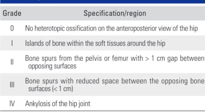
Clinical variables including the mean operation time, drain volume, the arc of range of motion (ROM), pre- and postoperative Harris Hip Score (HHS)9) were compared between the 2 groups. Radiological evaluations were per- formed on the angle of articular cup anteversion1) and in- clination, femoral stem slope, and pre- and postoperative leg length discrepancy (Figs. 1 and 2).10-12) Postoperative complications such as radiological evidence of aseptic or septic loosening, heterotopic ossification, and deep infec- tion were evaluated (Fig. 3).13)
Statistical Analysis
Demographic characteristics, clinical outcomes, radiologi- cal outcomes, and complications were compared between the 2 groups using the independent t-test. Kolmogorov- Smirnov test was used to determine normal distributions.
All data were analyzed using IBM SPSS ver. 20.0 (IBM Co., Armonk, NY, USA). Statistical significance was defined as p-value less than 0.05 (two-tailed).
RESULTS
The operation time was longer in the AS group than in the AVN group (120.2 ± 26.2 min vs. 79.5 ± 11.1 min;
p = 0.007). Postoperative drainage volume was 764.5 ± 355.4 mL and 510.5 ± 195.6 mL in the AS group and AVN group, respectively (p = 0.004).
Preoperative HHS was different between the AS group and AVN group (55.6 ± 13.8 vs. 59.2 ± 2.8; p = 0.001).
Similarly, postoperative HHS at the last follow-up was sig- nificantly lower in the AS group than in the AVN group
(92.8 ± 2.8 in AS vs. 97.4 ± 2.6 in AVN; p < 0.05).
Preoperative arc of ROM was 146.5° ± 13.2° in the AS group, which was improved to 254.7° ± 17.2° postop- eratively. In the AVN group, the value was improved from 182.6° ± 15.5° to 260.4° ± 13.7°.
No differences in the angle of articular cup antever- sion or inclination or femoral stem slope were detected between the groups in the radiological evaluation. Postop- erative leg length discrepancy was 2.3 ± 5.7 mm in the AS group and 3.4 ± 2.5 mm in the AVN group (p = 0.240).
The AS group had 3 cases of heteroscopic ossifica- tion, whereas 1 case was observed in the AVN group. Of the 4 heteroscopic ossification cases, 1 was Brooker clas- sification I, and 3 were Brooker classification III (Table 3).
One deep tissue infection and one femoral stem loosening (Fig. 2) were identified during follow-up in the AS group (Table 4).
DISCUSSION
AS is a chronic rheumatologic disease affecting 1% of the
A B C
Fig. 3. Radiographs of a patient with ankylosing spondylitis who presented with heterotopic ossification (A) Preoperative radiograph. (B) One-year postoperative radiograph. (C) Three-year postoperative radiograph. Heterotopic ossification of Brooker grade III was observed around the right hip joint.
Table 3. Brooker’s Classification of Heterotopic Ossification of the Hip Joint
Grade Specification/region
0 No heterotopic ossification on the anteroposterior view of the hip I Islands of bone within the soft tissues around the hip
II Bone spurs from the pelvis or femur with > 1 cm gap between opposing surfaces
III Bone spurs with reduced space between the opposing bone surfaces (< 1 cm)
IV Ankylosis of the hip joint
population. Hip involvement in AS is between 30% and 50%, and the disease may be bilateral in 47%–90% of pa- tients.14-17) However, most published studies have a small number of cases or a short follow-up.
Recent advances in implant surgery have resulted in improvement of techniques producing better outcomes after cementless THA. Still, THA in patients with AS is far more challenging due to the conditions accompanying osteoarthritic changes or secondary structural abnormali- ties. The lengthy immovable state of the joint induces soft tissue contracture and a distorted anatomical relationship, which produces deceptive anatomical landmarks leading to poor implant placement.14-16) Hence, long-term out- comes of THA are worse in patients with AS.17)
Several studies have reported clinical results after THA in patients with AS. Tang and Chiu11) reported a 63.6% survival rate at 11 years of follow-up of patients who
underwent cementless THA. Kim et al.12) also reported that 3 of 24 ankylosed hips that underwent cementless THA developed osteolysis and 2 developed aseptic loosen- ing. They concluded that fixation of the hip joints in an external position in patients with bilateral hip ankylosis resulted in poor positioning of the implant and poor long- term results.12) In contrast, Ye et al.14) reported good out- comes of THA performed in 15 hip ankylosis cases.
Treatment of AS is theoretically a modifiable risk factor. In this study, we considered AS a cause of anky- losed hips and analyzed the clinical and radiological re- sults after THA. Then, we compared the results with those from AVN of the femoral head cases. After a mean of 69 months (range, 50 to 118 months) of follow-up, only 1 case of femoral stem loosening was found in the AS group, without any cases of acetabular loosening or dislocation.
This can be attributed to implantation of the acetabular cup and femoral stem at proper angles, resulting in a nor- mal angle of articular cup anteversion and inclination and femoral stem slope. However, the operation time and post- operative drain volume increased significantly, suggesting that patients with AS must be closely followed for infec- tion. Postoperative HHS was different between the groups;
however, mean scores were > 90 in both groups.
As other studies have suggested, the AS group had a higher prevalence of heteroscopic ossification. Heterotopic ossification results from differentiation of mesenchymal cells into osteoprogenitor cells, although the precise path- way remains unknown. The natural setting of heterotopic ossification is extraarticular outside the joint capsule.18,19) Superficial or deep infections are a risk factor for hetero- topic ossification.2,20,21) Furthermore, preoperative hip an- kylosis and postoperative infection are major risk factors for heterotopic ossification.6,7,22-24) In the AS group, we only observed one deep infection in a patient who developed grade III heterotopic ossification. More studies are needed with different patient groups and longer follow-up times to verify this finding.
The lower incidence of heterotopic ossification in our patients was likely due to less muscle damage dur- ing the surgery using a minimally invasive technique.
Minimally invasive THA with the modified two-incision technique facilitated proper implantation of the acetabular cup and stem and minimized muscle damage and other postoperative complications, ultimately resulting in excel- lent outcomes in patient with AS.
This study has some limitations. The study was retrospective with the inherent potential for inaccurate medical records and information bias. The relatively small study population could have weakened the power of the Table 4. Comparison of Clinical and Radiological Outcomes between
the Groups
AS AVN p-value
Clinical
HHS* 55.6 ± 13.8 59.2 ± 2.8 0.001
HHS† 92.8 ± 2.8 97.4 ± 2.6 0.012
ROM (°)* 146.5 ± 13.2 182.6 ± 15.5 0.01
ROM (°)† 254.7 ± 17.2 260.4 ± 13.7 0.12 Operation time (min) 120.2 ± 26.2 79.5 ± 11.1 0.007 Total drain output (mL) 764.5 ± 355.4 510.5 ± 195.6 0.004 Radiological
Acetabular anteversion 21.9 ± 6.8 18.9 ± 6.2 0.079 Acetabular inclination 36.6 ± 5.8 37.6 ± 4.3 0.476 Femoral stem tilt 1.1 ± 2.1 0.4 ± 1.2 0.012
LLD (mm)* 3.1 ± 6.3 3.9 ± 5.7 0.357
LLD (mm)† 2.3 ± 5.7 3.4 ± 2.5 0.240
Heterotopic ossification
stage (case) II (1), III (2) III (1) 0.310 Complication
Deep infection 1 0 0.326
Aseptic loosening 1 (stem) 0 0.326
Values are presented as mean ± standard deviation.
AS: ankylosing spondylitis, AVN: avascular necrosis, HHS: Harris Hip Score, ROM: range of motion, LLD: leg length discrepancy.
*Preoperative. †Postoperative.
analysis. In addition, patient selection was not perfectly randomized because it was based on diagnosis and match- ing demographics. However, the strength of this study is that that all operations were performed by the same sur- geon. Moreover, as far as we are aware this is the first study comparing the outcomes of THA between patients with AS and those with AVN of the femoral head. Several re- ports have suggested that THA is the most preferred treat- ment for AS with severe hip involvement. However, there is a lack in comparison studies with other hip diseases containing subsequent surgical outcomes.
Surgeons may have some concerns about the long- term outcome in young patients who undergo THA.
Several reports have shown excellent functional results with long survival rates of the implant in AS.14-16) However, there is a paucity in the literature on systematic compari- son of AS with other inflammatory diseases involving the
hip joint. In our study, significant functional improvement could be achieved after THA in patients with AVN as well as AS.
In conclusions, cementless THA in patients with AS showed satisfactory mid-term results, despite the longer operation time, larger blood loss, and lower HHS than in patients with AVN of the femoral head. Thus, patients with AS must be closely followed with more intense post- operative care than those with AVN. The results of this study suggest that cementless THA is an effective and reli- able treatment in both AS and AVN.
CONFLICT OF INTEREST
No potential conflict of interest relevant to this article was reported.
REFERENCES
1. Joshi AB, Markovic L, Hardinge K, Murphy JC. Conversion of a fused hip to total hip arthroplasty. J Bone Joint Surg Am. 2002;84(8):1335-41.
2. Thilak J, Panakkal JJ, Kim TY, Goodman SM, Lee SS, Salvati EA. Risk factors of heterotopic ossification following total hip arthroplasty in patients with ankylosing spondylitis. J Arthroplasty. 2015;30(12):2304-7.
3. Yu MC, Jo YJ, Kim GI, Jeon YS, Lee YH, Ha JH. Conversion total hip arthroplasty in bony ankylosis of the hip joint. J Korean Hip Soc. 2003;15(1):1-9.
4. Wang W, Huang G, Huang T, Wu R. Bilaterally primary ce- mentless total hip arthroplasty in patients with ankylosing spondylitis. BMC Musculoskelet Disord. 2014;15:344.
5. Edwards DS, Barbur SA, Bull AM, Stranks GJ. Posterior mini-incision total hip arthroplasty controls the extent of post-operative formation of heterotopic ossification. Eur J Orthop Surg Traumatol. 2015;25(6):1051-5.
6. Downing MR, Knox D, Gibson P, Reid DM, Potter A, Ash- croft GP. Impact of trochanteric heterotopic ossification on measurement of femoral bone density following cemented total hip replacement. J Orthop Res. 2008;26(10):1334-9.
7. Higo T, Mawatari M, Shigematsu M, Hotokebuchi T. The incidence of heterotopic ossification after cementless total hip arthroplasty. J Arthroplasty. 2006;21(6):852-6.
8. Yoon TR, Bae BH, Choi MS. A modified two-incision mini- mally invasive total hip arthroplasty: technique and short- term results. Hip Int. 2006;16 Suppl 4:28-34.
9. Harris WH. Traumatic arthritis of the hip after dislocation
and acetabular fractures: treatment by mold arthroplasty: an end-result study using a new method of result evaluation. J Bone Joint Surg Am. 1969;51(4):737-55.
10. Choi IY, Kim YH, Roh WI, Cho WJ. Cementless total hip arthroplasty in bony ankylosed hip. J Korean Orthop Assoc.
2003;38(7):710-5.
11. Tang WM, Chiu KY. Primary total hip arthroplasty in patients with ankylosing spondylitis. J Arthroplasty. 2000;15(1):52-8.
12. Kim YM, Kim HJ, Rhyu KH, Oh HC, Lim CK, Park KW.
Total hip arthroplasty in bony fused hip. J Korean Orthop Assoc. 2000;35(6):873-8.
13. Brooker AF, Bowerman JW, Robinson RA, Riley LH Jr.
Ectopic ossification following total hip replacement: inci- dence and a method of classification. J Bone Joint Surg Am.
1973;55(8):1629-32.
14. Ye C, Liu R, Sun C, et al. Cementless bilateral synchronous total hip arthroplasty in ankylosing spondylitis with hip an- kylosis. Int Orthop. 2014;38(12):2473-6.
15. Bhan S, Eachempati KK, Malhotra R. Primary cementless total hip arthroplasty for bony ankylosis in patients with an- kylosing spondylitis. J Arthroplasty. 2008;23(6):859-66.
16. Gu M, Zhang Z, Kang Y, et al. Roles of sagittal anatomical parameters of the pelvis in primary total hip replacement for patients with ankylosing spondylitis. J Arthroplasty.
2015;30(12):2219-23.
17. Goodman SM, Zhu R, Figgie MP, Huang WT, Mandl LA.
Short-term total hip replacement outcomes in ankylosing spondylitis. J Clin Rheumatol. 2014;20(7):363-8.
18. Weng HK, Wu PK, Chen CF, et al. Total hip arthroplasty for patients who have ankylosing spondylitis: is postoperative irradiation required for prophylaxis of heterotopic ossifica- tion? J Arthroplasty. 2015;30(10):1752-6.
19. Zhu Y, Zhang F, Chen W, Zhang Q, Liu S, Zhang Y. Inci- dence and risk factors for heterotopic ossification after total hip arthroplasty: a meta-analysis. Arch Orthop Trauma Surg. 2015;135(9):1307-14.
20. Spinarelli A, Patella V, Petrera M, Abate A, Pesce V, Patella S. Heterotopic ossification after total hip arthroplasty: our experience. Musculoskelet Surg. 2011;95(1):1-5.
21. Okano K, Aoyagi K, Osaki M, Motokawa S, Matsumoto T.
Bone mineral density is not related to heterotopic ossification
after total hip arthroplasty. Int Orthop. 2012;36(6):1163-6.
22. Kilgus DJ, Namba RS, Gorek JE, Cracchiolo A 3rd, Amstutz HC. Total hip replacement for patients who have ankylosing spondylitis: the importance of the formation of heterotopic bone and of the durability of fixation of cemented compo- nents. J Bone Joint Surg Am. 1990;72(6):834-9.
23. Hug KT, Alton TB, Gee AO. Classifications in brief: Brooker classification of heterotopic ossification after total hip ar- throplasty. Clin Orthop Relat Res. 2015;473(6):2154-7.
24. Rama KR, Vendittoli PA, Ganapathi M, Borgmann R, Roy A, Lavigne M. Heterotopic ossification after surface replace- ment arthroplasty and total hip arthroplasty: a randomized study. J Arthroplasty. 2009;24(2):256-62.