Anatomical versus Non-Anatomical Single Bundle Anterior Cruciate Ligament Reconstruction: A Cadaveric Study of Comparison of Knee Stability
Hong-Chul Lim, MD, Yong-Cheol Yoon, MD, Joon-Ho Wang, MD*, Ji-Hoon Bae, MD
†Department of Orthopaedic Surgery, Korea University Guro Hospital, Korea University College of Medicine, Seoul,
*Department of Orthopaedic Surgery, Samsung Medical Center, Sungkyunkwan University School of Medicine, Seoul,
†Department of Orthopaedic Surgery, Korea University Ansan Hospital, Korea University College of Medicine, Ansan, Korea
Received November 28, 2011; Accepted April 9, 2012 Correspondence to: Ji-Hoon Bae, MD
Department of Orthopaedic Surgery, Korea University Ansan Hospital, Korea University College of Medicine, 123 Jeokgeum-ro, Danwon-gu, Ansan 425-707, Korea
Tel: +82-31-412-5043, Fax: +82-31-487-9502 E-mail: [email protected]
Transtibial single bundle anterior cruciate ligament (ACL) reconstruction with over the top femoral tunnel position generally provides successful clinical results. However,
Background: The purpose of this study was to compare the initial stability of anatomical and non-anatomical single bundle an- terior cruciate ligament (ACL) reconstruction and to determine which would better restore intact knee kinematics. Our hypothesis was that the initial stability of anatomical single bundle ACL reconstruction would be superior to that of non-anatomical single bundle ACL reconstruction.
Methods: Anterior tibial translation (ATT) and internal rotation of the tibia were measured with a computer navigation system in seven pairs of fresh-frozen cadaveric knees under two testing conditions (manual maximum anterior force, and a manual maxi- mum anterior force combined with an internal rotational force). Tests were performed at 0, 30, 60, and 90 degrees of flexion with the ACL intact, the ACL transected, and after reconstruction of one side of a pair with either anatomical or non-anatomical single bundle ACL reconstruction.
Results: Under manual maximal anterior force, both reconstruction techniques showed no significant difference of ATT when compared to ACL intact knee state at 30° of knee flexion (p > 0.05). Under the combined anterior and internal rotatory force, non- anatomical single-bundle ACL reconstruction showed significant difference of ATT compared to those in ACL intact group (p < 0.05).
In contrast, central anatomical single bundle ACL reconstruction showed no significant difference of ATT compared to those in ACL intact group (p > 0.05). Internal rotation of the tibia showed no significant difference in the ACL intact, the ACL transected, non- anatomical reconstructed and anatomical reconstructed knees.
Conclusions: Anatomical single bundle ACL reconstruction restored the initial stability closer to the native ACL under combined anterior and internal rotational forces when compared to non-anatomical ACL single bundle reconstruction.
Keywords: Anatomic, Non-anatomic, Anterior cruciate ligament, Transtibial, Single bundle
some patients still have residual knee instability and do not return to pre-injury activity level.1-3) Long-term clinical results have shown that 11% to 30% of the patients treated with traditional techniques had arthritic changes.4,5) A critical evaluation of ACL reconstruction techniques has revealed that single bundle grafts placed by conventional transtibial drilling cannot provide adequate restraint to translational and rotatory forces.6-8) One of the reasons for these unsatisfactory results may be a non-anatomic verti- cal graft position.7,9-14) With this transtibial technique, the tibia tunnel is in most cases placed near the posterolateral
Copyright © 2012 by The Korean Orthopaedic Association
This is an Open Access article distributed under the terms of the Creative Commons Attribution Non-Commercial License (http://creativecommons.org/licenses/by-nc/3.0) which permits unrestricted non-commercial use, distribution, and reproduction in any medium, provided the original work is properly cited.
Clinics in Orthopedic Surgery • pISSN 2005-291X eISSN 2005-4408
bundle insertion site and the femoral tunnel is placed in a higher position in the intercondylar notch.15,16)
The kinematics of the reconstructed knee can be affected by the location of the tibial and femoral tunnels.
Recommendations for tunnel placement have changed, and recently there has been an emphasis on anatomical graft placements to recreate normal physiologic graft ten- sion and more knee kinematics. Single bundle ACL grafts placed in the center of their anatomic insertions can pro- vide nearly normal knee kinematics comparable to double bundle procedures.6,7,17-19) However, there are incomplete in vitro data on the comparison of an anatomical single- bundle ACL reconstruction with non-anatomic single bundle ACL reconstruction. The purpose of this study was to compare the initial stability of anatomical and non-an- atomical single bundle ACL reconstruction and to deter- mine which would better restore intact knee kinematics.
Our hypothesis was that the initial stability of anatomical single bundle ACL reconstruction would be superior to that of non-anatomical single bundle ACL reconstruction.
To test our hypothesis, a navigation system was used to measure the kinematics of the knee, in particular anterior translation of the tibia and internal rotation of the tibia.
METHODS
Specimen Preparation
This study was approved by our Institutional Review Board. Seven fresh-frozen whole body human cadavers (Korean population; age, 63.4 ± 7.5 years; range, 59 to 83 years; stored at -20°C) from donors who had died of car- diopulmonary diseases were used in this study. Before test- ing, each specimen was thawed to room temperature. The lower extremities and pelvis were inspected; there were no gross abnormalities or deformities or ligamentous laxity or contracture. Both anteroposterior and lateral radiographs of the knee were obtained; if there were radiographic evidence of malalignment, deformity, or osteoarthritis, those specimens were excluded. Left and right limbs of each specimen were randomly assigned to 2 groups; non- anatomical single bundle ACL group (group A, n = 7) and anatomical single bundle ACL reconstruction group (group B, n = 7).
Testing Protocol
An imageless navigation system (OrthoPilot ACL ver. 2.1, B. Braun-Aesculap, Tuttlingen, Germany) was used to evaluate the kinematics of the knee, in particular anterior translation of the tibia and internal rotation of the tibia (Fig.
1).18-20) To track the position of the femur and tibia and to
digitize the knee anatomy, two rigid bodies with infra-red emitters were fixed to the proximal femur and distal tibia.
To measure the motion of femur and tibia, extra-articular landmarks (including the tibia tuberosity, the anterior edge of the tibia, and the medial and lateral points of the tibia plateau) were registered. The intra-articular landmarks were not registered because we used the navigation system only to collect the motion measurements with 6 degrees of freedom per bone and to provide real time calculation of translation and rotation at all degrees of knee flexion while the surgeon manipulated the knee joint.
In the cadaveric experimental set-up, the assessment for stability consisted of the following steps: 1) initial set- up of the femoral and tibial rigid bodies, 2) anatomical registration of intact knee, 3) kinematic tests on the intact knee, 4) ACL resection, 5) kinematic tests on the ACL- deficient knee, 6) ACL reconstruction according to the chosen technique, 7) kinematic tests on the reconstructed knee.
Tests were performed sequentially with 2 loading conditions. 1) Manual maximum anterior force, 2) com- bined manual maximum anterior and internal rotation of the tibia. The amount of anterior translation of tibia, inter- nal rotation of tibia were assessed at 0°, 30°, 60°, and 90° of knee flexion in each group. Values of anterior translation of the tibia were recorded by the navigation system and expressed in millimeters; values of internal rotation of the
Fig. 1. Navigation software system (OrthoPilot ACL ver. 2.1, B. Braun- Aesculap) allowing the surgeon to measure the anterior translation of the tibia in millimeters and internal and external rotation of the tibia in degrees. This picture showed 16 mm of anterior translaton of the tibia, 23° of internal rotation and 12° of external rotation of the tibia at 30° of knee flexion under manual maximal anterior force in ACL deficient knee specimen. ACL: anterior cruciate ligament.
tibia were expressed in degrees. The accuracy of navigation system is estimated to be 0 to 1 mm or 0° to 1° if the land- marks are registered correctly21) All of them were assessed by the same experienced surgeon who performed all sur- gical procedures to minimize inter-operator variability.
Surgical Procedure
Non-anatomical (tibia posterolateral to femoral high an- teromedial tunnel) reconstruction technique
All procedures were performed using medial parapatellar open approach. After 10 cm midline skin incision, medial parapatellar capsulotomy was made. We first investigated intra-articular pathology, and proceeded further if there was no significant pathology. The intact ACL was cut and knee kinematics were measured using navigation system.
After completion of testing for ACL deficient knee, the gracillis and semitendinosus tendons were harvested from each knee and prepared for a quadruple graft.
For the trans-tibial technique, a commercial tibial tunnel ACL guide (Acufex Director; Smith & Nephew Ar- throscopy, Andover, MA, USA) was set at 45° to prepare 7 to 9 mm tunnel determined by the size of quadrupled hamstring grafts. The intra-articular exit point of the guide pin was directed 5 mm lateral to the medial tibial spine and inner margin of the anterior horn of the lateral meniscus.22) The external starting point was placed at the anterior border of the medial collateral ligament (MCL) insertional fibers in all cases to allow for oblique orienta- tion of the guide of approximately 45° from the horizontal plane. The guide pin was placed and over-reamed to a corresponding graft size. After completion of the tibial tunnels, a commercially available 6-mm offset femoral tunnel aimer (Smith & Nephew Arthroscopy) was passed through the tibia tunnel and placed in the over-the-top position on the posterior cortex of the femur (10:30 o’clock on right knee, 1:30 o’clock on left knee). A beath pin was retrograded through the tibial tunnel to the selected point.
The femoral tunnel was then drilled to a depth of 30 mm.
The graft was introduced distal to proximal. After we confirmed that a minimum 25 mm length of the graft was placed in the femoral tunnel, the folded end of the ham- string graft was first secured at the lateral femoral cortex with the bioabsorbable interference screw (BioRCI, Smith
& Nephew Arthroscopy) 1 mm larger in diameter than the graft size and 25 mm in length. The graft was tensioned before fixation with 20 lb of force applied equally to all strands by use of the spring gauge force applicator while the knee was slowly cycled between 0° and 90° 10 times.
While 20 lb of graft tension was being maintained, fixa- tion on the tibial side was performed at 10° of knee flexion
with a bioabsorbable interference screw (BioRCI, Smith &
Nephew Arthroscopy) 1 mm larger in diameter than the graft size and 25 mm in length. The distal sutures in each of the grafts were tensioned manually. After completion of reconstruction, wound was closed layer by layer.
Anatomical reconstruction technique
Medial parapatellar approach was used to perform cen- tral anatomical single bundle ACL reconstruction. After completion of testing for ACL deficient knee and harvest- ing hamstring graft, the tibial and femoral footprints of the ACL were outlined with an indelible marker. The femoral tunnel center was placed approximately 25% from the pos- terior femoral cortex and 25% from the Blumensaat line.
The tibial tunnel center was placed in the center of the tibial insertion based on radiographic imaging of the ACL insertion; 45% from the medial tibial cortex and 45% from the anterior tibial cortex.
A commercial tibial tunnel ACL guide (Acufex Di- rector; Smith & Nephew Arthroscopy) with a 45° angle was directed to the center of tibial insertion. The external starting point was placed at the anterior border of the MCL insertional fibers in all cases to allow for oblique orientation. For the anatomical femoral tunnel, indepen- dent drilling with low anteromedial portal technique was used.6,23) After creating the tibial tunnel and performing soft tissue clearance of the notch, the knee was flexed to 120° and the guide pin was placed into the starting point previously marked as the center of the footprint of na- tive ACL bundle. The guide pin was drilled out the lateral femoral cortex and a cannulated acorn reamer was used to produce a femoral tunnel 30 mm in length. Graft passage, graft tension, and graft fixation technique were identical with non-anatomical single bundle ACL reconstruction.
Statistical Analysis
Statistical analysis was performed using SAS ver. 9.1 (SAS Institute Inc., Cary, NC, USA). Due to small sample sizes, a Wilcoxon signed ranks test was used to compare the kinematic data of the ACL intact knee with ACL- deficient, and to compare two different single-bundle ACL reconstructions. The normal left and right knees in each specimen were compared and no statistically significant differences were found. Similarly, the ACL deficient left and right knees were compared and no statistically signifi- cant differences were found. Therefore, normal knees were pooled and ACL deficient knees were pooled for compari- son between normal and ACL deficient knees. To compare two different reconstructions with normal state, kinematic data of the normal knees were randomly selected. Null
hypotheses of no difference were rejected if p-values were less than 0.05.
RESULTS
Normal Knees versus ACL Deficient Knees
Under manual maximal anterior force, after the ACL was sectioned, the anterior tibial translation (ATT) increased significantly at 30° of knee flexion (p < 0.05) (Table 1). Un- der the combined manual maximal anterior and internal rotatory force, the values increased significantly after sec- tioning of the ACL at 30° of knee flexion (p < 0.05) (Table 1). There was no significant change of the internal rotation of the tibia after sectioning of the ACL (Table 2).
Two Different Single Bundle Reconstructed Knees versus Normal Knees
Under manual maximal anterior force, both reconstruc- tion techniques showed no significant difference of ATT when compared to ACL intact knee state at 30° of knee flexion (p > 0.05) (Table 1). Under the combined anterior and internal rotatory force, non-anatomical single-bundle ACL reconstruction showed significant difference of ATT compared to those in ACL intact group (p < 0.05) (Table 1). In contrast, anatomical single bundle ACL reconstruc- tion showed no significant difference of ATT compared to those in ACL intact group at 30° of knee flexion (p >
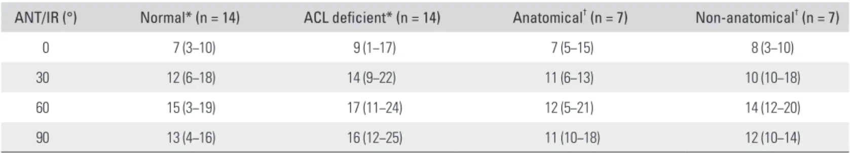
0.05) (Table 1). Internal rotation of the tibia showed no significant difference in the normal, non-anatomical re- constructed and anatomical reconstructed knees (Table 2).
Table 2. Internal Rotation of the Tibia (°) at Different Flexion Angles of the Knee
ANT/IR (°) Normal* (n = 14) ACL deficient* (n = 14) Anatomical† (n = 7) Non-anatomical† (n = 7)
0 7 (3−10) 9 (1−17) 7 (5−15) 8 (3−10)
30 12 (6−18) 14 (9−22) 11 (6−13) 10 (10−18)
60 15 (3−19) 17 (11−24) 12 (5−21) 14 (12−20)
90 13 (4−16) 16 (12−25) 11 (10−18) 12 (10−14)
Values are presented as internal rotation of the tibia (°) and median (range).
ANT/IR: combined manual maximal anterior and internal rotational force, ACL: anterior cruciate ligament.
*No signicant differences between normal and ACL deficient knees at each flexion angle. †No signicant differences between anatomical and non-anatomical reconstructed knees at each flexion angle.
Table 1. Anterior Tibial Translation (mm) at Different Flexion Angles of the Knee
Normal (n = 14) ACL deficient (n = 14) Anatomical (n = 7) Non-anatomical (n = 7) ANT (°)
0 2 (1−8) 5 (1−7) 3 (2−4) 4 (2−5)
30 6 (2−10) 12 (7−17)* 5 (1−8) 7 (5−11)
60 5 (1−9) 7 (3−11) 3 (1−10) 5 (3−9)
90 4 (1−6) 7 (3−8) 3 (1−9) 4 (2−8)
ANT/IR (°)
0 3 (2−9) 5 (2−8) 5 (2−6) 4 (2−6)
30 7 (2−13) 15 (9−19)* 6 (5−11) 10 (6−15)*,†
60 6 (1−9) 8 (6−13) 7 (2−9) 8 (4−9)
90 5 (2−7) 7 (4−9) 4 (3−9) 6 (4−11)
Values are presented as anterior tibial translation (mm) and median (range).
ACL: anterior cruciate ligament, ANT: manual maximal anterior force, ANT/IR: combined manual maximal anterior and internal rotational force.
*p < 0.05 compared with normal knees at same flexion angle. †p < 0.05 compared with anatomical reconstructed group at same flexion angle.
DISCUSSION
The present study demonstrated that the anatomic single bundle ACL reconstruction provides better initial stabil- ity when compared to the non-anatomic single bundle ACL reconstruction under the combined anterior and internal rotatory forces. No difference in knee stability was observed between the intact ACL and after anatomical single bundle ACL reconstruction. Several studies have extensively examined tunnel position in ACL reconstruc- tion and found that inappropriate graft placement had significant adverse effect on graft incorporation and knee function.24-28) Theoretically, anatomically located ACL can reproduce more closely normal knee kinematics.
Recent studies comparing an anatomical single bun- dle reconstruction with a double bundle reconstruction support the finding that the double bundle reconstruction may not be a better method to restore knee stability. Ho et al.17) compared the kinematics of a central anatomic single bundle ACL reconstruction with a double bundle ACL reconstruction. They found that anatomical single bundle and double bundle ACL reconstruction are equally effec- tive in restoring normal anterior translation to the knee under both anterior and rotational loads. Ferretti et al.19) conducted a comprehensive kinematic study using com- puter navigation. The addition of the posterolateral bundle to the anteromedial bundle did not significantly reduce internal and external rotation of the tibia. They concluded that the effective role of the anatomical double bundle procedure in better restoring knee kinematics and allow- ing better clinical outcomes is questionable. With this new attention, we focused our study on an anatomical single bundle ACL reconstruction.
The present study supports recent studies compar- ing anatomical single bundle ACL reconstruction with non-anatomical single bundle ACL reconstruction. Steiner et al.6) compared the knee laxity between a central anatom- ical and conventional non-anatomical single bundle ACL reconstruction. They concluded that a central anatomi- cal single bundle ACL reconstruction using independent drilling technique is superior to restore normal anterior and rotational knee laxity compared with non-anatomical single bundle ACL reconstruction using conventional transtibial drilling technique. Kato et al.8) investigated the effect of tunnel position for anatomic single bundle ACL reconstruction on knee biomechanics in a porcine model.
They found that the anatomic mid-mid (central) single bundle ACL reconstruction provided the better stabil- ity when compared to the non-anatomic single bundle ACL reconstruction (tibia posterolateral to femoral high
anteromedial tunnel) and more closely restored normal knee kinematics. In our study, under an anterior force, a combined anterior force plus internal rotation torque, cen- tral anatomic single bundle ACL reconstruction restored normal ATT at all knee flexion testing angles while non- anatomical single-bundle ACL reconstruction reduced ATT significantly when compared to the ACL deficient knee but not to ACL intact knee state.
ACL deficient knees result in increased internal tibial rotation relative to uninjured knees.6,11,17) In con- trast to these studies, the results of present study shows that cutting the ACL resulted in no significant increase in internal rotation of the tibia under manual maximal com- bined anterior + internal rotatory force. Two recent studies support the results of the current study. Diermann et al.29) investigated whether ACL deficiency leads to increased in- ternal rotation under a simulated pivot shift test in human cadaveric knees. They found that ACL deficiency does not increase the internal tibial rotation under a simulated piv- ot shift test. Monaco et al.30) evaluated the role of the ACL and its secondary restraint in controlling knee stability using a navigation system. The primary kinematic effect of an ACL injury is an increase in anterior tibial translation, but there is no significant change in internal or external rotation. It is difficult to compare our results with previous studies due to large variations (loading conditions, mea- surement methods, knee flexion angles) between examin- ers during testing the ACL deficient knees. The amount of internal tibial rotation elicited depends on the amount of the rotational force during the test. Thus it is difficult to obtain reproducible results with the coupled motions in cadaver knees. However, it is our belief that the increase in ATT is the primary abnormality in ACL deficient knees.
Cutting the ACL alone may produce a small increase (sig- nificant or not) in internal tibial rotation, but the increases are so small that they are clinically not relevant.
Rotational instability after ACL injury can be ex- plained by the combined motions of ATT and internal rotation of the tibia with shifting the axis medially. There- fore, it is very hard to quantify the objective amount of rotational instability following ACL reconstructions with different techniques clinically. In the present study, the internal rotation of tibia is not significantly changed by the integrity of the ACL. Instead, the addition of internal rota- tory force to the anterior force accentuated the amount of ATT in ACL deficient knees. In addition, significant differ- ence of ATT under the anterior and internal rotatory forc- es was detected between anatomical and non-anatomical single bundle ACL reconstruction groups. These findings are very important in clinical practice. When we com-
pare the rotational stability between anatomical and non- anatomical groups or single bundle or double bundle ACL reconstruction group, ATT under the combined anterior and internal rotatory forces rather than internal rotation of the tibia should be assessed.
Several limitations of the present study should be addressed. First, the age of human cadavers did not rep- resent the typical age for population that sustains ACL rupture. Inferior bone quality, low viscoelastic character of ligament and degenerative status of the meniscus may affect the results of this study. However, the number of young human donors is rare. Even though the cadaveric biomechanical model was not representative of in vivo clinical situation, we could obtain the comparative data between the intact, ACL deficient knee and ACL recon- structed knee using the same specimen. Second, it is a cadaveric model of time zero biomechanical stability and cannot simulate the dynamic contributions of the muscu- lature even though we used whole body human cadavers.
Third, the values of the ATT and rotation may be different in vivo setting. In addition, external loads were applied manually to measure the kinematic changes, so forces may not be consistent. However, all measurements were recorded under a manual maximum force applied by the same experienced surgeon, who made every effort to apply a similar loading to the knee. Fourth, we did not perform a true pivot shift test, which is a dynamic motion involv- ing knee flexion combined with an internal rotation and valgus torque. The pivot shift test is a complex multiplanar
maneuver. As such, quantitative kinematic analysis of the pivot shift examination is far more challenging to describe and track compared with uniplanar stress testing. Finally, the relatively limited number of human cadavers makes it difficult to draw major conclusions. However, it was diffi- cult to obtain enough human cadavers in our country. Our specimen number was similar with other biomechanical studies using human cadavers.
In conclusions, the present study demonstrated that both techniques were effective in controlling anterior translation, but anatomical reconstruction restored the stability closer to the native ACL under combined anterior and internal rotational forces. Therefore, we recommend that the anatomic single-bundle ACL reconstruction should be considered when single bundle ACL reconstruc- tion is performed.
CONFLICT OF INTEREST
No potential conflict of interest relevant to this article was reported.
ACKNOWLEDGEMENTS
We thank Dr. Soon-Sun Kwon for statistical analysis, Mr.
Young-Jin Kim and Mr. Min-Su Kim for their technical assistance. We also appreciate B. Braun Korea for provid- ing instruments and navigation system for this study. The authors did not receive any financial support.
REFERENCES
1. Kocher MS, Steadman JR, Briggs KK, Sterett WI, Hawkins RJ. Relationships between objective assessment of ligament stability and subjective assessment of symptoms and func- tion after anterior cruciate ligament reconstruction. Am J Sports Med. 2004;32(3):629-34.
2. Biau DJ, Tournoux C, Katsahian S, Schranz P, Nizard R.
ACL reconstruction: a meta-analysis of functional scores.
Clin Orthop Relat Res. 2007;458:180-7.
3. Liden M, Ejerhed L, Sernert N, Laxdal G, Kartus J. Patellar tendon or semitendinosus tendon autografts for anterior cruciate ligament reconstruction: a prospective, randomized study with a 7-year follow-up. Am J Sports Med. 2007;35(5):
740-8.
4. Aglietti P, Buzzi R, D'Andria S, Zaccherotti G. Long-term study of anterior cruciate ligament reconstruction for chronic instability using the central one-third patellar ten- don and a lateral extraarticular tenodesis. Am J Sports Med.
1992;20(1):38-45.
5. Boonriong T, Kietsiriroje N. Arthroscopically assisted ante- rior cruciate ligament reconstruction: comparison of bone- patellar tendon-bone versus hamstring tendon autograft. J Med Assoc Thai. 2004;87(9):1100-7.
6. Steiner ME, Battaglia TC, Heming JF, Rand JD, Festa A, Baria M. Independent drilling outperforms conventional transtibial drilling in anterior cruciate ligament reconstruc- tion. Am J Sports Med. 2009;37(10):1912-9.
7. Howell SM, Gittins ME, Gottlieb JE, Traina SM, Zoellner TM. The relationship between the angle of the tibial tunnel in the coronal plane and loss of flexion and anterior laxity after anterior cruciate ligament reconstruction. Am J Sports Med. 2001;29(5):567-74.
8. Kato Y, Ingham SJ, Kramer S, Smolinski P, Saito A, Fu FH.
Effect of tunnel position for anatomic single-bundle ACL reconstruction on knee biomechanics in a porcine model.
Knee Surg Sports Traumatol Arthrosc. 2010;18(1):2-10.
9. Arnold MP, Kooloos J, van Kampen A. Single-incision technique misses the anatomical femoral anterior cruciate ligament insertion: a cadaver study. Knee Surg Sports Trau- matol Arthrosc. 2001;9(4):194-9.
10. Loh JC, Fukuda Y, Tsuda E, Steadman RJ, Fu FH, Woo SL.
Knee stability and graft function following anterior cruciate ligament reconstruction: comparison between 11 o'clock and 10 o'clock femoral tunnel placement. 2002 Richard O'Connor Award paper. Arthroscopy. 2003;19(3):297-304.
11. Georgoulis AD, Papadonikolakis A, Papageorgiou CD, Mitsou A, Stergiou N. Three-dimensional tibiofemoral kinematics of the anterior cruciate ligament-deficient and reconstructed knee during walking. Am J Sports Med.
2003;31(1):75-9.
12. Scopp JM, Jasper LE, Belkoff SM, Moorman CT 3rd. The effect of oblique femoral tunnel placement on rotational constraint of the knee reconstructed using patellar tendon autografts. Arthroscopy. 2004;20(3):294-9.
13. Steiner ME, Murray MM, Rodeo SA. Strategies to improve anterior cruciate ligament healing and graft placement. Am J Sports Med. 2008;36(1):176-89.
14. Tashman S, Collon D, Anderson K, Kolowich P, Anderst W. Abnormal rotational knee motion during running after anterior cruciate ligament reconstruction. Am J Sports Med.
2004;32(4):975-83.
15. Forsythe B, Kopf S, Wong AK, et al. The location of femoral and tibial tunnels in anatomic double-bundle anterior cruci- ate ligament reconstruction analyzed by three-dimensional computed tomography models. J Bone Joint Surg Am.
2010;92(6):1418-26.
16. Kopf S, Forsythe B, Wong AK, et al. Nonanatomic tun- nel position in traditional transtibial single-bundle ante- rior cruciate ligament reconstruction evaluated by three- dimensional computed tomography. J Bone Joint Surg Am.
2010;92(6):1427-31.
17. Ho JY, Gardiner A, Shah V, Steiner ME. Equal kinematics between central anatomic single-bundle and double-bundle anterior cruciate ligament reconstructions. Arthroscopy.
2009;25(5):464-72.
18. Ferretti A, Monaco E, Labianca L, Conteduca F, De Carli A.
Double-bundle anterior cruciate ligament reconstruction: a computer-assisted orthopaedic surgery study. Am J Sports
Med. 2008;36(4):760-6.
19. Ferretti A, Monaco E, Labianca L, De Carli A, Maestri B, Conteduca F. Double-bundle anterior cruciate ligament re- construction: a comprehensive kinematic study using navi- gation. Am J Sports Med. 2009;37(8):1548-53.
20. Park JK, Song EK, Seon JK. Comparison of intraoperative stability in ACL reconstruction based on femoral tunnel po- sitions. Orthopedics. 2010;33(10):94-7.
21. Koh J, Koo SS, Leonard J, Kodali P. Anterior cruciate liga- ment (ACL) tunnel placement: a radiographic comparison between navigated versus manual ACL reconstruction. Or- thopedics. 2006;29(10 Suppl):S122-4.
22. Rue JP, Ghodadra N, Lewis PB, Bach BR Jr. Femoral and tibial tunnel position using a transtibial drilled anterior cruciate ligament reconstruction technique. J Knee Surg.
2008;21(3):246-9.
23. Lubowitz JH. Anteromedial portal technique for the ante- rior cruciate ligament femoral socket: pitfalls and solutions.
Arthroscopy. 2009;25(1):95-101.
24. Morgan CD, Kalman VR, Grawl DM. Definitive landmarks for reproducible tibial tunnel placement in anterior cruciate ligament reconstruction. Arthroscopy. 1995;11(3):275-88.
25. Friedman RL, Feagin JA Jr. Topographical anatomy of the intercondylar roof: a pilot study. Clin Orthop Relat Res.
1994;(306):163-70.
26. Yaru NC, Daniel DM, Penner D. The effect of tibial attach- ment site on graft impingement in an anterior cruciate liga- ment reconstruction. Am J Sports Med. 1992;20(2):217-20.
27. Topliss C, Webb J. An audit of tunnel position in anterior cruciate ligament reconstruction. Knee. 2001;8(1):59-63.
28. Ekdahl M, Nozaki M, Ferretti M, Tsai A, Smolinski P, Fu FH. The effect of tunnel placement on bone-tendon healing in anterior cruciate ligament reconstruction in a goat model.
Am J Sports Med. 2009;37(8):1522-30.
29. Diermann N, Schumacher T, Schanz S, Raschke MJ, Peters- en W, Zantop T. Rotational instability of the knee: internal tibial rotation under a simulated pivot shift test. Arch Or- thop Trauma Surg. 2009;129(3):353-8.
30. Monaco E, Maestri B, Labianca L, et al. Navigated knee ki- nematics after tear of the ACL and its secondary restraints:
preliminary results. Orthopedics. 2010;33(10):87-93.