Effects of Lumbar Stabilization Using Pressure Biofeedback Unit During Hip Abduction in Side-Lying in Patients With Low Back Pain
Young-taek Seo, MSc, PT, Seung-chul Chon, PhD, PT
Dept. of Physical Therapy, College of Medical Science, Konyang UniversityAbstract
Background: Lumbar stabilization (LS) improve the thickness of the quadratus lumborum (QL) muscle and muscle activity of the gluteus medius (GM) muscle during hip abduction in a side-lying position in patients with low back pain (LBP).
Objects: The purpose of this study was to assess the effects of LS on muscle thickness of QL and muscle activity of GM during hip abduction in side-lying in patients with LBP.
Methods: The study included 32 patients with LBP, who were randomly divided into the control group and experimental group, each with 16 patients. All subjects performed 35° preferred hip abduction (control group) and 35° hip abduction with LS (experimental group) during side-lying. An ultrasonography and a surface electromyography were used to measure the thickness of the QL muscle, and the muscle activities of the GM muscle respectively. Independent t-test was used to compare the muscle thickness of the QL and the muscle activity of the GM muscle, respectively.
Results: Anterio-posterior diameter in the muscle thickness of QL muscle was decreased significantly in hip abduction with LS more than in preferred hip abduction (p<.001), but medio-lateral diameter in the muscle thickness of QL muscle was not significantly different between in preferred hip abduction and in hip abduction with LS (p=.06). The muscle activity of GM was increased significantly in hip abduction with LS more than in preferred hip abduction (p<.001).
Conclusion: These findings suggest that hip abduction with LS could be recommended as a hip abduction for LS and a prevention unwanted compensatory pelvic lateral tilting movement.
Key Words: Gluteus medius; Hip abduction; Quadratus lumborum; Ultrasonography.
Introduction
One of the causes of the low back pain (LBP) is an instability of the lumbar segment (Panjabi, 2003).
This instability can cause disequilibrium in the move- ment of lumbar system (Comerford and Mottram, 2001). The disequilibrium with decreasing joint mobi- lity of the lower extremity in LBP could be observed during the activities of sitting, standing and walking.
And, the improper control of joint mobility in the spine and lower extremities, could cause the pain of the lumbo-pelvic-hip complex (Hoffman et al, 2011).
The dysfunctional movement of the gluteus medius (GM) muscle is related to several musculoskeletal
disorders of the lumbo-pelvic-hip complex (O’Sullivan et al, 2010). According to previous research, the GM muscle in patients with chronic LBP was weak and rapidly fatigued (Arab and Nourbakhsh, 2010). For example, the height difference in hip joint and pain around the hip joint in patients with LBP is caused by weakness of the GM muscle (Sahrmann, 2002).
Additionally, weakness of the GM muscle can cause
the lateral bending of the trunk during one leg stand-
ing (Neumann, 2010). Thus, rehabilitation management
of the muscles around the hip joint should be consid-
ered a strategic approach in lumbar stabilization
(Nelson-Wong and Callaghan, 2010). Specifically, im-
proving the function of the GM muscle will improve
Corresponding author: Seung-chul Chon [email protected]
Parameters Control group (n
1=16) Experimental group (n
2=16) t p
Age (year) 28.9±4.3
a30.6±10.4 .622 .539
Height (㎝) 168.3±7.2 163.3±7.2 -1.961 .059
Weight (㎏) 64.7±15.7 58.1±10.3 -1.410 .169
VAS
b2.5±.5 2.6±.6 .307 .761
ODI
c10.4±1.5 10.3±1.1 -.399 .693
a
mean±standard deviation,
bvisual analogue scale,
cOswestry disability index.
Table 1. Demographic and clinical characteristics of the patients (N=32) neuromuscular coordination of the lumbo-pelvic-hip
complex and prevent LBP (Fredericson et al, 2000).
Janda et al (1996) report that compensatory move- ments such as hip flexion, hip external rotation and pelvic lateral tilting should not occur until the hip joint reaches 40° of abduction. When the GM muscle is weakened, a synergist such as the quadratus lumbo- rum (QL) muscle can be used to compensate and pel- vic lateral tilting can occur (Comerford and Mottram, 2001). Similarly, the muscle activity of the QL muscle is increased due to excessive use, it may cause lumbar lateral flexion by pelvic elevation, which results in in- stability or impairment of the lumbo-pelvic-hip com- plex system (Sahrmann, 2002).
Most of the studies related to core stability meas- urements have been conducted with the subjects in a supine position. In previous studies, hip abduction measurements in a side-lying position using pressure biofeedback unit (PBU) have been finitely presented in patients with LBP. Therefore, research related to the hip abduction mechanisms underpinning the ef- fects of core stability to the selective recruitment of the GM muscle associated excluding the compensa- tory movement of the QL muscle in a side-lying po- sition is much needed.
This study was undertaken to determine the addi- tive effect of core stability and hip abduction in a side-lying position for LBP patients. To quantita- tively investigate the mechanism of core stability on muscle thickness and associated muscle activity, our study has been undertaken for patients with LBP using the real-time ultrasound (US) imaging techni- que and electromyography (EMG) technique. Our ba-
sic hypothesis was that the decrease in size of the QL muscle and increase in amplitude in the GM mus- cle would significantly improve in the experimental group (which performed both the hip abduction and core stability exercises) than the control group (which performed the hip abduction alone).
Methods
Subjects
A convenience sample of thirty-two patients with LBP was recruited from local private center (Daejeon, South Korea). All the procedures were explained to the subjects, and each subject signed an informed consent form. General characteristics of the subjects are pre- sented (Table 1). The inclusion criteria were as follows:
(1) persistent LBP for at least 3 months (Airaksinen et al, 2006), (2) average pain intensity over the last 2 weeks
<3 point on 10 point of Visual Analogue Scale, (3) 10~
25 point on 50 point of Oswestry Disability Index, (4)
failure of the abdominal draw in maneuver (ADIM) for-
mal test (considered “failure” when the participant was
unable to reduce and maintain a 5~10 ㎜Hg difference
in the PBU during 10 seconds in the side-lying ADIM
formal test) (von Garnier et al, 2009). The exclusion
criteria were as follows: (1) below good grade of GM
muscle on manual muscle testing, (2) diagnosis of other
neurologic disorders that may affect this study, (3) se-
vere cardiovascular diseases, (4) osteoporosis, structural
deformity, systemic inflammatory disease, and nerve
root compression that could affect the experimental
tests (Powers et al, 2008).
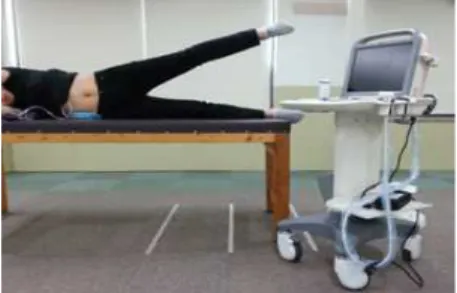
Figure 1. Preferred hip abduction. Figure 2. Hip abduction with lumbar stabilization.
Procedure
Each subject was positioned in a side-lying position with their non-dominant knee joints slightly flexed. All subjects were right leg dominant. They were positioned straight on a manual table. The lumbar spine was in a neutral position. Two positions were assessed: (1) pre- ferred hip abduction (PHA) (Figure 1) and hip abduc- tion with lumbar stabilization (HALS) (Figure 2). The PHA position involves a 35° abduction of the dominant hip while side-lying. The HALS position is an ADIM using a PBU with a 35° abduction of the dominant hip while side-lying (Cynn et al, 2006). The EMG activity and US thickness were measured in the GM and QL muscles, respectively, while performing the hip abduc- tion of the dominant lower extremity while side-lying.
The non-dominant lower extremity could be slightly flexed at the knee joints. The subject was asked to perform a hip abduction while side-lying with the dom- inant lower extremity in the preferred condition, with and without the PBU. A digital inclinometer (Dualer IQ Inclinometer, J-Tech Medical, Heber city, USA) was used to determine the 35° angle for hip abduction. To control the individual variability in hip abduction range, a target bar was placed at the level of 35° of hip ab- duction (Cynn et al, 2006). In the lumbar stabilization condition, the PBU was placed between the therapeutic table, which has a firm mattress, and the subject’s lum- bar spine in the side-lying position. The PBU was in- flated until the pressure level was approximately 40 ㎜Hg, at which point the target pressure was determined.
Pressure changes of 5 ㎜Hg were allowed to account for changes induced by respiration (Cynn et al, 2006).
Prior to testing, all subjects were trained for ap-
proximately 10 min to familiarize themselves with the PHA and HALS positions, until they demonstrated proficiency. When the hip was placed at 35° of abduction, the participants maintained the ADIM posture and normal respiration. At 2/3 of the participant’s normal expira- tion they were asked to hold the position for 5 seconds.
US images of the QL muscle were collected during this 5 seconds period. There was a 5 min rest between each trial. The subjects were not provided with any biofeed- back regarding their performance or results for any trials.
After a 30 min rest, an electrode was attached to collect EMG data from the GM muscle. The subjects were asked to perform the PHA and HALS positions in the same manner. All examinations were conducted by the same researcher.
Ultrasonography and data processing This study used US equipment (Achievo CST, V2U Healthcare, Pte, Ltd. Midview city, Singapore).
Using a 3.5 ㎒ convex transducer, the thicknesses of
the QL muscle on the dominant side was measured
during the PHA and HALS periods. The thickness of
the QL muscle was measured by US imaging due to
muscle depth in this study. To measure the QL
muscle, the transducer was moved laterally from the
transverse plane at the L3 level until an image was
obtained (Reeve and Dilley, 2009). The thickness of
the QL muscle was measured at the medio-lateral
(M-L) and anterio-posterior (A-P) diameters at the
widest point (Desmoulin and Millner, 2007) (Figure
3). The location of the transducer head was marked
so that the identical placement would be used for all
measurements. All US images were collected at the
Figure 3. Measurement of quadratus lumborum
muscle thickness. Figure 4. Placement of EMG electrodes on
gluteus medius.
end of the posture while the subject was holding his or her breath after expiration, and the images were stored. Each measurement was repeated thrice and the average was used.
Surface EMG recording and data processing EMG was recorded using the TeleMyo 2400T Direct Transmission System (DTS), and analysis was completed using Myo-research software (Noraxon Inc., Scottsdale, AZ, USA). EMG channel recorded the muscle activity of the dominant side GM muscle.
The surface EMG measurement for the GM muscle was used due to the superficial location in this study.
The skin was shaved with a razor and cleaned with rubbing alcohol to minimize the effects of contamination.
Disposable Ag-AgCl surface electrodes were posi- tioned at an inter-electrode distance of 2 ㎝. EMG data were collected over the proximal third of the distance between the iliac crest and the greater tro- chanter ipsilateral to the dominant side GM muscle (Criswell, 2010) (Figure 4). The sampling frequency of raw EMG signals was 1000 ㎐. A band-pass filter (20∼500 ㎐) and notch filter (60 ㎐) were used.
EMG data were converted to root mean square (RMS) values. The mean RMS at the maximal vol- untary isometric contraction (MVIC) was recorded from each subject for 5 seconds, thrice, with a 5 min rest interval. The GM muscle was contracted as hard as possible in a manual muscle-testing position (Kendall et al, 2005). The data were expressed as a percentage of the MVIC (%MVIC), and the mean val- ue of three trials was used for data analysis.
Statistical analysis
The descriptive statistics including the means and standard deviations were used in general characteristics.
Independent t-test was used to determine significant differences in muscle thicknesses of the QL muscle and muscle activity of the GM muscle between PHA (control group) and HALS (experimental group). The alpha level of statistical significance was set at .05.
All the data were analyzed using the SPSS version 18.0 (SPSS Inc., Chicago, IL, USA).
Results
The independent t-tests revealed that the A-P thickness of QL muscle was significantly decreased in experimental group (HALS) compared with control group (PHA) (p<.001), however, the M-L thickness of QL muscle was not significantly decreased in ex- perimental group (HALS) compared with control group (PHA) (p=.06) (Table 2).
The independent t-tests showed that the muscle ac- tivity of GM muscle was significantly increased in ex- perimental group (HALS) compared with control group (PHA) (p<.001) (Table 3).
Discussion
This study demonstrated whether core stabilization
could improve the thickness of the QL muscle and
muscle activity of the GM muscle during hip abduc-
Muscle thickness Control group (n
1=16) Experimental group (n
2=16) t p
QL (A-P)
a1.53±.34
b1.38±.31 6.35 <.001*
QL (M-L)
c3.91±.65 3.77±.70 1.96 .06
a
quadratus lumborum (anterio-posterior),
bmean±standard deviation,
cquadratus lumborum (medio-lateral), *p<.05.
Table 2. Comparison of muscle thicknesses of quadratus lumborum muscle between control group (preferred hip abduction) and experimental group (hip abduction with lumbar stabilization) (Unit=㎝)
Muscle Control group (n
1=16) Experimental group (n
2=16) t p
GM
a38.10±12.55
b47.90±12.44 -5.28 <.001*
a