ORIGINAL ARTICLE
위점막 순응 및 소장 손상으로 인한 만성 비스테로이드성 소염제 유발 위염증 백서 모델 수립의 어려움
이병환1,3, 김나영1,4, 남령희1, 이주엽1, 이혜승2, 이창희3, 박지현4, 이동호1,4
분당서울대학교병원 내과1, 병리과2, 인천사랑병원 내과3, 서울대학교 의과대학 내과학교실 간연구소4
Difficult Establishment of a Chronic Nonsteroidal Anti-inflammatory Drugs Induced Gastric Inflammation Rat Model due to Gastric Adaptation and Small Bowel Damage
Byoung Hwan Lee1,3, Nayoung Kim1,4, Ryoung Hee Nam1, Ju Yup Lee1, Hye Seung Lee2, Chang Hee Lee3, Ji Hyun Park4 and Dong Ho Lee1,4
Departments of Internal Medicine1 and Pathology2, Seoul National University Bundang Hospital, Seongnam, Department of Internal Medicine, Incheon Sarang Hospital, Incheon3, Department of Internal Medicine and Liver Research Institute, Seoul National University College of Medicine, Seoul4, Korea
Background/Aims: The prevalence of peptic ulcer disease has not decreased mainly due to an increase in the use of NSAIDs.
This study was conducted in order to determine whether a chronic NSAID-induced gastric inflammation model could be established by repeated administration of NSAID.
Methods: Indomethacin (10 mg/kg) was administered once per week for six weeks in 8- and 26-week rats and animals were sacrificed every week after administration. Gross ulcer index, histologic damage index, myeloperoxidase (MPO) activity, and mucus (glucosamine) levels were measured. Small bowel damage was also evaluated.
Results: Gross gastric damage index showed a peak level at three weeks and then decreased slowly in the 26-week indomethacin group. Gastric mucosal glucosamine level increased in both the 8-week (p=0.038) and 26-week groups (p=0.007). In addition, gastric mucosal MPO level decreased in the 8-week group (p=0.018) but did not show a decrease in the 26-week group.
Small bowel damage began to occur at three weeks during the schedule and eight of 36 rats (22.2%) died due to perforation or peritonitis of the small bowel in the 8- and 26-week indomethacin groups, respectively.
Conclusions: Due to gastric adaptation and small bowel damage, repeated administration of NSAID to experimental animals may not be an adequate method for establishment of the chronic gastric inflammation model. (Korean J Gastroenterol 2014;63:341-347) Key Words: Adaptation; Glucosamine; Gastric; Nonsteroidal anti-inflammatory agents; Small intestine
Received February 20, 2014. Revised March 22, 2014. Accepted April 2, 2014.
CC This is an open access article distributed under the terms of the Creative Commons Attribution Non-Commercial License (http://creativecommons.org/licenses/
by-nc/3.0) which permits unrestricted non-commercial use, distribution, and reproduction in any medium, provided the original work is properly cited.
교신저자: 김나영, 463-707, 성남시 분당구 구미로 173번길 82, 분당서울대학교병원 소화기내과
Correspondence to: Nayoung Kim, Department of Internal Medicine, Seoul National University Bundang Hospital, 82 Gumi-ro 173beon-gil, Bundang-gu, Seongnam 463-707, Korea. Tel: +82-31-787-7008, Fax: +82-31-787-4051, E-mail: [email protected]
Financial support: This work was supported by a grant (No. 06-2013-099) from the Seoul National University Bundang Hospital Research fund. Conflict of interest:
None.
INTRODUCTION
Peptic ulcer disease (PUD) has remained as one of the main problems in upper gastrointestinal tract diseases. As progressed to an aging society, complicated PUD was more prevalent, despite decreased prevalence of Helicobacter py-
lori infection.1 The main cause of such a phenomenon was considered the result of increased usage of NSAID, partic- ularly in the aged population.2 Proton pump inhibitor (PPI) has been used for prevention of NSAID associated PUD3; however, several problems have been reported with chronic PPI use, including risk of hip fracture in aged individuals.4
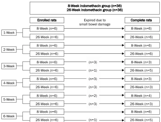
Fig. 1. Schematic diagram of study design.
Therefore, more physiologic drugs other than PPI may be needed in chronic NSAID users.
To test the effect of gastroprotective drugs, many trials were conducted for establishment of a chronic ulcer animal model. Acetic acid model has been recommended as a chronic ulcer model mainly due to the consistency of ulcer for- mation and prolonged duration of ulcer maintenance.5 However, this model causes some artificial defaults, such as leakage peritonitis, subserosal necrosis containing nerves and vessels, which are not observed in human PUD.6,7 In ad- dition, this model did not reflect the pathogenesis of NSAID-induced PUD, which occurs by repeated exposure to NSAID. To date, only a few studies regarding the formation of an NSAID-induced chronic gastric inflammation model have been reported.8 In addition, other reports have suggested the difficulty of this model due to adaptive mechanism.9
From this background we attempted to determine whether establishment of a chronic NSAID-induced gastric inflam- mation rat model is possible by repeated administration of NSAID. In addition, the effect of NSAID-induced damage on the small bowel was evaluated along with the gastric damage.
SUBJECTS AND METHODS
1. Animals
The animals used in this study were 6- and 24-week-old male Sprague-Dawley rats, which were purchased (Orient Co.
Ltd., Seoul, Korea) and housed in wire-bottom cages. They were maintained at 20-26oC, with 35-75% humidity and in a 12/12 hour light/dark cycle (lights on, 8:00-20:00) under pathogen-free conditions. After two weeks of adaptation, 8-week-old (weighting 260-290 g), 26-week-old (weighting 600-800 g) rats were used for this study. All experimental pro- cedures were approved by the Institutional Animal Care and Use Committee (IACUC) of Seoul National University Bundang Hospital (BA0903-040/013-02).
2. Experimental design
The time schedule of this study is shown in Fig. 1. A total of 72 rats (36 rats of 8-week-old and 26-week-old, re- spectively) were assigned to the indomethacin group, in which NSAIDs were administered on a weekly basis and were sacrificed at one week after administration. In each NSAID group, indomethacin (10 mg/kg body weight; Sigma-Aldrich Co., St. Louis, MO, USA) was administrated via gavage feed- ing through a metal tube attached to a 5- or 10-mL syringe ev-
ery week, respectively. The indomethacin solution was dis- solved in 0.5% (wt/vol) carboxy methyl cellulose (CMC) with 10% ethanol, pH ranging from 1.3 to 1.5, mainly because of the solubility of the indomethacin. The 10% ethanol was test- ed on the control rat and no significant lesion was observed when compared to 0.5% CMC control rats.
3. Gross ulcer index and histological index
In this chronic experiment, starvation was not maintained due to the possibility of stress with recurrent starvation every week. After sacrifice, small bowel perforation or peritonitis was carefully evaluated in the indomethacin group. Next, the isolated stomach was cut open along the greater curvature and washed with ice-cold saline. To investigate the degree of gross mucosal damage, the mucosal sides of the stomachs were photographed, using a digital camera. Gross damage of the gastric mucosa was assessed by two experienced gastro- enterologists, who were blinded to the experiment, using a previously described gross ulcer index10 defined as the sum of (number of type I lesions)+(number of type II le- sions)×2+(number of type III lesions)×3. The lesion type was classified as follows: type I was defined as the presence of edema, hyperemia, or a single submucosal punctuated form of hemorrhage; type II was defined as the presence of sub- mucosal hemorrhagic lesions with small erosions; and type III was defined as the presence of a deep ulcer with erosions.
The stomach was cut longitudinally to a width of 5 mm from the cardia to the pylorus in the anterior aspect after opening the stomach along the greater curvature, regardless of the damaged area or inflammation, and was fixed in 10%
formalin. The tissue specimens were dehydrated and em- bedded in paraffin. The longest part of the specimen was sec- tioned into 6 μm fragments, and the sections were stained with hematoxylin and eosin. All tissues were examined by an experienced pathologist, who was blinded to the experiment.
The mucosal damage then was graded by assigning a pre- viously described index of histological injury11 defined as (%
type I damage)×1+(% type II damage)×2+(% type III dam- age)×3. The types of damage were defined as follows: type 0 damage meant that all gastric mucosal cells appeared in- tact and had a normal shape, location, appearance, and den- sity; type I damage indicated that surface epithelial cells and the uppermost two or three cells lining the glands were dam- aged; type II damage described damage greater than type I
but involving <50% of the thickness of the gastric mucosa;
and type III damage indicated damage involving >50% of the gastric mucosa depth.
In the case of a rat that had expired or was in a dying con- dition before the sacrifice schedule, autopsy was performed quickly in order to determine the cause of death. If a small bowel perforation and/or peritonitis were detected without other definite cause of death then small bowel damage was interpreted as the cause of death. The small bowel, in whole, was extracted in non-peritonitis rats. In case of peritonitis, small bowel could not be obtained because of necrosis. The entire small bowel was grossly examined for the existence of perforation. For evaluation of myeloperoxidase (MPO), dam- aged small bowel segment was obtained. In case of no small bowel damage by gross finding, small bowel tissue within 5 cm from pylorus was used.
4. Measurement of mucosal myeloperoxidase
An assay of gastric mucosal MPO concentration was used to quantify the degree of neutrophil infiltration.12,13 Three hundred milligrams of scraped mucosa from the stomach and the small intestine were homogenized for 30 sec using a polytron homgenizer in 1.0 mL of ice-cold 0.5% hexadecyl- trimethylammonium bromide in 50 mM phosphate buffer (pH 6.0). Hexadecyltrimethylammonium bromide was used to negate the pseudoperoxidase activity of hemoglobin and to solubilize membrane-bound MPO. The homogenate was sonicated for 10 sec, freeze-thawed three times, and centri- fuged for 20 min at 18,000×g. The supernatant was isolated and the MPO concentration was determined using an ELISA kit (Immunodiagnostik AG, Bensheim, Germany).
5. Measurement of glucosamine
After the excised stomach was washed extensively with ice-cold saline, the gastric mucosa was scraped with a slide glass and then frozen at −70oC, until it was used. A stock sol- ution of glucosamine hydrochloride (Fluca, Buchs, Switzer- land) was prepared fresh, daily, and stored at 5oC; 0.3 mL (Sigma-Aldrich) of acetylacetone was added to 10 mL of car- bonate buffer with pH 10 (8 g of sodium bicarbonate and 2.1 g of sodium bicarbonate in 100 mL); the solution was pre- pared immediately before use. Ehrlich’s reagent was pre- pared immediately before use by dissolving 0.4 g of p-dime- thylaminobenzaldehyde (Sigma-Aldrich) in 1.5 mL of con-
Table 1. Occurrence of Small Bowel Damage in the Indomethacin Group during Six Weeks in 8- or 26-Week Rats
Rats’ age (week) Perforation or peritonitisa
1 Week 2 Week 3 Week 4 Week 5 Week 6 Week Total
8 0/6 2/6 2/3 2/3 1/4 0/6 7/28 (25.0%)
16 0/6 0/6 2/5 2/3 0/3 0/5 4/28 (14.3%)
aPerforation or peritonitis number/total sacrifice rat number.
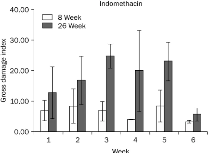
Fig. 2. Gross gastric damage index during the study period in the 8-week and 26-week indomethacin groups. Data are shown with mean±standard error.
Fig. 3. Histological index of gastric mucosal damage during the study period in the 8-week and 26-week indomethacin groups. Data are shown with mean±standard error.
centrated hydrochloric acid (HCl) and 13.5 mL of isoamyl alcohol. Gastric mucosal mucin extracted with Triton X-100, was hydrolyzed with 1 mL of 3 N HCl. Finally, hexosamine ob- tained from hydrolyzed mucin was assayed.14
6. Statistical analysis
All statistical calculations were performed using SPSS software (version 12.0; SPSS Inc., Chicago, IL, USA). The time trend of the results was compared using regression analysis.
Statistical significance was set at p<0.05
RESULTS
1. Gastric gross and histological damage index In 26-week rats, gross gastric damage index showed a peak level at three weeks and then showed a slow decrease in the indomethacin gavaged group. However, gross damage index was very small and no tendency was observed in 8-week rats in the indomethacin group (Fig. 2). Histological damage index was rather small and no constant trend was observed (Fig. 3). This could be explained by the fact that
NSAID-induced gross damage was usually not even and 5 mm width gastric tissue for histologic examination was col- lected from the anterior aspect of the antrum regardless of gross gastric damage.
2. Small bowel damage
Perforation began to occur from three weeks in the 8-week-rat and 26-week-rat groups. Death rate was 22.2%
(8/36) in both the 8-week and the 26-week indomethacin groups (Fig. 1). In addition, small bowel perforation or peri- tonitis was observed in the 8-week group (25%, 7/28) com- pared with the 26-week indomethacin group (14.3%, 4/28) in survived rats observed during sacrifice (Table 1). In cadav- er autopsy, all rats showed peritonitis, thus, small bowel dam- age was considered the cause of death.
3. Gastric mucosal and intestinal mucosal myeloper- oxidase levels during the course of study
Gastric mucosal MPO showed a decreasing trend after four weeks with repeated administration in the 8-week in- domethacin group (p=0.018). However, this was not a sig-
Table 2. Time Trend of Gastric Mucosal Myeloperoxidase (MPO) and Glucosamine Levels in the Indomethacin Groups
1 Week 2 Week 3 Week 4 Week 5 Week 6 Week p-valuea Indomethacin
8-week-rat group
MPO (ng/mg protein) 0.68±0.19 0.54±0.09 0.64±0.12 0.26±0.03 0.31±0.06 0.22±0.05 0.018 Glucosamine (mg/g mucosa) 8.36±1.09 8.18±0.59 8.01±1.15 9.31±1.35 9.59±2.10 12.24±0.91 0.038 Indomethacin
26-week-rat group
MPO (ng/mg protein) 0.51±0.07 0.72±0.11 0.71±0.07 0.36±0.06 0.31±0.02 0.48±0.11 0.270 Glucosamine (mg/g mucosa) 7.74±1.35 6.49±1.70 10.54±1.31 11.19±1.07 11.73±2.53 14.06±1.95 0.007 Data are presented as mean±standard error.
aBy regression analysis.
Fig. 4. Myeloperoxidase levels in the small bowel during the study period in 8- and 26-week groups. No statistically significant trend was observed. Data are shown with mean±standard error.
nificant trend in the 26-week indomethacin group (p=0.270).
This trend could be matched with less significant in- flammation in the 8-week indomethacin group than in the 26-week indomethacin group (Table 2). In the small bowel, MPO did not show any specific trend in both the 8-week and the 26-week indomethacin groups (Fig. 4).
4. Gastric mucosal glucosamine levels during the course of study
In both groups, the levels of glucosamine showed a steady increase from four weeks after administration of indome- thacin in the 8-week group (p=0.038) and three weeks after in the 26-week group (p=0.007) (Table 2).
DISCUSSION
Chronic NSAID induced gastric inflammation model was not well-established in previous animal studies. In addition, the causes of difficulty in chronic NSAID induced gastric dam- age formation have not been clearly identified. A few reports have suggested that an adaptation mechanism, with re-
peated NSAID exposure, could be one of the causes in the chronic NSAID induced gastric damage model.9,15,16 The mechanisms of such adaptation were suggested as an up-regulation of COX-2 enzyme17 or enhancement of a de- fense mechanism.9 In the current study, no increases of the gross damage scale and MPO in 8-week rats also suggest the possibility of an adaptation to repeated NSAID exposure.
Gastric glucosamine, which reflects secreted mucin as a strong mucosal protecting marker, was increased in the in- domethacin rat groups during the study period. In addition, gastric glucosamine level showed an increasing trend, which is quite a contrast with the decrease of MPO in the 8-week in- domethacin group. In the 26-week group, the gross ulcer scale showed a peak level at three weeks and then a slow de- crease, and an increasing trend was observed in gastric glu- cosamine level but not in MPO level. These results suggest enhanced mucosal protection as an adaptive mechanism in case of repeated administration of indomethacin.
Another difficult point in formation of a chronic NSAID-in- duced gastric inflammation model was found to originate from the small bowel damage. Small bowel damage could not be avoided if a sufficient dose of NSAID was administered in order to produce gastric damage. We thought that adult rats may have thicker small bowel wall and greater resistance to NSAID than young rats. Therefore, we compared small bowel damage between 8-week young rats and 26-week adult rats.
In this study, critical small bowel damage was also observed in the indomethacin groups, as death occurred in 16/72 (22.2%) and perforation or peritonitis in 11/56 (19.6%), when the rate was calculated in the total rats regardless of 8- or 26-week rats. As a result, no significant difference in small bowel damage was observed between the 8- and 26-week age groups. Furthermore, in our preliminary study, small bowel damage was not avoidable with repeated admin- istration of 5 mg/kg indomethacin daily (data not shown).
Previous reports also demonstrated that 7.5-15 mg/kg was
a sufficient dose in the indomethacin-induced acute enteritis rat model.18-20 This low dose of indomethacin might be in- sufficient to induce a satisfactory gastric damage over- whelming adaptation mechanism. Taken together, small bowel damage may be the most serious problem in establish- ment of a chronic NSAID-induced gastric inflammation model.
Acetic acid model, with maintenance dose of NSAID, may be an alternative method in formation of a chronic NSAID-as- sociated ulcer model and a few studies have demonstrated this possibility.21-23 However, this method is somewhat artifi- cial and it may induce leakage peritonitis and subserosal necrosis. A recent study suggested that calcitonin gene-re- lated peptide was an important mechanism in gastric protection.7 Therefore, subserosal and muscular damage in an acetic acid model may differ from those in the human gas- tric ulcer induced by chronic NSAID usage. Furthermore, an adaptive mechanism might also have an effect on the main- tenance dose of NSAID. Refed NSAID model could be possi- ble as a chronic NSAID-induced antral ulcer model.24,25 Diffuse antral ulcer was documented in these study models.
However, 25-30 mg/kg indomethacin used in this study could induce small bowel perforation. Therefore, another method is required for maintenance of a chronic model. As a result, the model in the combination of refed NSAID and in- ducer, such as HCl, could be attempted in the next study. In this model, NSAID dosage must be lowered as much as possi- ble in order to minimize damage to the small bowel.
The limitations of our study could be that we administered only one dose of indomethacin (10 mg/kg) once per week.
Polat et al.,9 who administered various doses of in- domethacin, found a decrease of ulcerous area at the dose of 1-3 mg/kg/day, whereas the ulcerous area started to in- crease at the dose of 4-5 mg/kg/day. They concluded that the best adaptation was observed in rats administered 3 mg/kg/day. We speculated that reduced dosing and longer administration would not cause small bowel perforation, fib- rosis, or stenosis as well as chronic strictures in the NSAID-in- duced chronic model. Contrary to our expectation, we found perforation of the small intestine in approximately 20% of cases. In addition, it is quite puzzling that intestinal MPO lev- els in the survived rats did not show any relationships with small bowel damage. The possibility that severe inflam- matory response occurred in some rats, resulting in death,
could not be excluded; on the other hand, the other rats that survived indomethacin-induced damage were not affected by NSAIDs. In the future, these issues could be explained when we succeed in establishment of this chronic NSAID-in- duced gastric damage rat model with several doses and ad- ministration intervals of several kinds of NSAID such as in- domethacin, diclofenac, and aspirin.
In conclusion, establishment of a chronic NSAID-induced gastric damage model was difficult due to an adaptive mech- anism of the stomach and small bowel damage. Gastric mu- cus, a defensive mechanism, could be suggested as a cause of adaptive mechanism. Conduct of further studies will be needed in order to establish a chronic NSAID-induced gastric damage model.
REFERENCES
1. Wong GL, Wong VW, Chan Y, et al. High incidence of mortality and recurrent bleeding in patients with Helicobacter pylori-negative idiopathic bleeding ulcers. Gastroenterology 2009;137:525- 531.
2. Gisbert JP, Calvet X. Review article: Helicobacter pylori-negative duodenal ulcer disease. Aliment Pharmacol Ther 2009;30:791- 815.
3. Sung JJ, Chan FK, Chen M, et al; Asia-Pacific Working Group.
Asia-Pacific Working Group consensus on non-variceal upper gastrointestinal bleeding. Gut 2011;60:1170-1177.
4. Yang YX, Lewis JD, Epstein S, Metz DC. Long-term proton pump inhibitor therapy and risk of hip fracture. JAMA 2006;296:
2947-2953.
5. Okabe S, Pfeiffer CJ. Chronicity of acetic acid ulcer in the rat stomach. Am J Dig Dis 1972;17:619-629.
6. Kang JM, Kim N, Kim B, et al. Enhancement of gastric ulcer heal- ing and angiogenesis by cochinchina Momordica seed extract in rats. J Korean Med Sci 2010;25:875-881.
7. Evangelista S. Role of sensory neurons in restitution and healing of gastric ulcers. Curr Pharm Des 2006;12:2977-2984.
8. Fiorucci S, Distrutti E, de Lima OM, et al. Relative contribution of acetylated cyclo-oxygenase (COX)-2 and 5-lipooxygenase (LOX) in regulating gastric mucosal integrity and adaptation to aspirin. FASEB J 2003;17:1171-1173.
9. Polat B, Suleyman H, Alp HH. Adaptation of rat gastric tissue against indomethacin toxicity. Chem Biol Interact 2010;186:
82-89.
10. Nam SY, Kim N, Lee CS, et al. Gastric mucosal protection via en- hancement of MUC5AC and MUC6 by geranylgeranylacetone.
Dig Dis Sci 2005;50:2110-2120.
11. Lacy ER, Ito S. Microscopic analysis of ethanol damage to rat gas- tric mucosa after treatment with a prostaglandin. Gastroenterology 1982;83:619-625.
12. Bradley PP, Priebat DA, Christensen RD, Rothstein G. Measure-
ment of cutaneous inflammation: estimation of neutrophil con- tent with an enzyme marker. J Invest Dermatol 1982;78:206- 209.
13. Grisham MB, Benoit JN, Granger DN. Assessment of leukocyte involvement during ischemia and reperfusion of intestine.
Methods Enzymol 1990;186:729-742.
14. Nishida K, Ohta Y, Ishiguro I. Teprenone, an anti-ulcer agent, in- creases gastric mucosal mucus level via nitric oxide in rats. Jpn J Pharmacol 1998;78:519-522.
15. Alderman BM, Cook GA, Familari M, Yeomans ND, Giraud AS.
Resistance to apoptosis is a mechanism of adaptation of rat stomach to aspirin. Am J Physiol Gastrointest Liver Physiol 2000;278:G839-G846.
16. Konturek SJ, Brzozowski T, Stachura J, Dembinski A, Majka J.
Role of gastric blood flow, neutrophil infiltration, and mucosal cell proliferation in gastric adaptation to aspirin in the rat. Gut 1994;35:1189-1196.
17. Konturek PC, Brzozowski T, Pierzchalski P, et al. Activation of genes for spasmolytic peptide, transforming growth factor alpha and for cyclooxygenase (COX)-1 and COX-2 during gastric adapta- tion to aspirin damage in rats. Aliment Pharmacol Ther 1998;
12:767-777.
18. Kuroda M, Yoshida N, Ichikawa H, et al. Lansoprazole, a proton pump inhibitor, reduces the severity of indomethacin-induced rat enteritis. Int J Mol Med 2006;17:89-93.
19. Saud B, Nandi J, Ong G, Finocchiaro S, Levine RA. Inhibition of TNF-alpha improves indomethacin-induced enteropathy in rats by modulating iNOS expression. Dig Dis Sci 2005;50:1677- 1683.
20. Stadnyk AW, Dollard C, Issekutz TB, Issekutz AC. Neutrophil mi- gration into indomethacin induced rat small intestinal injury is CD11a/CD18 and CD11b/CD18 co-dependent. Gut 2002;50:
629-635.
21. Ogihara Y, Okabe S. Mechanism by which indomethacin delays gastric ulcer healing in the rat: inhibited contraction of the ulcer base. Jpn J Pharmacol 1993;61:123-131.
22. Penney AG, Malcontenti-Wilson C, O'Brien PE, Andrews FJ.
NSAID-induced delay in gastric ulcer healing is not associated with decreased epithelial cell proliferation in rats. Dig Dis Sci 1995;40:2684-2693.
23. Wang JY, Yamasaki S, Takeuchi K, Okabe S. Delayed healing of acetic acid-induced gastric ulcers in rats by indomethacin.
Gastroenterology 1989;96:393-402.
24. Onodera S, Tanaka M, Aoyama M, et al. Antiulcer effect of lafuti- dine on indomethacin-induced gastric antral ulcers in refed rats.
Jpn J Pharmacol 1999;80:229-235.
25. Satoh H, Inada I, Hirata T, Maki Y. Indomethacin produces gastric antral ulcers in the refed rat. Gastroenterology 1981;81:719- 725.