with a subepithelial connective tissue graft in advanced recession
Chun-Teh Lee1, Po-Chun Chang2, Nawar Touchan3, Daniel Royzman4,*
1Division of Periodontology, Department of Oral Medicine, Infection and Immunity, Harvard School of Dental Medicine, Boston, MA, USA
2Graduate Institute of Clinical Dentistry, School of Dentistry, National Taiwan University, Taipei, Taiwan
3Private Practice, Ottawa, ON, Canada
4Division of Periodontics, Section of Oral and Diagnostic Sciences, College of Dental Medicine, Columbia University, New York, NY, USA
Case Report
J Periodontal Implant Sci 2014;44:300-306 http://dx.doi.org/10.5051/jpis.2014.44.6.300
Purpose: A laterally positioned flap (LPF) combined with a subepithelial connective tissue graft (SCTG) is one of the conventional approaches for resolving gingival recession defects, with the advantages of flap flexibility and extended coverage of the tissue graft. However, thus far, evidence is lacking for the use of this technique for the treatment of advanced gingival recession defects. This report discusses three Miller class III cases with interproxi- mal bone loss and wide and deep defects treated with a combination procedure of a modi- fied laterally positioned flap (mLPF) and SCTG.
Methods: mLPF combined with SCTG was performed for each case. The defect size and the degree of hypersensitivity at baseline and the final appointment in each case were docu- mented.
Results: The three cases had a mean initial defect of 7.7±1.5 mm and a mean residual de- fect of 1.7±1 mm at the 6-, 3-, and 36-month follow-up, respectively, after the root cov- erage surgery. The symptom of hypersensitivity was improved, and the patients were satis- fied with the clinical outcomes.
Conclusions: The results demonstrated that the combination of mLPF with SCTG is promis- ing for treating these advanced cases with respect to obtaining the expected root coverage with the gingival tissue.
Keywords: Case reports, Gingival recession, Periodontics.
Received: Oct. 18, 2014 Accepted: Dec. 24, 2014
*Correspondence:
Daniel Royzman
Division of Periodontics, Section of Oral and Diagnostic Sciences, College of Dental Medicine, Columbia University, 630 West 168th Street, PH7E-111, New York, NY 10032, USA E-mail: [email protected] Tel: +1-917-515-0007 Fax: +1-212-308-6325
INTRODUCTION
According to the reports of the National Health and Nutrition Examination Survey (NHANES III, 1988–1994) in the United States [1], gingival recession defects, characterized by the expo- sure of the tooth root surface and the symptom of dentinal hypersensitivity, affected more than 50% of the population. To resolve these complications, covering the tooth root with the gingival tissue by performing periodontal plastic surgery, namely the ‘root coverage’ proce- dure, was proposed. The coronally advanced flap (CAF) and laterally positioned flap (LPF) were the two main techniques used in this root coverage procedure [2]. These techniques, combined with the use of tissue grafts or biomaterials, were proposed to increase the predictability of clinical outcomes by providing extra tissue, increasing flap thickness, and guiding tissue growth [3,4].
CAF has been widely used for treating gingival recession for fifty years now [5], and there
This is an Open Access article distributed under the terms of the Creative Commons Attribution Non-Commercial License (http://creativecommons.org/licenses/by-nc/3.0/).
is plenty of clinical evidence supporting the predictability of CAF or CAF combined with a tissue graft [4]. The LPF, also called the sliding flap, lateral pedicle flap, or rotated flap, was first introduced by Grupe and Warren Jr [6] in 1956, and either combination with [7,8]
or without subepithelial connective tissue graft (SCTG) placement under specific circumstances was suggested [9-11]. The combination of LPF and SCTG can improve root coverage, reduce the chances of gingival recession of the flap elevation site [12], and retain the ad- vantages of the LPF technique, such as flap flexibility and obtaining predictable keratinized gingiva [13].
Of all the root coverage procedures, CAF combined with SCTG is the most commonly used one and is regarded as the most predict- able technique for Miller class I or II gingival recession defects [2-4]
(Miller’s classification) [14]. However, the therapeutic effect of this procedure on advanced cases, such as Miller class III gingival reces- sion with a wide and deep defect, is still inconclusive. Although ac- ceptable clinical results for treating Miller class III cases by using CAF+SCTG have been reported, most of these cases originally had just mild interproximal bone loss and minor-to-moderate gingival recession defects [15]. On the other hand, although LPF was not in- vestigated quite as often as CAF, the clinical outcomes of these two procedures were comparable [4]. In certain clinical circumstances,
such as the limited amount of keratinized tissue apical to the reces- sion defect and presence of a very shallow vestibule, LPF can be an alternative to CAF to obtain predictable root coverage [12].
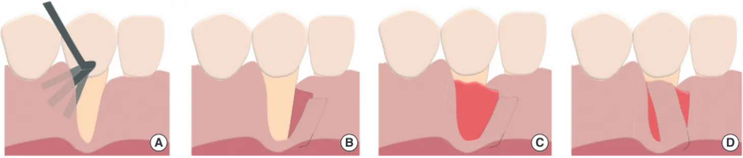
The three cases with advanced gingival recession discussed in this report were treated with a modified laterally positioned flap (mLPF), characterized by an oblique incision beyond the mucogingival junc- tion with a partial thickness flap to increase the flap flexibility (Fig.
1A, B). SCTG was placed to augment tissue thickness and obtain pre- dictable biological attachment, thereby increasing the root coverage rate (Fig. 1C, D) [4]. All patients were treated in the periodontal clinic of the College of Dental Medicine, Columbia University. The percent- age of root coverage as well as self-reported hypersensitivity before and after surgery was assessed, and the results are summarized in Table 1.
CASE DESCRIPTION
Case I History
A 50-year-old female, who had undergone orthodontic treatment in private practice in 2007, presented with generalized severe root resorption and buccal gingival recession (Fig. 2A, B), which caused
A B C D
Figure 1. Surgical technique of modified laterally positioned flap combined with subepithelial connective tissue graft placement. (A) Use the instrument (e.g., Dr. Allen intrasulcular knife) to partially elevate the interproximal gingiva without opening the flap. The partially elevated area should be larger than the area where the tissue graft will be placed. (B) Incise and elevate the modified laterally positioned flap. The incision starts 2 mm below the zenith of the interproxi- mal papilla and should cross the mucogingival junction in vertical and oblique directions to increase the flexibility. The flap is split in thickness, and the perios- teum underneath the gingiva is intact. (C) Insert the subepithelial connective tissue graft underneath the partially and fully elevated gingiva. The upper border of the graft is positioned about 2–3 mm above the interproximal crestal bone level. (D) The flap is laterally positioned and sutured. The flap with sufficient flex- ibility should be positioned about 1–2 mm above the expected level of the future gingival margin.
Table 1. Summary of clinical results in cases 1, 2, and 3.
Case Follow-up period (month) Defect deptha) (mm)
Root coverage rateb) (%) Self-evaluation of hypersensitivity symptomc)
Initial Final Initial Final
1 6 9 2 78 2 1
2 3 6 2.5 58 1 0
3 36 8 0.5 94 0 0
Mean±SD - 7.7±1.5 1.7±1 77±18 - -
SD: standard deviation.
a)Defect depth was measured as the longest distance between the buccal cementoenamel junction and gingival margin. b)Root coverage rate was calculated as follows: [(initial defect depth)–(final defect depth)/(initial defect depth)]×100%. c)Hypersensitivity symptom was reported by the patient on the basis of his/her own experience: 0, no symptom; 1, mild hypersensitivity that usually does not bother the patient; 2, moderate hypersensitivity that makes the patient feel sensitive sometimes while eating or drinking; 3, severe hypersensitivity that makes the patient feel pain frequently while eating or drinking.
esthetic concerns and root hypersensitivity, in a consultation ap- pointment. The gingiva of the mandibular right canine was inflamed, with insufficient width of the keratinized gingiva. The mandibular right canine exhibited significant root prominence and a Miller class III gingival recession defect (Fig. 2B). The tooth was vital as deter- mined by a cold test and grade I mobility [16]. The patient was in- formed that the tooth had questionable prognosis due to severe periodontal attachment loss. Further, complete root coverage could not be expected due to the significant interproximal bone loss.
Treatment and outcome
The exposed root surface of the mandibular right canine was scaled and root planed before flap elevation, and no chemical re-
agent or drug was utilized to condition or clean the root surface.
The distal interproximal gingiva was partially elevated by using the tunneling technique [17], and the mesial interproximal gingiva of the mandibular right canine was included in the mLPF. Vertical and oblique incisions were made (Fig. 2C), and then, a split thickness flap was elevated. After flap elevation, the mandibular right canine revealed a significant loss of the buccal alveolar bone and severe root resorption (Fig. 2D). SCTG was harvested from the palate by using the two-incision flap design. The selection of the size of tis- sue graft (dimensions: 16 mm×8 mm×2 mm) was based on the areas of the recession defect and flap elevation sites. The graft, whose position was 1 mm below the cementoenamel junction (CEJ) level and 3 mm above the interproximal bone level, was
A B C
D E F
Figure 2. Case 1 clinical photographs. (A) The radiographic image of tooth #43; Tooth #43 had severe root resorption and significant interproximal bone loss. (B) The patient had generalized gingival recession and an open bite. Tooth #43 had a Miller class III gingival recession defect which was wide and deep (length: 8–9 mm). (C) A modified laterally positioned flap was made at the mesial interproximal gingiva with vertical and oblique incision. (D) The buccal alveolar bone of tooth #43 was missing and the resorption of the root apex was significant. (E) A clinical photograph on the date of completing surgery. (F) A clinical photo- graph at the six-month follow-up.
placed underneath the mesial and distal interproximal gingiva.
Stabilization of the tissue graft was achieved with a chromic gut 5-0 suture. The split thickness flap was distally rotated to cover the exposed tissue graft and was sutured with chromic gut 5-0 (Fig. 2E).
The patient was instructed to take ibuprofen 800 mg every eight hours as needed for resolving postoperative pain or discomfort, and rinse the mouth for 30 seconds twice a day with half an ounce of 0.12% chlorhexidine to maintain the hygiene of the surgical area during the first two weeks. The sutures were removed two weeks after the procedure, and the regular oral hygiene regimen was re- sumed.
The gingival level of the mandibular right canine moved apically during the first two months by about 1–2 mm as compared to the level just after surgery, particularly on the mesial side. Seventy-eight percentage of the defect was covered (Table 1), and the probing depth was within 3 mm in the 6-month follow-up (Fig. 2F). The incomplete defect coverage was caused by the low interproximal bone level. The patient was satisfied with the outcome of root coverage, and the hy- persensitivity symptom was improved.
Case 2 History
A 46-year-old female patient presented with generalized buccal gingival recession with hypersensitivity from the exposed roots. She was also concerned about the esthetics of the mandibular right central incisor, which had minimal buccal keratinized gingiva and an advanced gingival recession defect (Miller class III) (Fig. 3A). The mandibular right central incisor also showed interproximal bone loss radiographically (Fig. 3B).
Treatment and outcome
The procedure was similar to that in case 1. A split thickness flap was elevated at the mesial site of the mandibular right central inci- sor to preserve the integrity of the distal interproximal papillae. The SCTG (dimensions: 20 mm×10 mm×1.5 mm) was placed under- neath the partially elevated gingiva and the exposed root surface.
Distally, the flap was rotated to cover the tissue graft and sutured (Fig. 3C). The postoperative instructions were the same as those of the first patient in case 1. The gingival recession defect had 58%
defect coverage, and the probing depths of all sites were within 3 mm in the 3-month follow-up (Fig. 3D) (Table 1); long-term follow- up was scheduled.
Case 3 History
A 40-year-old male complained of unacceptable esthetics of the mandibular right canine. The probing depth of this canine was with- in 3 mm, and mild interproximal alveolar bone loss was found on the radiographs (Fig. 4A, B) and upon clinical sounding. One-millimeter buccal keratinized gingiva was present at the mandibular right ca- nine, and the recession was diagnosed as Miller class III (Fig. 4C).
Treatment and outcome
The procedure was similar to that of case 1. Flap flexibility was as- sessed (Fig. 4D), and the tissue graft (dimensions: 15 mm×7 mm×1.5 mm) was sutured to the alveolar periosteum of the mandibular right canine (Fig. 4E). The flap was rotated mesially and stabilized 1 mm above the CEJ level and 3 mm above the interproximal bone level (Fig. 4F). The gingival recession defect of the mandibular right canine had 94% root coverage in the 6-month follow-up (Fig. 4G), and the gingival level was still stable after 3 years (Fig. 4H) (Table 1).
DISCUSSION
A review published in 1996 [2] demonstrated a mean root cover- age rate of 63% of the rotational flap, including 15 studies utilizing LPF and 2 studies utilizing a double papilla flap (DPF), which is a modification of LPF. These results provoked many clinicians to ques- tion the predictability of the LPF technique at that time. Conversely, from the results of studies published after 1996, LPF had a mean root coverage rate ranging from 74% to 96% (Table 2), which is comparable to the root coverage rate of CAF or CAF+SCTG [4]. Re-
A B
C D
Figure 3. Case 2 clinical photographs. (A) Tooth #41 had a Miller class III gin- gival recession defect (length: 7–8 mm) and 0.5–1 mm wide buccal keratin- ized gingiva. (B) Tooth #41 had 1–2 mm of interproximal bone loss. (C) Modi- fied laterally positioned flap was sutured and most of the subepithelial tissue graft was covered underneath the flap. (D) A clinical photograph at the three-month follow-up.
A B C
D
E F G H
Figure 4. Case 3 clinical photograhs. (A) Periapical radiograph at the initial visit. (B) Bitewing radiograph at the initial visit. (C) Tooth #43 had a Miller class III gingival recession defect and the interproximal soft tissue had mild loss. (D) The flap was rotated mesially to assess the flexibility. (E) The subepithelial tissue graft was placed and sutured with Vicryl 5-0. (F) A clinical photograph on the date of completing surgery. (G) A clinical photograph at the six-month follow- up. (H) A clinical photograph of case 3 at the three-year follow-up.
Table 2. Summary of clinical studies utilizing laterally positioned flap for root coverage.
Studya) Study design Groups Surgical sites & criteria No. of subjects/
No. of treated teeth
Follow-up period (month)
Mean initial recession defect (mm)
Mean root coverage
(%) Zucchelli et al. (2004) [12] Case series LPF Max or Man incisors, canines,
premolars, molars (Miller class I or II) 100/120 12 4.4 95
Chambrone et al. (2009) [24] Prospective cohort LPF Max or Man teeth (Miller class I or II) 32/32
(Max, 16; Man,16) 24 Max: 4.6
Man: 4.8 94
94 Santana et al. (2010) [13] RCT LPF
CAF Max incisors, canines, premolars
(Miller class I) 18/18
18/18 6 3.4
3.2 96
97 Zucchelli et al. (2012) [25] RCT LPF
CAF+SCTG Max or Man first molars (Miller class I
or II) 25/25
25/25 12 5.0
4.6 74
89
Yilmaz et al. (2014) [26] RCT LPF
LPF (+LA) Man incisors (Miller class II) 15/15
16/16 6 4.7
5.0 74
96 LPF: laterally positioned flap, Max: maxillary, Man: mandibular, RCT: randomized controlled trial, CAF: coronally advanced flap, SCTG: subepithelial connective tissue graft, LA: laser- assisted with an external vestibular releasing incision made by a diode laser.
a)These studies were published after 1996 and reported the clinical results of root coverage by using a laterally positioned flap. Case reports were not included.
garding the technique of LPF combined with SCTG, some articles have reported the clinical outcomes of several cases [7,18]. In Nel- son [7], DPF combined with SCTG was utilized to treat the defect of a single tooth, and LPF combined with SCTG was utilized to treat multiple defects. The results demonstrated successful clinical out- comes even in advanced recession cases (88% average root cover- age rate in cases with 7- to 10-mm-long defects) during a 6- to
42-month follow-up. Ricci et al. [18] utilized the same technique as Nelson [7]. The procedure had root coverage rates for treating Mill- er class I or II cases similar to those of the guided tissue regenera- tion technique at the 1-year follow-up (80.88% vs. 77.08%; the mean initial defect was 4.88 mm or 5.88 mm, respectively).
The root coverage rates in the present study, utilizing mLPF com- bined with SCTG, were approximately 60%–95% and were in agree-
ment with the results of previous studies. Additionally, the cases se- lected in the present report are the Miller class III cases with signifi- cant interproximal bone loss (case 1) or deep, wide defects (cases 2 and 3). The involved teeth also had a limited amount of keratinized gingiva, thus causing difficulty in coronally repositioning the flap to fully cover the tissue graft. Although it has been documented that the tissue graft can be left exposed in certain situations [19], there still remain the clinical risks of having an uneven gingival margin [20]. Further, leaving a significant area of connective tissue graft exposed on the avascularized root surface may increase the chance of partial necrosis where the graft cannot receive a suffi- cient blood supply [21]. Thus, LPF appeared to be a superior option to CAF in these advanced gingival recession cases. LPF can ensure that the SCTG is covered by a gingival flap, which provides a suffi- cient blood supply laterally to increase the plasmatic circulation during initial healing [22].
In all three cases discussed in this report, the SCTG was placed about 2–3 mm above the interproximal bone level based on the concept of biological width around the periodontium, instead of solely following the CEJ level, which is the expected level of the complete root coverage, because complete root coverage is not gen- erally expected in Miller class III cases. A retrospective study demon- strated that the complete root coverage of Miller class III recession could only be achieved in sites under certain conditions, including complete integrity of the interproximal gingiva, graft thickness greater than 2 mm, interproximal bone loss not exceeding 3 mm, and an initial recession defect width not greater than 3 mm [23].
However, our reported cases did not fulfill these requirements.
Additionally, case 3, in which the interproximal bone loss was less than that in cases 1 and 2, demonstrated the greatest root coverage rate among these cases (Table 1), indicating that the level of inter- proximal bone is associated with the extent of root coverage in the Miller class III cases. To elucidate the exact association between the interproximal bone level and the success of root coverage, further well-designed clinical studies are needed.
mLPF combined with SCTG placement could be a promising tech- nique for treating Miller class III cases with significant interproximal bone loss and wide and deep recession defects. Future controlled studies are needed to provide more clinical evidence.
CONFLICT OF INTEREST
No potential conflict of interest relevant to this article was re- ported.
ACKNOWLEDGEMENTS
We would like to thank Dr. Yu-Cheng Lin for preparing the illus- trations.
ORCID
Chun-Teh Lee http://orcid.org/0000-0001-7812-5637 Po-Chun Chang http://orcid.org/0000-0002-4493-6922 Nawar Touchan http://orcid.org/0000-0002-6390-5793 Daniel Royzman http://orcid.org/0000-0002-0079-7676
REFERENCES
1. Albandar JM, Kingman A. Gingival recession, gingival bleeding, and dental calculus in adults 30 years of age and older in the United States, 1988-1994. J Periodontol 1999;70:30-43.
2. Wennström JL. Mucogingival therapy. Ann Periodontol 1996;1:
671-701.
3. Oates TW, Robinson M, Gunsolley JC. Surgical therapies for the treatment of gingival recession: a systematic review. Ann Peri- odontol 2003;8:303-20.
4. Cairo F, Nieri M, Pagliaro U. Efficacy of periodontal plastic surgery procedures in the treatment of localized facial gingival reces- sions: a systematic review. J Clin Periodontol 2014;41 Suppl 15:
S44-62.
5. Harvey PM. Management of advanced periodontitis. I. Preliminary report of a method of surgical reconstruction. N Z Dent J 1965;
61:180-7.
6. Grupe HE, Warren RF Jr. Repair of gingival defects by a sliding flap operation. J Periodontol 1956;27:92-5.
7. Nelson SW. The subpedicle connective tissue graft: a bilaminar re- constructive procedure for the coverage of denuded root surfaces.
J Periodontol 1987;58:95-102.
8. Borghetti A, Louise F. Controlled clinical evaluation of the subped- icle connective tissue graft for the coverage of gingival recession.
J Periodontol 1994;65:1107-12.
9. Smukler H. Laterally positioned mucoperiosteal pedicle grafts in the treatment of denuded roots: a clinical and statistical study. J Periodontol 1976;47:590-5.
10. Caffesse RG, Guinard EA. Treatment of localized gingival reces- sions. Part IV. Results after three years. J Periodontol 1980;51:
167-70.
11. Caffesse RG, Alspach SR, Morrison EC, Burgett FG. Lateral sliding flaps with and without citric acid. Int J Periodontics Restorative Dent 1987;7:42-57.
12. Zucchelli G, Cesari C, Amore C, Montebugnoli L, De Sanctis M.
Laterally moved, coronally advanced flap: a modified surgical ap- proach for isolated recession-type defects. J Periodontol 2004;75:
1734-41.
13. Santana RB, Furtado MB, Mattos CM, de Mello Fonseca E, Dibart S.
Clinical evaluation of single-stage advanced versus rotated flaps in the treatment of gingival recessions. J Periodontol 2010;81:
485-92.
14. Miller PD Jr. A classification of marginal tissue recession. Int J Peri- odontics Restorative Dent 1985;5:8-13.
15. Cairo F, Cortellini P, Tonetti M, Nieri M, Mervelt J, Cincinelli S, et
al. Coronally advanced flap with and without connective tissue graft for the treatment of single maxillary gingival recession with loss of inter-dental attachment. A randomized controlled clinical trial. J Clin Periodontol 2012;39:760-8.
16. Miller SC. Textbook of periodontia. Philadelphia: Blakiston Co.;
1938.
17. Allen AL. Use of the supraperiosteal envelope in soft tissue graft- ing for root coverage. II. Clinical results. Int J Periodontics Restor- ative Dent 1994;14:302-15.
18. Ricci G, Silvestri M, Rasperini G, Cattaneo V. Root coverage: a clin- ical/statistical comparison between subpedicle connective tissue graft and laterally positioned full thickness flaps. J Esthet Dent 1996;8:66-73.
19. Han JS, John V, Blanchard SB, Kowolik MJ, Eckert GJ. Changes in gingival dimensions following connective tissue grafts for root coverage: comparison of two procedures. J Periodontol 2008;79:
1346-54.
20. Bouchard P, Malet J, Borghetti A. Decision-making in aesthetics:
root coverage revisited. Periodontol 2000 2001;27:97-120.
21. Langer B, Langer L. Subepithelial connective tissue graft technique for root coverage. J Periodontol 1985;56:715-20.
22. Gordon HP, Sullivan HC, Atkins JH. Free autogenous gingival grafts.
II. Supplemental findings: histology of the graft site. Periodontics 1968;6:130-3.
23. Esteibar JR, Zorzano LA, Cundin EE, Blanco JD, Medina JR. Complete root coverage of Miller Class III recessions. Int J Periodontics Re- storative Dent 2011;31:e1-7.
24. Chambrone LA, Chambrone L. Treatment of Miller class I and II lo- calized recession defects using laterally positioned flaps: a 24-month study. Am J Dent 2009;22:339-44.
25. Zucchelli G, Marzadori M, Mele M, Stefanini M, Montebugnoli L.
Root coverage in molar teeth: a comparative controlled random- ized clinical trial. J Clin Periodontol 2012;39:1082-8.
26. Yilmaz E, Ozcelik O, Comert M, Ozturan S, Seydaoglu G, Teughels W, et al. Laser-assisted laterally positioned flap operation: a ran- domized controlled clinical trial. Photomed Laser Surg 2014;32:
67-74.