대한소화기학회지 2010;56:273-275 □ IMAGE OF THE MONTH □ DOI: 10.4166/kjg.2010.56.5.273
연락처: 김광하, 602-739, 부산시 서구 아미동 1가 10번지 부산대학교 의학전문대학원 내과학교실
Tel: (051) 240-7869, Fax: (051) 244-8180 E-mail: [email protected]
Correspondence to: Gwang Ha Kim, M.D.
Department of Internal Medicine, Pusan National University School of Medicine, 1-10, Ami-dong, Seo-gu, Busan 602-739, Korea
Tel: +82-51-240-7869, Fax: +82-51-244-8180 E-mail: [email protected]
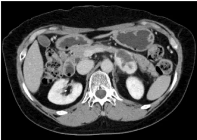
Fig. 1. Abdominal CT showed a 4.5×3.4 cm sized solid mass with heterogenous enhancement between pancreatic tail and left kidney.
췌장 미부 종양으로 오인된 부신 혈관종
부산대학교 의학전문대학원 내과학교실
정재훈ㆍ김광하
A Case of Adrenal Hemangioma Misdiagnosed as a Pancreatic Tail Tumor
Jae Hoon Cheong, M.D. and Gwang Ha Kim, M.D.Department of Internal Medicine, Pusan National University School of Medicine, Busan, Korea
증례: 평소 건강하였던 66세 여자가 전신피로감을 주소로 개인병원에서 시행받은 복부초음파검사에서 췌장 미부에 낭성 종괴가 발견되어 본원에 방문하였다. 다른 병력이나 약제 복용력은 없었으며, 가족력, 사회력에서도 특이소견은 없었다. 활력징후는 정상이었으며 신체검사에서 특별한 이 상은 보이지 않았다. 본원에서 시행한 말초혈액검사에서 백 혈구 6,310/mm3, 혈색소 11.7 g/dL, 혈소판 219,000/mm3, AST 20 IU/L, ALT 17 IU/L, 총 빌리루빈 0.38 mg/dL, alkaline phosphatase 186 IU/L, 크레아티닌 0.9 md/dL, CEA 2.62 ng/mL, CA 19-9 25.27 U/mL였으며, 다른 검사는 모두 정상 이었다.
복부전산단층촬영 및 자기공명영상검사를 시행했고 췌장 미부와 좌측 부신 사이에 4.5×3.4 cm 크기의 경계가 명확한 낭성 종괴가 관찰되어, 췌장 미부의 췌도세포종이 가장 의 심되었으며, 기타 부신선종, 갈색세포종 등과의 감별이 필 요하였다(Fig. 1). 종괴의 원발 부위를 좀 더 알기 위해 추가 로 시행한 내시경초음파검사에서 좌측 부신의 바깥쪽 테두 리와 연결되며 내부에 낭성 변화를 가진 저에코성 종괴가 확인되었다(Fig. 2). 결국 우연히 발견된 부신 종양(inciden- taloma)이었고 호르몬 분비능의 이상은 없었으나, 크기가 크 고 임상적으로 악성을 배제할 수 없어 환자는 비뇨기과에서 복강경하 좌측 부신제거술을 받았고, 조직검사 결과 정상적 인 부신의 선조직과 함께 확장된 혈관들이 증식되어 있는 소견을 보였다(Fig. 3).
진단: 부신의 혈관종
부신절제술 후 조직검사 결과 좌측 부신에서 발생한 3.5×
1.0 cm의 혈관종으로 진단되었다. 악성을 시사하는 소견은 보이지 않았고, 수술 후 특별한 합병증이나 증상 없이 환자 는 외래에서 경과 관찰 중이다.
혈관종은 대개 피부나 간에 흔하게 발생하며, 부신에서 생기는 경우는 매우 드물다. 대다수가 무증상이며 비특이적 인 증상이나 막연한 복통을 호소하며 내원하기도 한다. 사 후 부검이나 다른 질환과 관련되어 시행한 영상검사에서 우
274 대한소화기학회지: 제56권 제5호, 2010
Fig. 2. EUS showed that a well- circumscribed heterogeneous hypo- echoic mass with internal cystic change (A) originateed from the lateral rim of the left adrenal gland (B, arrow).
Fig. 3. Microscopically, the mass was composed of various sized blood vessels (H&E, orig. mag. ×200). Normal adrenal gland pa- renchyma was noted on the surface of the mass. There was no histologic evidence of malignancy.
연히 발견되는 경우가 대부분으로, 1955년 Johnson과 Jeppesen에 의해 처음으로 발표된 이후1 현재까지 55예가 보 고되고 있다. 부신의 우연종은 복부전산단층촬영에서 1-5%
의 빈도로 보고되지만, 이 중에서 혈관종은 매우 드물며 술 전에는 감별하기가 힘들다. 대개 해면상(cavernous) 혈관종 이며, 일측성이고 50-60대에 흔하며 남녀 발생비율은 2:1 정 도로 여성에서 흔하다.2-4 직경은 2-25 cm로 다양하며 10 cm 보다 큰 경우가 흔하다.5-7 부신의 혈관종은 대부분이 비기 능성 종양으로 호르몬을 분비하는 종양은 현재까지 3예에 서 보고되었을 뿐이며,8-10 대개 무증상이지만 인접장기 압박 에 의해 복통을 호소할 수 있고, 심각한 자발성 후복강출혈 을 야기하는 경우도 있다.3,11 부신은 타장기의 종양이 흔히 전이하는 부위 중 하나이기 때문에, 악성흑색종이나 폐암, 유방암, 소화기계의 악성종양의 전이일 가능성을 고려해야
한다. 비소세포폐암과 총담관암, 부인과종양이 혈관종과 공 존했던 예들도 보고되었고,12-14 부신의 혈관종이 유방암과 함께 골수 외 조혈조직이 공존했던 예도 보고되고 있다.15 부신의 혈관종을 다른 종양, 특히 악성종양과 감별하기는 매우 힘들며, 대부분 술 후 조직검사로 최종 진단된다. 혈관 종을 의심해볼 수 있는 영상학적 소견으로는, 복부전산단층 촬영과 자기공명영상에서 혈관종의 비교적 흔한 소견인 종 괴 테두리의 조영증강을 보일 수 있으며,16,17 늘어난 혈관에 생긴 phlebolith로 인해 종양 전체에 얼룩덜룩한 석회화가 관 찰될 수도 있다.18,19 그러나 이런 소견들은 갈색세포종이나 다른 선종 또는 부신암에서도 나타날 수 있어 혈관종에 특 이적이라고 할 수는 없다.
종양의 크기가 수술적 절제의 적응증이 되며, 직경 6 cm 이상은 악성일 확률이 35-98%까지 예상되기 때문에 절제가 원칙이다. 4-6 cm의 종양은 다른 영상검사 소견이나 타장기 의 악성종양의 병력, 환자의 나이나 동반질환, 일상 활동도 를 고려하여 결정한다. 부신절제술을 받거나, 영상검사를 반복하며 경과관찰하는 것도 가능하다.3 지금까지 대부분의 부신 혈관종은 크기가 커서 수술로 치료했으며, 주변장기의 압박 증상을 유발하는 경우나 출혈 등의 합병증 등으로 인 해 수술이 필요한 경우도 있었다. 부신절제는 6 cm 이하의 경우 복강경으로 절제가 가능하며, 더 큰 종양은 술기의 문 제와 함께 악성의 가능성을 고려하여 개복술이 적절하며, 주로 전방 또는 후방, 흉복부 접근법이 적용된다.7,20 내시경초음파검사는 현재까지 주로 소화기관의 점막하 종양의 감별진단이나 췌담도계의 영상 및 여러 조직의 세침 흡인검사에 이용되었지만, 이번 증례와 같이 좌측 부신과 췌장주위의 해부학적 구조를 명확히 볼 수 있는 검사이다.
비록 우측 부신은 소화관강과 멀리 떨어져 있어 일정한 영 상을 얻기 힘들고 다소 부정확하지만, 좌측 부신과 주변의 장기는 다른 영상검사와 비교하여 충분히 높은 해상력의 영
정재훈 외 1인. 췌장 미부 종양으로 오인된 부신 혈관종 275
상을 실시간으로 볼 수 있는 장점이 있다. 실제로 이번 증례 에서는 복부전산단층촬영 및 자기공명영상검사에서 종괴의 정확한 기시부를 알 수 없었지만, 내시경초음파검사에서 좌 측 부신에서 기원한 병변임을 알 수 있어 복강경하 수술 시 도움을 주었다. 그러므로 췌장 미부 근처에 병변이 있는 경 우, 복부전산단층촬영 및 자기공명영상검사 후에도 병변의 기시부를 명확히 알 수 없을 때에는 내시경초음파검사가 진 단과 치료 방침 설정에 유용한 정보를 제공할 수 있을 것으 로 생각된다.
참고문헌
1. Johnson CC, Jeppesen FB. Hemangioma of the adrenal. J Urol 1955;74:573-577.
2. Heis HA, Bani-Hani KE, Bani-Hani BK. Adrenal cavernous haemangioma. Singapore Med J 2008;49:e236-237.
3. Forbes TL. Retroperitoneal hemorrhage secondary to ruptured cavernous hemangioma. Can J Surg 2005;48:78-79.
4. Sabanegh E Jr, Harris MJ, Grider D. Cavernous adrenal hemangioma. Urology 1993;42:327-330.
5. Makiyama K, Fukuoka H, Kawamoto K, Suwa Y. Surgical removal of adrenal haemangioma after five years of fol- low-up: a case report. Hinyokika Kiyo 1998;44:579-581.
6. Hisham AN, Samad SA, Sharifah NA. Huge adrenal hae- mangioma. Australas Radiol 1998;42:250-251.
7. Nigri G, Bellagamba R, Giaccaglia V, et al. Minimally in- vasive adrenalectomy for incidentally discovered cavernous hemangioma. Minim Invasive Ther Allied Technol 2008;17:
255-258.
8. Stumvoll M, Fritsche A, Wehrmann M, Dammann F, Becker HD, Eggstein M. A functioning adrenocortical haemangioma.
J Urol 1996;155:638.
9. Oh BR, Jeong YY, Ryu SB, Park YI, Kang HK. A case of adrenal cavernous haemangioma. Int J Urol 1997;4:608-610.
10. Ng AC, Loh HL, Shum CF, Yip SK. A case of adrenal cav- ernous hemangioma presenting with progressive enlargement and apparent hormonal hypersecretion. Endocr Pract 2008;14:
104-108.
11. Boraschi P, Campatelli A, Di Vito A, Perri G. Hemorrhage in cavernous hemangioma of the adrenal gland: US, CT and MRI appearances with pathologic correlation. Eur J Radiol 1995;21:41-43.
12. Alcázar J, Márquez A, Rosales M. An unusual cause of adre- nal mass in a patient with operable non-small-cell pulmonary carcinoma. Arch Bronconeumol 1998;34:513-514.
13. Päivänsalo M, Siniluoto T, Seppänen U. Cavernous hae- mangioma of the adrenal gland. Diagn Imaging Clin Med 1986;55:168-171.
14. Chudácek Z, Kohoutek V. Simultaneous occurrence of a cav- ernous adrenal gland hemangioma and a bile duct-liver carcinoma. Rofo 1980;132:460-462.
15. Arkadopoulos N, Kyriazi M, Yiallourou AI, et al. A rare co- existence of adrenal cavernous hemangioma with extra- medullar hemopoietic tissue: a case report and brief review of the literature. World J Surg Oncol 2009;7:13.
16. Yamada T, Ishibashi T, Saito H, et al. Two cases of adrenal hemangioma: CT and MRI findings with pathological correlations. Radiat Med 2002;20:51-56.
17. Marotti M, Sučić Z, Krolo I, et al. Adrenal cavernous he- mangioma: MRI, CT, and US appearance. Eur Radiol 1997;
7:691-694.
18. Rothberg M, Bastidas J, Mattey WE, Bernas E. Adrenal he- mangiomas: angiographic appearance of a rare tumor. Radio- logy 1978;126:341-344.
19. Thiele JW, Bodie B. Adrenal hemangioma. Surgery 2001;129:
373-374.
20. Trupka A, Hallfeldt K, Schmidbauer S. Laparoscopic adrena- lectomy with lateral approach--a comparison with the conven- tional dorsal technique. Chirurg 2001;72:1478-1484.