In complex primary total knee arthroplasty (TKA), when a surgeon faces problems of imbalance of mediolateral gap or improper flexion-extension gap balance, a varus-valgus constrained (VVC) prosthesis is commonly used instead of posterior-stabilized (PS) TKA.1-4) The VVC prosthesis can provide substantial varus-valgus constraint and limit
When to Use a Condylar Constrained Insert in Non-Stemmed Posterior-Stabilized Total Knee
Arthroplasty
Satit Thiengwittayaporn, MD, Natthapong Hongku, MD, Umaporn Uawisetwathana, PhD*, Pichai Sansawat, MD
Department of Orthopaedics, Faculty of Medicine Vajira Hospital, Navamindradhiraj University, Bangkok,
*National Center for Genetic Engineering and Biotechnology (BIOTEC), National Science and Technology Development Agency, Pathum Thani, Thailand
Background: The constrained insert with non-stemmed tibial and femoral components can be used in the modern total knee arthroplasty (TKA) when soft-tissue balance and adequate stability from a posterior-stabilized (PS) insert cannot be achieved. This study aimed to identify the prevalence and predictive factors associated with the constrained insert use during primary TKA for varus deformity.
Methods: From August 2016 to March 2019, 554 primary TKAs were consecutively performed by one surgeon. The choice of using a conventional PS polyethylene insert versus a constrained insert was made by the surgeon, depending on the stability detected after an attempt to balance the soft tissue. The decision to convert to a constrained liner was made if the ligament could not be balanced, if flexion-extension gaps were mismatched, or if the varus-valgus opening was 3 mm or more when varus and valgus stress tests at 0° were applied. We retrospectively investigated the preoperative, intraoperative, and postoperative factors associ- ated with the constrained insert use. Multiple logistic regression analysis was used to identify predictive factors of constrained insert use, and a receiver operating characteristic curve analysis was used to pinpoint a cutoff value of tibiofemoral varus angle.
Results: Constrained inserts were used in 130 of 497 varus knees (26.1%). A multivariate analysis revealed that the factors asso- ciated with an increased adjusted risk of constrained insert use included preoperative severe varus deformity (odds ratio [OR], 5.78;
95% confidence interval [CI], 2.75–12.16; p < 0.001) and severe release of soft tissue through the superficial medial collateral liga- ment (OR, 6.38; 95% CI, 2.94–13.85; p < 0.001). A preoperative anatomic tibiofemoral varus angle of > 19.8° was associated with the use of a constrained articulation with an area under the curve of 0.7 (95% CI, 0.4–0.8).
Conclusions: Prevalence of 26.1% for constrained insert use was found in this study. Preoperative anatomic tibiofemoral varus angle of > 19.8° and severe release of soft tissue through the superficial medial collateral ligament were associated with the use of a constrained articulation. The findings from this study will help surgeons to improve efficiency of surgical sequence planning and provide counseling to patients regarding the associated cost.
Keywords: Total knee arthroplasty, Posterior-stabilized, Varus-valgus constrained prosthesis, Varus
Copyright © 2020 by The Korean Orthopaedic Association
This is an Open Access article distributed under the terms of the Creative Commons Attribution Non-Commercial License (http://creativecommons.org/licenses/by-nc/4.0) which permits unrestricted non-commercial use, distribution, and reproduction in any medium, provided the original work is properly cited.
Clinics in Orthopedic Surgery • pISSN 2005-291X eISSN 2005-4408 Received December 9, 2019; Accepted February 14, 2020
Correspondence to: Satit Thiengwittayaporn, MD
Department of Orthopaedics, Faculty of Medicine Vajira Hospital, Nava- mindradhiraj University, 681 Samsen Rd, Dusit, Bangkok 10300, Thailand Tel: +66-22443376, Fax: +66-22436106
E-mail: [email protected]
torsion moments due to the enlarged tibial post insert in the tibial tray that fits within the intercondylar notch of the femoral component. Most surgeons use intramedullary stems with constrained components for complex primary TKA to provide load-sharing mechanisms over the di- aphyseal bone of the femur and tibia.5) However, the use of these stem extensions has a number of disadvantages. For instance, it may increase the risk of embolization from the invasion of the intramedullary canal, increase operating time and cost from the steps of procedures, possibly result in postoperative end-of-stem pain, and cause difficulty for revision if required.6)
Recently, the constrained insert with non-stemmed tibial and femoral components (stemless VVC) has been used in some modern PS-TKA designs with reportedly acceptable outcomes in the early and mid-term periods when properly used.4,6-8) The stemless constrained inserts can be switched from the PS insert intraoperatively, al- lowing an instant surgical solution in the operation room.
Although a short-term retrieval study reported an increase in the wear around the region of the post of stemless con- strained inserts compared to the PS inserts, the increased damage and deviation of the post surfaces were minimal and likely clinically insignificant.9) However, studies on the prevalence and indication of constrained insert use in non-stemmed PS-TKA are very limited.
Therefore, this study aimed to answer the following questions: (1) What is the prevalence rate of constrained inserts used in primary TKA for varus deformity? (2) Are there any predictive factors associated with the use of con- strained inserts in primary TKA for varus deformity?
METHODS
Study Design
This retrospective study was approved by the Ethical Committee of our institution (IRB No. COA61/2560).
From August 2016 to March 2019, 554 minimally invasive total knee arthroplasties (MIS-TKAs) were consecutively performed on 529 patients by a single surgeon (ST) using a fixed bearing PS-TKA (Legion Total Knee System; Smith
& Nephew, Memphis, TN, USA). TKA procedures were performed with the possibility of using a conventional PS insert or a constrained insert with the implants depend- ing on the stability detected after an attempt to balance the soft tissue (Fig. 1). All patients with knee osteoarthritis whose symptoms could not be treated with a conservative measure were admitted under the care of one surgeon (ST) for a primary MIS-TKA. Patients were excluded from the study if they had valgus deformity. A total of 497 knees met the inclusion criteria, excluding 52 knees with valgus deformity and 5 knees with incomplete data (Fig. 2).
Clinical Protocol
A standard procedure of a minimally invasive technique without patellar resurfacing was performed. Regional an- esthetics were administered to all patients. The tourniquet pressure was at 280 mmHg in all cases. The incision was typically less than 9 cm long, which represented no more than twice the length of the patella. A mini-midvastus approach was performed to allow exposure of the knee without everting the patella. The distal femoral resection was performed using the intramedullary technique at 6°
of valgus. The femoral rotation was determined by the posterior femoral condylar axis and Whiteside’s line10) and using the transepicondylar axis in patients with ana-
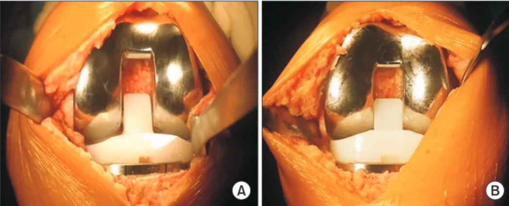
Fig. 1. Comparison between a posterior-stabilized (PS) insert (A) and constrained insert (B). A constrained insert has a thicker and wider post than a PS insert. The constrained insert provides substantial varus-valgus constraint and limits torsion moments due to the enlarged tibial post insert that fits within the intercondylar notch of the femoral component. With this modern total knee arthroplasty design, surgeons can switch from a PS insert to a constrained insert intraoperatively without the use of a stem extension.
A B
tomical abnormalities. The femoral component size was determined by the posterior referencing instrumentation system. The proximal tibial resection was performed us- ing the extramedullary technique. Tibial components with asymmetric tibial trays were rotated using the functional alignment method. Flexion and extension gap balancing was performed with the use of spacer blocks to obtain equally symmetric gaps. We performed a subperiosteal release of the deep medial collateral ligament (MCL) or medial capsule as the first step in the medial release. Fur- ther release of the semimembranosus tendon and postero- medial capsule may have been needed in some cases and was performed as the second step of medial release when necessary. The superficial MCL was the final step of the sequential medial release. We used a periosteal elevator to perform a careful elevation of the superficial MCL posteri- or to the pes insertion. Both complete and partial releases were included at this final step.11,12)
After implanting the components, a standard PS insert was used in the well-balanced knees if symmetrical
rectangular gaps were achieved and if the medial/lateral opening did not exceed 3 mm when varus and valgus stress tests at 0° were applied.13) The decision to convert to a constrained liner was made if the ligament could not be balanced, if flexion-extension gaps were mismatched or if the varus-valgus opening was 3 mm or more when varus and valgus stress tests at 0° were applied (Fig. 3). Femoral and tibial components were cemented in all cases. Both groups received the same postoperative pain control and rehabilitation methods, which consisted of a multimodal approach to avoid parenteral narcotics and provide early postoperative mobilization.
Associated Factor and Data Analysis
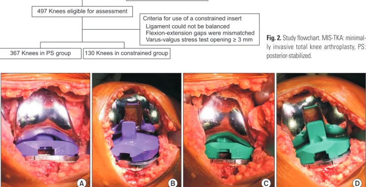
To identify the predictive factors associated with the use of a constrained insert in primary TKA, the following factors relevant to pre-, intra-, and postoperative steps were con- sidered as variables in the analysis. Briefly, the analyzed preoperative variables included the following: (1) Quan- titative variables: patient’s age, body mass index, range of Fig. 3. Decision-making procedure to use the constrained insert. When a conventional posterior-stabilized insert was used, the knee was well-balanced in full extension (A), but unstable in full flexion (B). After replacing with a constrained insert with the same thickness, the knee was well-balanced in both full extension (C) and full flexion (D).
A B C D
Criteria for use of a constrained insert Ligament could not be balanced
Flexion-extension gaps were mismatched Varus-valgus stress test opening > 3 mm 57 Knees excluded
52 Knees valgus deformity 5 Knees incomplete data 554 Knees from 529 patients
underwent MIS-TKA between August 2016 and March 2019
497 Knees eligible for assessment
367 Knees in PS group 130 Knees in constrained group
Fig. 2. Study flowchart. MIS-TKA: minimal- ly invasive total knee arthroplasty, PS:
posterior-stabilized.
motion, Knee Society score (KSS),14) and degree of poste- rior tibial slope.15) (2) Qualitative variables: sex, Kellgren and Lawrence radiographic grading (KL grade),16) align- ment measured on anteroposterior (AP) weight-bearing hip-to-ankle standing radiographs, severity of anatomic varus angle quantified by tibiofemoral angle (mild: < 10°, moderate: 10°−15°, and severe: > 15°),17) severity of coro- nal plane laxity (mild: < 6°, moderate: 6°−9°, and severe:
> 9°),14) and severity of sagittal plane laxity (mild: < 5 mm, moderate: 5−10 mm, and severe: > 10 mm).14) The sever- ity of the coronal plane and sagittal plane laxities were assessed by a single surgeon (ST) at the time of admission and prior to surgery in all cases. The analyzed intraopera- tive variables included degree of soft-tissue release in varus knees (step 1−3: mild, moderate, and severe).
The postoperative variables that were analyzed at 6 weeks included the outliers of the hip-knee-ankle angle (outlier: > ±3°), the outliers of the posterior tibial slope (outlier: < 0° posterior tibial slope or > 7° posterior tibial slope), and the outliers of the femoral flexion angle (outlier:
< 0° femoral flexion angle or > 5° femoral flexion angle).
For the anteroposterior and lateral weight-bearing standing radiographs, the feet were placed apart with the knees in maximum extension and the toes pointing straight. The angles were calculated using a digital vec- toral goniometer and the radiographs. Two independent investigators (TW and NK) performed the radiographic measurements. The mean of the 2 results was used in the analysis.
Statistical Analysis
Descriptive statistics included means and standard de- viations for continuous variables and frequencies and percentages for categorical variables. Demographic and clinical variables were compared between the PS and con- strained groups using paired 2-tailed Student t-tests for continuous variables and chi-square test or Fisher exact test for categorical variables.
A univariate analysis of the predictive factors for stemless constrained insert use in primary TKA with varus deformity for all variables was initially performed with a logistic regression for each variable. A multivariate analysis was subsequently performed on the variables that showed a statistically significant correlation in the univari- ate analysis (p < 0.05) with a logistic regression. The re- sults of the regression analyses are presented as odds ratios (ORs) with accompanying 95% confident intervals (CIs).
The results were considered to be statistically significant when the null value (1.00) was absent from the CI or the p-value was < 0.05. A receiver operating characteristic
(ROC) curve analysis was performed to calculate the sen- sitivity and specificity, and the area under the curve (AUC) to identify the cutoff point for a preoperative degree of an anatomic tibiofemoral varus angle needing use of a con- strained articulation. The optimal cutoff point was defined as the concentration with the highest sum of sensitivity and specificity.18) The variable with the greatest AUC was defined as the most effective tool for classifying patients into the 2 groups. Data were analyzed using IBM SPSS ver. 23.0 (IBM Corp., Armonk, NY, USA). The computed power was achieved with an alpha of 0.05, a sample size of 286, and each OR and R-squared attributed to 14 indepen- dent variables using Z test with a significance level of 0.05.
RESULTS
Demographic and Clinical Variables Data
A total of 554 knees were identified but 52 were excluded owing to valgus deformity and 5 owing to incomplete data. Thus, a total of 497 knees were included in this study. The constrained insert was used in 130 of 497 knees (26.1%). The preoperative, intraoperative, and postopera- tive demographics and clinical variables between the PS and constrained groups were compared, which revealed that the following variables were significantly different between the groups: ROM, KSS, severity of varus angle (mild, moderate, severe), severity of coronal plane laxity (mild and severe), severity of sagittal plane laxity (moderate and severe), posterior tibial slope, and degree of soft-tissue release (mild and severe) (Table 1).
Predictive Factors for Constrained Insert Use
Table 2 examines the ORs of the predictive factors for constrained insert use through the multivariate regression analysis. Preoperative, intraoperative, and postoperative clinical factors associated with the increased adjusted risk of constrained insert use were severe varus deformity (OR, 5.78; 95% CI, 2.75–12.16; p < 0.001) and severe release of soft tissue (OR, 6.38; 95% CI, 2.94–13.85; p < 0.001). No meaningful differences were found in the other factors.
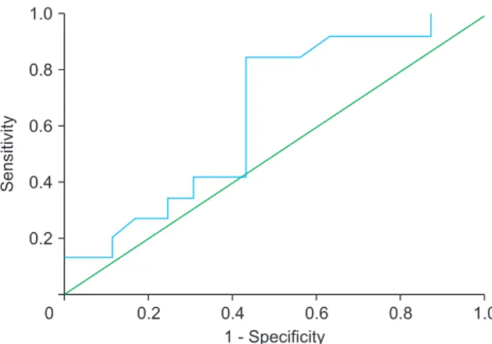
An ROC curve was constructed to determine the optimal cut-off value of 19.8° for the tibiofemoral angle that neces- sitates the use of a constrained insert with an AUC of 0.7 (95% CI, 0.4–0.8) (Fig. 4).
DISCUSSION
To identify the prevalence and predictive factors associ- ated with an increased constrained insert use, a retrospec- tive study was conducted. In this study, the prevalence of
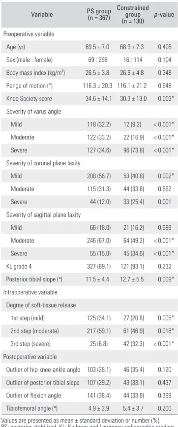
Table 1. Comparison of Demographic and Clinical Variables between the PS and Constrained Groups
Variable PS group
(n = 367)
Constrained group
(n = 130) p-value Preoperative variable
Age (yr) 69.5 ± 7.0 68.9 ± 7.3 0.408
Sex (male : female) 69 : 298 16 : 114 0.104 Body mass index (kg/m2) 26.5 ± 3.8 26.9 ± 4.8 0.348 Range of motion (°) 116.3 ± 20.3 116.1 ± 21.2 0.948 Knee Society score 34.6 ± 14.1 30.3 ± 13.0 0.003*
Severity of varus angle
Mild 118 (32.2) 12 (9.2) < 0.001*
Moderate 122 (33.2) 22 (16.9) < 0.001*
Severe 127 (34.6) 96 (73.8) < 0.001*
Severity of coronal plane laxity
Mild 208 (56.7) 53 (40.8) 0.002*
Moderate 115 (31.3) 44 (33.8) 0.662
Severe 44 (12.0) 33 (25.4) 0.001
Severity of sagittal plane laxity
Mild 66 (18.0) 21 (16.2) 0.689
Moderate 246 (67.0) 64 (49.2) < 0.001*
Severe 55 (15.0) 45 (34.6) < 0.001*
KL grade 4 327 (89.1) 121 (93.1) 0.232
Posterior tibial slope (°) 11.5 ± 4.4 12.7 ± 5.5 0.009*
Intraoperative variable Degree of soft-tissue release
1st step (mild) 125 (34.1) 27 (20.8) 0.005*
2nd step (moderate) 217 (59.1) 61 (46.9) 0.018*
3rd step (severe) 25 (6.8) 42 (32.3) < 0.001*
Postoperative variable
Outlier of hip-knee-ankle angle 103 (28.1) 46 (35.4) 0.120 Outlier of posterior tibial slope 107 (29.2) 43 (33.1) 0.437 Outlier of flexion angle 141 (38.4) 44 (33.8) 0.399 Tibiofemoral angle (°) 4.9 ± 3.9 5.4 ± 3.7 0.200 Values are presented as mean ± standard deviation or number (%).
PS: posterior-stabilized, KL: Kellgren and Lawrence radiographic grading.
*Indicates significant difference (p < 0.05).
Table 2. Multivariable Logistic Regression Analysis to Identify Predictive Factors for the Use of Constrained Insert in Varus TKA
Characteristic Odds ratio 95% CI p-value
Preoperative variable Age (yr)
≤ 65 1 (Reference)
> 65 0.71 0.42−1.18 0.182
Sex
Male 1 (Reference)
Female 1.60 0.81−3.15 0.174
Body mass index (kg/m2)
Underweight to normal (≤ 22.9) 1 (Reference)
Overweight to obese (≥ 23.0) 1.02 0.54−1.95 0.945 Range of motion (°)
≤ 100 1 (Reference)
> 100 0.87 0.48−1.58 0.645
Knee Society score
≤ 50 1 (Reference)
> 50 0.58 0.22−1.52 0.271
Severity of varus angle
Mild 1 (Reference)
Moderate 1.54 0.70−3.40 0.286
Severe 5.78 2.75−12.16 < 0.001*
Severity of coronal plane laxity
Mild 1 (Reference)
Moderate 0.95 0.55−1.64 0.860
Severe 0.84 0.41−1.72 0.642
Severity of sagittal plane laxity
Mild 1 (Reference)
Moderate 0.53 0.28−1.01 0.052
Severe 0.87 0.41−1.84 0.714
KL grading
3 1 (Reference)
4 0.74 0.31−1.80 0.509
Posterior tibial slope (°)
≤ 12 1 (Reference)
> 12 1.17 0.73−1.87 0.515
constrained insert use in primary TKA with varus defor- mity was found to be 26.1%, while preoperative severe varus deformity and severe release of soft tissue through the superficial MCL were found to be the key factors for the use of the constrained insert in primary varus TKA. A preoperative anatomic tibiofemoral varus angle of > 19.8°
was associated with the use of a constrained articulation and could be utilized prospectively to predict implant se- lection.
Stability is essential for successful TKA. Increasing the constraint to achieve stability in complex primary TKA often requires the use of the VVC system to provide wider polyethylene posts and larger femoral component boxes to limit the varus-valgus and torsional movements. However, several concerns with the VVC design persist. One is that a high degree of post-box constraint can increase the load to bone-implant interface, and stem extension is invasive, which can increase the complexity and cost of surgery.
The modern TKA design tries to address these pitfalls of the VVC system by providing stemless constrained inserts that can be switched from the PS insert intraoperatively.
The use of the stemless insert in a primary TKA may be preferred to the VVC system because the bone defect in
primary TKA is still minimal. Selective use of the stemless constrained insert is recommended. Therefore, it would be helpful for surgeons prior to the operation to be able to identify the knees with the possibility of using the stemless constrained insert.
To the best of our knowledge, there has never been any study reporting precise factors that can be used pre- operatively to help predict the use of the constrained ar- ticulation to provide stability of the knee intraoperatively.
Previously, a retrospective study found high prevalence of constrained insert use in the patients with severe varus deformity undergoing TKA using the extensive medial soft-tissue release technique.13) While the severe varus de- formity and severe release of soft tissue might be deemed to play roles in the increased risk for using constrained inserts, there was no direct evidence on this.
Our study therefore aimed to identify predictive factors for the use of the stemless constrained insert in pri- mary TKA to help the surgeon prepare the implant preop- eratively. We found that severe varus deformity or severe release of soft tissue was associated with an increased risk of constrained insert use in primary varus TKA without stem extensions. In the current study, we found that the patients with severe varus deformity measured with tib- iofemoral angle from radiography had a 5.78-fold higher risk of needing constrained insert use than the patients with mild varus deformity (adjusted OR, 5.78; 95% CI, 2.75–12.16; p < 0.001). We identified the optimal value of Table 2. Continued
Characteristic Odds ratio 95% CI p-value
Intraoperative variable Degree of soft-tissue release
1st step (mild) 1 (Reference)
2nd step (moderate) 1.40 0.79−2.47 0.252
3rd step (severe) 6.38 2.94−13.85 < 0.001*
Postoperative variable Outlier of hip-knee-ankle angle
No outlier 1 (Reference)
Outlier 1.22 0.74−2.01 0.436
Outlier of posterior tibial slope
No outlier 1 (Reference)
Outlier 1.28 0.78−2.11 0.320
Outlier of flexion angle
No outlier 1 (Reference)
Outlier 0.73 0.45−1.20 0.219
TKA: total knee arthroplasty, CI: confidence interval, KL: Kellgren and Lawrence radiographic grading.
*Indicates significant difference (p < 0.05).
0 0.2 0.4 0.6 0.8
1.0
0.8
0.6
0.4
0.2
1.0
Sensitivity
1 - Specificity
Fig. 4. The receiver operating characteristic (ROC) curve was used to determine the threshold level of the tibiofemoral varus angle above which the risk of constrained insert use was significantly elevated.
Diagonal line represents a reference line. The inflection point of the curve corresponds to the preoperative tibiofemoral varus angle of 19.8°, which represents the value with the highest sensitivity and specificity.
The area under the curve of 0.7 (95% confidence interval, 0.4–0.8) is the area beneath the ROC.
19.8° tibiofemoral varus angle that could be utilized pro- spectively to predict implant selection. This information is important for surgeons to determine the severity of the deformity preoperatively so that implants with the appro- priate constraint are made available.
In addition to severe varus deformity factor, this current study performing the classic Insall medial soft- tissue release technique on varus deformity patients found that patients with severe release of the soft tissue or super- ficial MCL release had an increased risk of constrained inserts used (adjusted OR, 6.38; 95% CI, 2.94–13.85; p
< 0.001) compared to the patients with mild soft-tissue release. Insufficient medial soft-tissue release resulted in unsatisfactory deformity correction, whereas excessive superficial MCL release relatively increased the flexion- extension gap on the medial side, which could eventually lead to instability of the knee. When faced with this prob- lem, surgeons have 2 options to manage. The first option is to use lateral soft-tissue release for gap balancing and a thick polyethylene often needs to be inserted, which can result in restricted ROM due to higher joint line, patel- lofemoral maltracking, and extension restriction. The second option, which was used in this study, is to switch to a constrained insert that provides stability and retain the joint line and patellofemoral kinematics. Unlike our study that examined only primary TKAs with varus deformi- ties, an equivalent retrospective study examining primary TKAs with valgus deformities found that medial laxity was the sole independent factor associated with the higher frequency of implantation of constrained prostheses with an OR of 1.9 (1.2−2.7).19) However, this study did not ex- amine the intraoperative factors on the prosthetic switches to VVC; thus, the severity of soft-tissue release was not considered in the study.
Our study revealed 26.1% prevalence of constrained insert use in primary TKA with varus deformity. Previous- ly, Goudarz Mehdikhani et al.13) conducted a similar retro- spective study focusing on the patients with severe varus deformity undergoing TKA using the extensive medial soft-tissue release technique and found a higher incidence (> 40%) of constrained insert use in severe varus deformi- ties. However, another study with valgus deformity report- ed a similar prevalence (26/93 implantations, 27.96%) of constrained prostheses use to ours.19) Considering the high incidences from our and previous reports, the surgeon should have a constrained prosthesis available, especially
in the cases with the risk factors.
The present study has several strengths. The se- ries of patients were consecutive, and all operations were performed by a single surgeon. Therefore, the surgical technique and decision-making with respect to the use of a PS or a constrained insert were identical. While most previous studies were concerned only with the reliable results of constrained insert use in primary TKA without stem extensions, this present study employed a multivari- ate logistic regression analysis to estimate the association between constrained insert use and independent predic- tive factors including various clinical and radiographic parameters.
There are also several limitations of this study. First, the retrospective nature of this study allows demonstra- tion of correlation, not the true causation. Although we adjusted for potential confounders, there may have been additional confounders that we could not control. Second, sample sizes in some categories were relatively limited.
Third, clinical and radiological outcomes were not inves- tigated; however, the key objectives of this study were to identify the prevalence and predictive factors associated with the constrained insert use during primary TKA for varus deformity.
In conclusion, the prevalence of 26.1% for con- strained insert use was found in our study. Preoperative anatomic tibiofemoral varus angle of > 19.8° and severe re- lease of soft tissue through the superficial MCL were asso- ciated with the use of constrained articulation during sur- gery. Such information is useful for surgeon’s counseling to patients regarding the associated cost, improved efficiency of surgical sequence planning, and most importantly, hav- ing appropriate prosthetic parts and instruments available prior to surgery.
CONFLICT OF INTEREST
No potential conflict of interest relevant to this article was reported.
ACKNOWLEDGEMENTS
We would like to thank Ms Thikampa Wichai, BSc, and Nunnapat Kangkano, BSc (Na vamindradhiraj University), for their help in collecting and organizing the data in this study.
REFERENCES
1. Easley ME, Insall JN, Scuderi GR, Bullek DD. Primary con- strained condylar knee arthroplasty for the arthritic valgus knee. Clin Orthop Relat Res. 2000;(380):58-64.
2. Lachiewicz PF, Soileau ES. Ten-year survival and clinical results of constrained components in primary total knee ar- throplasty. J Arthroplasty. 2006;21(6):803-8.
3. Luque R, Rizo B, Urda A, Garcia-Crespo R, Moro E, Lopez- Duran L. Primary modular total knee replacement in severe and unstable osteoarthritis: predictive factors for failure. Int Orthop. 2015;39(11):2125-33.
4. King BR, Gladnick BP, Lee YY, Lyman S, Della Valle AG.
Range of motion and function are not affected by increased post constraint in patients undergoing posterior stabilized total knee arthroplasty. Knee. 2014;21(1):194-8.
5. Bugbee WD, Ammeen DJ, Engh GA. Does implant selection affect outcome of revision knee arthroplasty? J Arthroplasty.
2001;16(5):581-5.
6. Anderson JA, Baldini A, MacDonald JH, Tomek I, Pellicci PM, Sculco TP. Constrained condylar knee without stem ex- tensions for difficult primary total knee arthroplasty. J Knee Surg. 2007;20(3):195-8.
7. Anderson JA, Baldini A, MacDonald JH, Pellicci PM, Sculco TP. Primary constrained condylar knee arthroplasty without stem extensions for the valgus knee. Clin Orthop Relat Res.
2006;(442):199-203.
8. Deshmukh AJ, Rathod PA, Moses MJ, Snir N, Marwin SE, Dayan AJ. Does a non-stemmed constrained condy- lar prosthesis predispose to early failure of primary total knee arthroplasty? Knee Surg Sports Traumatol Arthrosc.
2016;24(10):3194-9.
9. Konopka J, Weitzler L, Westrich D, Wright TM, Westrich GH. The effect of constraint on post damage in total knee arthroplasty: posterior stabilized vs posterior stabilized con- strained inserts. Arthroplast Today. 2017;4(2):200-4.
10. Vanin N, Panzica M, Dikos G, Krettek C, Hankemeier S.
Rotational alignment in total knee arthroplasty: intraopera-
tive inter- and intraobserver reliability of Whiteside's line.
Arch Orthop Trauma Surg. 2011;131(11):1477-80.
11. Matsueda M, Gengerke TR, Murphy M, Lew WD, Gustilo RB. Soft tissue release in total knee arthroplasty: cadaver study using knees without deformities. Clin Orthop Relat Res. 1999;(366):264-73.
12. Mullaji AB, Padmanabhan V, Jindal G. Total knee arthro- plasty for profound varus deformity: technique and ra- diological results in 173 knees with varus of more than 20 degrees. J Arthroplasty. 2005;20(5):550-61.
13. Goudarz Mehdikhani K, Morales Moreno B, Reid JJ, de Paz Nieves A, Lee YY, Gonzalez Della Valle A. An algorithmic, pie-crusting medial soft tissue release reduces the need for constrained inserts patients with severe varus defor- mity undergoing total knee arthroplasty. J Arthroplasty.
2016;31(7):1465-9.
14. Insall JN, Dorr LD, Scott RD, Scott WN. Rationale of the Knee Society clinical rating system. Clin Orthop Relat Res.
1989;(248):13-4.
15. Faschingbauer M, Sgroi M, Juchems M, Reichel H, Kappe T.
Can the tibial slope be measured on lateral knee radiographs?
Knee Surg Sports Traumatol Arthrosc. 2014;22(12):3163-7.
16. Kellgren JH, Lawrence JS. Radiological assessment of osteo- arthrosis. Ann Rheum Dis. 1957;16(4):494-502.
17. Rajasekaran RB, Palanisami DR, Natesan R, Rajasekaran S.
Minimal under-correction gives better outcomes follow- ing total knee arthroplasty in severe varus knees-myth or reality? Analysis of one hundred sixty two knees with varus greater than fifteen degrees. Int Orthop. 2020;44(4):715-23.
18. Hajian-Tilaki K. Receiver operating characteristic (ROC) curve analysis for medical diagnostic test evaluation. Cas- pian J Intern Med. 2013;4(2):627-35.
19. Girard J, Amzallag M, Pasquier G, et al. Total knee arthro- plasty in valgus knees: predictive preoperative parameters influencing a constrained design selection. Orthop Trauma- tol Surg Res. 2009;95(4):260-6.