Advantage of Minimal Anterior Knee Pain and Long-term Survivorship of Cemented Single Radius Posterior-Stabilized Total Knee
Arthroplasty without Patella Resurfacing
Hyung-Min Ji, MD, Yong-Chan Ha, MD*, Ji-Hoon Baek, MD*, Young-Bong Ko, MD*
Department of Orthopedic Surgery, Ajou University Medical Center, Seoul,
*Department of Orthopaedic Surgery, Chung-Ang University College of Medicine, Seoul, Korea.
Total knee arthroplasty (TKA) is a well-established treat- ment for end stage degenerative knee joints. Recently, excellent long-term survivorships of 91% to 100% have been reported for TKA.1,2) However, patellofemoral com-
plications are among the most common causes of revision surgery following primary TKA.3) In 1996, the Scorpio single radius total knee system was introduced with the advantage of reduced patellar symptoms, as compared to conventional knee prosthesis. To date mid-term follow- up results for Scorpio total knee system have shown a 95% to 99% survival rate and reduced the patellofemoral complications.4-9) The only study of long-term outcomes for this device indicated favorable clinical outcomes at 5 to 9.5 years.9) However, the results focused on survival alone without addressing patellofemoral complications.
Background: The single radius total knee prosthesis was introduced with the advantage of reduced patellar symptoms; however, there is no long-term follow-up study of the same. The purpose of this study was to determine the survival rate of single radius posterior-stabilized total knee arthroplasty and patellofemoral complication rates in a consecutive series.
Methods: Seventy-one patients (103 knees) who underwent arthroplasty without patellar resurfacing using a single radius posterior-stabilized total knee prosthesis were followed up for a minimum 10 years. Clinical evaluation using Knee Society knee and function scores and radiologic evaluation were performed at regular intervals. Anterior knee pain as well as patellofemoral complications were evaluated with a simple questionnaire. The Kaplan-Meier product-limit method was used to estimate survival.
Results: Seventeen patients (23 knees) were excluded due to death (12 knees) or lost to follow-up (11 knees). Of the 80 knees en- rolled, all femoral components and 78 tibial components were well fixed without loosening at final follow-up. Two revisions were performed because of tibial component loosening and periprosthetic joint infection. One patient with tibial component loosening refused to have revision surgery. No obvious tibial insert polyethylene wear was observed. The survivorships at 132 months were 96.7% using revision or pending revision as end points. Anterior knee pain was present in 6 patients (6 knees, 7.5%) at the latest follow-up. No patellofemoral complication requiring revision was encountered.
Conclusions: The single radius posterior-stabilized total knee prosthesis demonstrated an excellent minimum 10-year survivor- ship. The low rates of implant loosening and 7.5% of anterior knee pain as a patellofemoral complication are comparable with those reported for other modern total knee prosthesis.
Keywords: Arthroplasty, Replacement, Knee, Single-radius, Survival rate
Copyright © 2015 by The Korean Orthopaedic Association
This is an Open Access article distributed under the terms of the Creative Commons Attribution Non-Commercial License (http://creativecommons.org/licenses/by-nc/3.0) which permits unrestricted non-commercial use, distribution, and reproduction in any medium, provided the original work is properly cited.
Clinics in Orthopedic Surgery • pISSN 2005-291X eISSN 2005-4408 Received August 7, 2013; Accepted April 7, 2014
Correspondence to: Yong-Chan Ha, MD
Department of Orthopaedic Surgery, Chung-Ang University College of Medicine, 102 Heukseok-ro, Dongjak-gu, Seoul 156-755, Korea
Tel: +82-2-6299-1577, Fax: +82-2-822-1710 E-mail: [email protected]
Patellar resurfacing during TKA is the most com- mon method used. However, patellar resurfacing related complications include chronic pain, component wear, loosening, patellar fracture, patellar clunk syndrome, and osteonecrosis. Thus, patellar resurfacing during TKA is still questionable. Therefore the purpose of this long-term follow-up study was to determine the survival rate of Scor- pio single radius posterior-stabilized TKA and to docu- ment patellofemoral complication rates in a consecutive series.
METHODS
This retrospective observational study was approved by the Institutional Review Board of Chung-Ang University Hospital and all patients provided informed consent. Be- tween January 2000 and April 2001, a consecutive series of 103 TKAs were performed in 71 patients using the Scorpio (Stryker Orthopaedics, Mahwah, NJ, USA) single radius posterior-stabilized total knee prosthesis by one surgeon.
The initial diagnoses were primary osteoarthritis in 98 knees (66 patients), posttraumatic arthritis in 3 knees (3 patients), and rheumatoid arthritis in 2 knees (2 patients) (Fig. 1). Of these 71 patients, 17 patients (23 knees) were excluded from the study; 9 patients (12 knees) died of un- related causes with the index TKA in situ at a mean of 5.9
years postoperatively (range, 1.5 to 8.5 years). Six patients (9 knees) could not be located and were considered lost to follow-up, and 2 patients (2 knees) could only be accessed by questionnaire administered by their primary care physi- cian. Complete radiographic and clinical follow-up details were available for a minimum of 10 years (range, 10 to 11 years) for the remaining 54 patients (80 knees; 50 women and 4 men) (Fig. 1). The mean age of patients at the time of index surgery was 66 years (range, 48 to 77 years) (Table 1).
All surgical procedures were performed by a single surgeon using the standard medial parapatellar approach, with sacrifice of anterior and posterior cruciate ligaments in all patients. After lateral subluxation of the patella, the intramedullary canal was accessed by drilling a hole located about 1 cm anterior to the center of the intercon- dylar notch. An epicondylar reference guide was placed in the intercondylar notch hole to adjust rotational align- ment and femoral alignment guide was inserted into the intramedullary hole. After adjusting external rotation alignment, anterior skim cut, distal femoral resection and anterior, posterior and chamfer resections were performed to remove a thickness of bone equal to that of the femoral component to be inserted. Neutral tibial cuts with a 0° to 5° posterior slope in the sagittal plane were made with an extramedullary tibial guide. Stability and alignment were assessed using the trial components. None of the patella was replaced; only osteophytes were excised in all patients.
All implants were inserted with cement. In cases with poor patellar tracking, lateral release was performed using a
‘no thumb’ technique. Eleven knees had positive finding of no thumb technique and lateral release was performed intraoperatively. Patients were mobilized immediately with weight-bearing as tolerated and active exercises were started under the supervision of a physiotherapist. During follow-up evaluations, the patients that did not return for scheduled visits were contacted by telephone. Two nurses and one private doctor found and visited nonresponders.
Clinical evaluations were performed using the Knee Society rating system.10) Results were classified as excel- lent (80–100), good (70–79), fair (60–69), or poor (< 60).
Radiographic analysis included long leg standing radiog- raphy from the pelvis to ankle joint for evaluating the axis, weight-bearing anteroposterior view, non-weight-bearing anteroposterior view, lateral view at 30° flexion, and sky- line view of the patella. Each radiograph was assessed us- ing the Knee Society evaluation system.11) Range of motion was measured as the arcs of maximal non-weight-bearing passive flexion using a goniometer.
We defined anterior knee pain as a persistent com- plaint on standing or stair-climbing, after TKA.12,13) At the Fig. 1. A flowchart showing the flow of patients through the study.
Death due to unrelated causes (12%) 9 Patients 12 Knees
Lost to follow-up (9%) 6 Patients
9 Knees
Only available for the questionnaire (2%) 2 Patients
2 Knees
Total 71 Patients 103 Knees
Included (78%) 54 Patients
80 Knees Excluded (22%)
17 Patients 23 Knees
time of initiating the study, no scoring system for measur- ing anterior knee pain had been validated. To determine the presence and severity of anterior knee pain, we asked a simple question to the patients “Do you have pain around the patella during daily living activity including sitting, walking, and climbing up and down stairs?” The patients were asked to describe their level of pain on a scale of 1 to 10 using the visual analog scale (VAS). We described VAS as none (0–2), mild (3–4), moderate (5–7), and severe (8–10). Moderate to severe pain was considered meaning- ful. Preoperative osteoarthritic changes of patellae were classified by International Cartilage Repair Society method.14) We compared Kellgren-Lawrence radiologic grade of patel- lofemoral joint using skyline view at the immediate postop- erative time point and latest follow-up state, respectively, to evaluate changes of patellofemoral osteoarthritis.
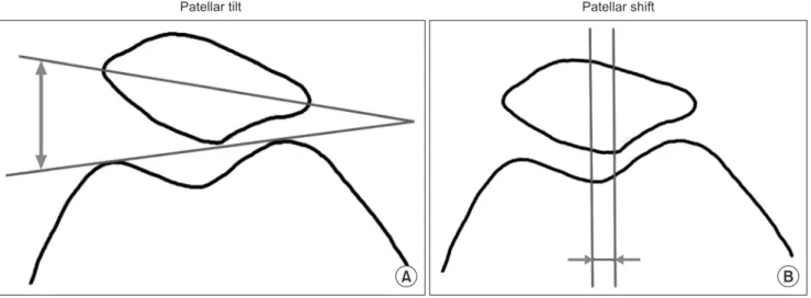
The patellar tilt was measured in the skyline view of the patella by measuring the angle of the line connecting the highest point of the medial and lateral articular surfaces of femoral prostheses (Fig. 2A). This angle is described as (+) if opened medially and as (–) if opened laterally. Lateral tilt was defined as ≥ +5°, and medial tilt as ≤ –5°. Interme- diate values were defined as neutral.15) Patellar shift was evaluated by measuring the distance between the vertical line drawn through the center of patella and the vertical line crossing the center of the femoral trochlear surface (Fig. 2B). Displacement was defined as a distance between 5 mm and 10 mm. A shift of ≥ 10 mm was considered subluxation. Dislocation was defined as complete loss of contact with the joint surface.16) Patellofemoral complica- tions such as anterior knee pain, patellar clunk syndrome, symptomatic subluxation, distal pole avulsion fracture, and lateral facet fracture were evaluated during follow-up periods.17,18)
Statistical Analysis
Potential relationships between anterior knee pain and age, body mass index (BMI), Knee Society knee and function scores, postoperative flexion contracture, the postoperative range of motion, postoperative alignment between the fe- mur and tibia, postoperative patellar tilt, and postoperative patellar displacement at final follow-up and gender were investigated using the Mann-Whitney U-test or Fisher ex- act test. A p-value of < 0.05 was considered significant.
Kaplan-Meier survival analysis with 95% confidence intervals (CIs) was performed on all knees with a mini- mum 10-year follow-up using radiographic failure, revi- sion or pending revision for any reason as a primary end point.19) We conducted 2 analyses: a best-case scenario (in Table 1. Demographic Data
Characteristic Value
No. of patients 54 (80 knees)
Gender (male:female) 4 (85.7) : 50 (14.3)
Age (yr) 66.0 ± 7.05 (48–77)
Body mass index (kg/m2) 24.7 ± 3.14 (17.1–32.9)
Follow-up (mo) 124.6 ± 3.37 (120–132)
Diagnosis
Primary OA 52 (78 knees) (97.5)
Post-traumatic OA 1 (1 knee) (1.25)
Rheumatoid arthritis 1 (1 knee) (1.25) Preoperative OA change of patellae
(ICRS classification)
Grade 2 5 (6 knees)
Grade 3 27 (37 knees)
Grade 4 22 (37 knees)
Preoperative radiologic OA change of patellofemoral joint (K-L radiologic grade)
Grade 1 10 knees
Grade 2 39 knees
Grade 3 26 knees
Grade 4 5 knees
Final follow-up radiologic OA change of patellofemoral joint (K-L radiologic grade)
Grade 1 1 knee
Grade 2 34 knees
Grade 3 38 knees
Grade 4 7 knees
Preoperative
Knee score 27.2 ± 1.28 (0–50)
Function score 33.4 ± 1.52 (0–55)
Flexion contracture (°) 8.4 ± 9.40 (0–30)
Further flexion (°) 121.8 ± 21.29 (30–150)
Range of motion (°) 113.4 ± 27.20 (30–150)
Postoperative
Flexion contracture (°) 1.3 ± 2.70 (0–10) Further flexion (°) 125.9 ± 15.52 (50–145) Range of motion (°) 124.6 ± 16.68 (50–145) Values are presented as number (%) or mean ± standard deviation (range).
OA: osteoarthritis, ICRS: International Cartilage Repair Society, K-L: Kellgren- Lawrence.
which all patients lost to follow-up were assumed to have a good outcome); and a worst one assuming that those lost to follow-up were failures. For all analyses we used SPSS ver. 15.0 (SPSS Inc., Chicago, IL, USA).
RESULTS
The mean Knee Society knee score improved from 27.2 points preoperatively (range, 0 to 50 points) to 88.5 points at the final follow-up (range, 45 to 100 points) in the 54 patients (80 knees) with a minimum follow-up of 10 years.
Clinical outcomes were classified as excellent or good for 72 (90%), fair for 3, and poor for 4. Mean preoperative function score improved from 35.9 points (range, 0 to 55 points) to 81.6 points (range, 40 to 100 points) at final fol- low-up. At the time of the most recent follow-up, the mean range of motion after TKA was improved from 113.4° to 124.6° (p < 0.001). Functional outcomes were classified as excellent or good for 66 (82.5%), fair for 8 (10%), and poor for 6 (7.5%). All patients had varus deformity preop- eratively when the hip-knee-ankle angle was measured on the long standing radiograph. At the latest postoperative follow-up, 70 knees were in the neutral position, 7 knees had varus angle of < 5° and 3 knees had valgus angle of < 5°.
Anterior knee pain was observed in 6 of the 80 knees (6/54 patients; 7.5%) at final follow-up. However, all 6 patients with patellar pain were able to climb up and down stairs slowly. No significant relation between the anterior knee pain group and no-pain group was found in parameters including age (p = 0.438), gender (p = 0.561), BMI (p = 0.146), preoperative and postoperative Knee Society knee score (p = 0.142, p = 0.103, respectively),
preoperative range of motion (p = 0.760), postoperative range of motion (p = 0.664), and all radiographic param- eters at final follow-up. However, preoperative (p = 0.023) and postoperative (p = 0.048) Knee Society knee function score was significantly decreased in the anterior knee pain group (Table 2).
A non-progressive radiolucent line (a radiographic demarcation of ≥ 2 mm) was observed in 1 femoral (1.2%) and 2 tibial (2.5%) components during serial follow-up.
A small osteolytic lesion of the femoral metaphysis was observed in 1 knee, but did not progress on serial radio- graphs. All femoral components and 78 tibial components were well fixed without loosening at the final follow-up.
No obvious tibial insert polyethylene wear was observed.
Two tibial components failed due to loosening at 5 years and 7 years, respectively. One of 2 patients underwent revision surgery at 7.5 years after index surgery, but the other patient refused revision surgery for personal reasons at 5.3 years postoperatively
Postoperative complications included 2 knees with periprosthetic fracture and 1 knee with periprosthetic joint infection. Two periprosthetic comminuted fractures of the femoral shaft were sustained at 2 years and 5 years, respec- tively; 1 patient was treated with a flexible intramedullary nail and the other with a plate and screws. The fractures united uneventfully and femoral components were still well-fixed at the final follow-up. One low-grade infection at 11 months postoperatively was treated by 2-stage reim- plantation. In the best-case scenario, survival at 10 years was 96.7% (95% CI, 93.3 to 99.9), and in the worst-case scenario, it was 86.0% (95% CI, 79.2 to 92.8) (Fig. 3).
Patellar shift Patellar tilt
A B
Fig. 2. Patellar tilt (A) and patella shift (B) were measured from the skyline view.
DISCUSSION
Single radius TKA was introduced in 1996 to reduce patel- lofemoral joint complications and to reproduce more physiologic kinematics. To date, excellent mid-term sur- vival outcomes of 95% to 99% were reported for single radius TKA.4-8) However, there are no reports of long-term survivorship and patellofemoral joint complications. To the best of our knowledge, this was the first study to ad-
dress long-term survivorship of this prosthesis, the preva- lence of anterior knee pain for this patella friendly implant, and to determine the relationship between anterior knee pain and specific radiological factors. We found 7.5% of anterior knee pain, no patellofemoral complications, and 96.7% of survivorship at the minimum 10 year follow-up.
Patellofemoral complications are an important type of mechanical failure after TKA, because they can impact Table 2. Clinical and Radiologic Parameters in Patients with or without Anterior Knee Pain
Characteristic No pain group Anterior knee pain group p-value
No. of knees (%) 74 (92.5) 6 (7.5) -
Gender (male:female) 4:70 0:6 0.561
Age (yr) 68.3 ± 3.40 65.4 ± 7.10 0.438
Body mass index (kg/m2) 21.8 ± 3.32 24.9 ± 3.08 0.146
Preoperative
Knee Society score 41.3 ± 7.50 26.5 ± 12.66 0.142
Function score 46.3 ± 1.44 32.7 ± 14.99 0.023
Varus angle (°) 13.3 ± 4.34 13.0. ± 6.49 0.045
Range of motion (°) 113.1 ± 28.00 116.7 ± 15.06 0.760
Postoperative
Knee Society score 97.0 ± 4.76 88.5 ± 11.94 0.103
Function score 95.0 ± 5.77 83.3 ± 13.36 0.115
Varus angle (°) –3.2 ± 2.31 –3.1 ± 2.48 0.115
Range of motion (°) 125.6 ± 15.92 129.2 ± 9.70 0.664
Preoperative OA change of patellae 0.421
Grade 2 5 1
Grade 3 33 4
Grade 4 36 1
Postoperative patella tilt 4.75 ± 2.06 6.32 ± 3.47 0.360
Lateral tilt 35 3
Neutral position 39 3
Medial tilt 0 0
Postoperative patella shift 2.60 ± 1.74 2.63 ± 1.58 0.419
No shift 63 6
Subluxation 11 0
Displacement 0 0
Values are presented as number (%) or mean ± standard deviation.
OA: osteoarthritis.
functional activities, such as, stair climbing and squat- ting.18,20) Although the causal factors of patellofemoral complications were multifactorial, the design of the Scor- pio knee system such as a single radius femoral compo- nent and a deep patella groove to reduce patellofemoral contact stress might be an advantage of patellofemoral problems.6) Kolisek and Barnes8) evaluated anterior knee pain and patellofemoral complications following the Scor- pio single radius posterior-stabilized TKA with patellar re- surfacing, and experienced 1 patellar fracture in 103 knees.
Among the 86 knees included with a minimum follow-up of 4 years, the incidence of anterior knee pain was 5%. No patellofemoral complication, such as dislocation or patel- lar fracture were observed in our study. Six patients (7.5%) complained of mild anterior knee pain after a minimum follow-up of 10 years. However, we could not find definite
mechanical causal factors such as soft tissue impingement, implant malrotation, patellar subluxation, and displace- ment in these 6 patients. The worse preoperative and post- operative function score in the anterior knee pain group is not causal but consequential. Therefore, it was difficult to explain the reasons of anterior knee pain in this study.
Prevalence of anterior knee pain and revision rate in our cohort is similar to that reported by Kolisek and Barnes8) and lower than those in studies using multi-radius total knee system with non-resurfaced patella (Table 3).21-26)
Patellar resurfacing during TKA remains controver- sial. Recently, Li et al.27) found that overall rates of postop- erative anterior knee pain of 12.9% and 24.1% for those that underwent and for those who did not undergo patel- lar resurfacing, respectively, among a total 1,421 knees.
However Garneti et al.28) found rates of anterior knee pain of 25% and 7% in their resurfaced and non-resurfaced groups in 121 patients that underwent Scorpio single radi- us cruciate retaining TKA. Liu et al.29) reported no advan- tage of patellar resurfacing. They performed a prospective randomized trial on patellar reshaping versus resurfacing in TKA in 133 patients with a minimum 7 years follow- up, and found rates of anterior knee pain of 12.4% and 14.7% in their reshaping and resurfacing groups, respec- tively. Although we did not perform patellar resurfacing surgery, the 7.5% rate of anterior knee pain was similar or lower than published data.27,28) Several factors probably contribute to the development of anterior knee pain, but our results suggested differences in cohort characteristics.
The current study population was predominantly female, with preoperative varus deformity, and less posttraumatic and rheumatoid arthritis than previously published study populations, which might be a positive protective factor.
Regarding clinical outcomes of single radius total knee prostheses, Abbas and Gunn4) performed 125 poste-
Table 3. Rates of Anterior Knee Pain and Reoperation due to Patellofemoral Complications after TKA Using Multi-Radius Total Knee System with Non-Resurfaced Patella
Study TKA implant type No. of cases Follow-up (yr) Pain (%) Revision (%)
Wood et al. (2002)21) MG-II 128 4.0 31.0 12
Waters and Bentley (2003)22) PFC 231 5.3 25.1 4.8
Burnett and Bourne (2004)23) AMK 48 10.8 25.0 6.0
Campbell et al. (2006)24) MG-II 54 10.0 43.0 3.7
Smith et al. (2008)25) Profix 86 4.4 21.0 1.2
Burnett et al. (2009)26) MG-II 60 10.0 16.0 12
TKA: total knee arthroplasty.
Fig. 3. Kaplan-Meier survival rate in the best-case scenario (with implant revision or loosening as endpoints) was 96.7% (95% confidence intervals [CI], 93.3 to 99.9) and in the worst-case scenario was 86.0% (95% CI, 79.2 to 92.8).
0 100
80
60
40
20
Survivalrate(%)
Months since operation 0
132
20 40 60 80 100 120
Revision Loosening
Worst-case scenario
} Best-case scenario
rior-stabilized Scorpio TKAs and only performed 1 revi- sion surgery, in which no aseptic loosening was apparent.
The survival rate at 8 years was 99.3%.4) Borrione et al.5) reported the results of a multicenter study that included 602 knees treated with a Scorpio TKA. They performed 18 prosthetic component revisions, which included 10 knees for infection, 6 knees for prosthesis loosening, 1 knee for an oversized tibial component, and 1 knee for painful stiff- ness. They reported survival rates at 6 years of 95.2% with revision for any reason, and 98.3% with revision for me- chanical failure such as aseptic loosening, oversized tibial component, and painful stiffness. In the present study, the survival rate at a minimum follow-up of 10 years was 96.7% with revision surgery for aseptic loosening and in- fection as the end point, which demonstrated that the ce- mented single radius posterior-stabilized total knee pros- thesis showed excellent radiologic stability during mid- to long-term follow-up.
This result was consistent with the survival rates of other modern design of cemented posterior-stabilized to- tal knee prostheses after long-term follow-up. Rand et al.2) evaluated 11,606 primary TKA at 10 years after index sur- gery and found that the survival rate was 91%. Rasquinha et al.30) evaluated 105 knees after posterior-stabilized modular knee arthroplasty with a minimum follow-up of 10 years. Revision surgeries were performed in 5 knees because of infection in 2, dislocation in 1, and femoral osteolysis in 2. They reported a 94.6% survival rate after a mean of 12 years with failure for any reason as the end point.
The present study had several notable limitations.
First, our cohort was rather small, and of the 71 patients (103 knees) initially considered, 12 (15 knees) died dur- ing follow-up and 12 (14 knees) were lost or refused to undergo regular radiologic evaluations. Thus, the analysis was performed on only 78% of eligible patients, which may have influenced our results. Nevertheless, our ob- served survival rate was comparable with published values.
Second, our data has some limits to generalization because the study cohort was very selective with predominantly women and all subjects had preoperative varus deformity.
It is known that sores for males and valgus knee result in inferior clinical outcomes. Third, this study had no con- trol group making it impossible to compare the clinical outcome regarding patellofemoral complications; further- more, we did not evaluate anterior knee pain in this study.
Therefore, the study has limited value of the status of ante- rior knee pain.
In summary, the 96.7% survival rate of Scorpio single radius posterior-stabilized TKA without patellar resurfacing at a minimum follow-up of 10 years, is en- couraging. The low rates of implant loosening and 7.5%
of anterior knee pain as a patellofemoral complications are comparable with those reported for other current total knee prosthesis.
CONFLICT OF INTEREST
No potential conflict of interest relevant to this article was reported.
REFERENCES
1. Archibeck MJ, White RE Jr. What's new in adult reconstruc- tive knee surgery. J Bone Joint Surg Am. 2006;88(7):1677- 86.
2. Rand JA, Trousdale RT, Ilstrup DM, Harmsen WS. Factors affecting the durability of primary total knee prostheses. J Bone Joint Surg Am. 2003;85(2):259-65.
3. Callaghan JJ, Rosenberg AG, Rubash HE, Simonian PT, Wickiewicz TL, eds. The adult knee. Philadelphia: Lippin- cott Williams & Wilkins; 2003.
4. Abbas D, Gunn RS. Medium-term results of the Scorpio to- tal knee replacement. Knee. 2006;13(4):307-11.
5. Borrione F, Bonnevialle P, Mabit C, et al. Scorpio single ra- dius total knee arthroplasty: a minimal five-year follow-up multicentric study. Int Orthop. 2011;35(12):1777-82.
6. D'Lima DD, Poole C, Chadha H, Hermida JC, Mahar A,
Colwell CW Jr. Quadriceps moment arm and quadriceps forces after total knee arthroplasty. Clin Orthop Relat Res.
2001;(392):213-20.
7. Gomez-Barrena E, Fernandez-Garcia C, Fernandez-Bravo A, Cutillas-Ruiz R, Bermejo-Fernandez G. Functional per- formance with a single-radius femoral design total knee arthroplasty. Clin Orthop Relat Res. 2010;468(5):1214-20.
8. Kolisek FR, Barnes CL. Scorpio posterior-stabilized knee system: 5-year clinical and functional results. J Arthroplasty.
2006;21(8):1187-92.
9. Mahoney OM, Kinsey TL. 5- to 9-year survivorship of sin- gle-radius, posterior-stabilized TKA. Clin Orthop Relat Res.
2008;466(2):436-42.
10. Insall JN, Dorr LD, Scott RD, Scott WN. Rationale of the Knee Society clinical rating system. Clin Orthop Relat Res.
1989;(248):13-4.
11. Insall JN, Hood RW, Flawn LB, Sullivan DJ. The total con- dylar knee prosthesis in gonarthrosis: a five to nine-year fol- low-up of the first one hundred consecutive replacements. J Bone Joint Surg Am. 1983;65(5):619-28.
12. Baldini A, Anderson JA, Zampetti P, Pavlov H, Sculco TP.
A new patellofemoral scoring system for total knee arthro- plasty. Clin Orthop Relat Res. 2006;452:150-4.
13. Meftah M, Jhurani A, Bhat JA, Ranawat AS, Ranawat CS.
The effect of patellar replacement technique on patellofem- oral complications and anterior knee pain. J Arthroplasty.
2012;27(6):1075-80.e1.
14. Outerbridge RE. The etiology of chondromalacia patellae. J Bone Joint Surg Br. 1961;43(4):752-7.
15. Grelsamer RP, Bazos AN, Proctor CS. Radiographic analysis of patellar tilt. J Bone Joint Surg Br. 1993;75(5):822-4.
16. Keblish PA, Varma AK, Greenwald AS. Patellar resurfacing or retention in total knee arthroplasty: a prospective study of patients with bilateral replacements. J Bone Joint Surg Br.
1994;76(6):930-7.
17. Harwin SF. Patellofemoral complications in symmetrical total knee arthroplasty. J Arthroplasty. 1998;13(7):753-62.
18. Yau WP, Wong JW, Chiu KY, Ng TP, Tang WM. Patellar clunk syndrome after posterior stabilized total knee arthro- plasty. J Arthroplasty. 2003;18(8):1023-8.
19. Kaplan EL, Meier P. Nonparametric estimation from incom- plete observations. J Am Stat Assoc. 1958;53(282):457-81.
20. Ip D, Wu WC, Tsang WL. Comparison of two total knee prostheses on the incidence of patella clunk syndrome. Int Orthop. 2002;26(1):48-51.
21. Wood DJ, Smith AJ, Collopy D, White B, Brankov B, Bul- sara MK. Patellar resurfacing in total knee arthroplasty:
a prospective, randomized trial. J Bone Joint Surg Am.
2002;84(2):187-93.
22. Waters TS, Bentley G. Patellar resurfacing in total knee arthroplasty: a prospective, randomized study. J Bone Joint Surg Am. 2003;85(2):212-7.
23. Burnett RS, Bourne RB. Indications for patellar resurfacing in total knee arthroplasty. Instr Course Lect. 2004;53:167- 86.
24. Campbell DG, Duncan WW, Ashworth M, et al. Patellar re- surfacing in total knee replacement: a ten-year randomised prospective trial. J Bone Joint Surg Br. 2006;88(6):734-9.
25. Smith AJ, Wood DJ, Li MG. Total knee replacement with and without patellar resurfacing: a prospective, randomised trial using the profix total knee system. J Bone Joint Surg Br.
2008;90(1):43-9.
26. Burnett RS, Boone JL, Rosenzweig SD, Steger-May K, Bar- rack RL. Patellar resurfacing compared with nonresurfacing in total knee arthroplasty: a concise follow-up of a random- ized trial. J Bone Joint Surg Am. 2009;91(11):2562-7.
27. Li S, Chen Y, Su W, Zhao J, He S, Luo X. Systematic review of patellar resurfacing in total knee arthroplasty. Int Orthop.
2011;35(3):305-16.
28. Garneti N, Mahadeva D, Khalil A, McLaren CA. Patellar resurfacing versus no resurfacing in Scorpio total knee ar- throplasty. J Knee Surg. 2008;21(2):97-100.
29. Liu ZT, Fu PL, Wu HS, Zhu Y. Patellar reshaping versus re- surfacing in total knee arthroplasty: results of a randomized prospective trial at a minimum of 7 years' follow-up. Knee.
2012;19(3):198-202.
30. Rasquinha VJ, Ranawat CS, Cervieri CL, Rodriguez JA. The press-fit condylar modular total knee system with a poste- rior cruciate-substituting design: a concise follow-up of a previous report. J Bone Joint Surg Am. 2006;88(5):1006-10.
![Fig. 3. Kaplan-Meier survival rate in the best-case scenario (with implant revision or loosening as endpoints) was 96.7% (95% confidence intervals [CI], 93.3 to 99.9) and in the worst-case scenario was 86.0% (95% CI, 79.2 to 92.8)](https://thumb-ap.123doks.com/thumbv2/123dokinfo/5218297.122716/6.909.83.827.915.1104/survival-scenario-revision-loosening-endpoints-confidence-intervals-scenario.webp)
