pISSN 1598-9992 eISSN 2233-6869
CASE REPORT
진단적 대장 내시경 시행 후 발생한 공기후복막, 종격동기종, 기흉, 피하공기증
이희성, 박환희, 김주석, 강선형, 문희석, 성재규, 이병석, 정현용
충남대학교 의과대학 ․ 의학전문대학원 내과학교실
Pneumoretroperitoneum, Pneumomediastinum, Pneumothorax, and Subcutaneous Emphysema after Diagnostic Colonoscopy
Hee Sung Lee, Hwan Hee Park, Ju Seok Kim, Sun Hyung Kang, Hee Seok Moon, Jae Kyu Sung, Byung Seok Lee and Hyun Yong Jeong
Department of Internal Medicine, Chungnam National University School of Medicine, Daejeon, Korea
Colonoscopy is a commonly performed endoscopic procedure. Although it is generally considered to be safe, serious complications, such as colorectal perforation, can occur. Most colonic perforations are intraperitoneal and cause pneumoperitoneum with acute abdominal pain as the initial symptom. However, extraperitoneal perforations with pneumoretroperitoneum may happen, albeit rarely, with atypical initial symptoms. We report a rare case of rectosigmoid perforation occurring after diagnostic colonoscopy that devel- oped into pneumoretroperitoneum, pneumomediastinum, pneumothorax, and subcutaneous emphysema, with a change in voice and neck swelling as the initial symptoms. The patient was successfully treated with endoscopic closure of the perforation and con- servative management. (Korean J Gastroenterol 2017;70:145-149)
Key Words: Perforation; Pneumoretroperitoneum; Pneumomediastinum; Pneumothorax; Subcutaneous emphysema
Received July 7, 2017. Revised August 15, 2017. Accepted August 28, 2017.
CC This is an open access article distributed under the terms of the Creative Commons Attribution Non-Commercial License (http://creativecommons.org/licenses/
by-nc/4.0) which permits unrestricted non-commercial use, distribution, and reproduction in any medium, provided the original work is properly cited.
Copyright © 2017. Korean Society of Gastroenterology.
교신저자: 문희석, 35015, 대전시 중구 문화로 282, 충남대학교 의과대학 ․ 의학전문대학원 내과학교실
Correspondence to: Hee Seok Moon, Department of Internal Medicine, Chungnam National University School of Medicine, 282 Munwha-ro, Jung-gu, Daejeon 35015, Korea. Tel: +82-42-280-7143, Fax: +82-42-257-5753, E-mail: [email protected]
Financial support: None. Conflict of interest: None.
INTRODUCTION
Colonoscopy is a commonly performed endoscopic proce- dure to evaluate and treat various colorectal diseases.1 Although it is generally considered to be a safe procedure, it does have a risk of a potentially life-threatening complication, colorectal perforation.2
Colorectal perforation usually appears as pneumoperito- neum, if it occurs in the intraperitoneal portion of the organ.
Rarely, however, colorectal perforation may cause an air leak into the retroperitoneal space, causing pneumoretroperitoneum
if the perforation site is located in the regions attached to the extraperitoneal space, such as the posterior walls of the sig- moid, rectosigmoid, rectum, ascending, or descending colon.3 Pneumoretroperitoneum can further progress to pneumo- mediastinum, pneumothorax, and subcutaneous emphyse- ma, especially if there is a large amount of air leakage through the perforation site.4
When colorectal perforation occurs, the patient typically complains of abdominal pain as an initial symptom. However, in case of extraperitoneal perforation, atypical symptoms, such as subcutaneous swelling with crepitus, chest dis-
Fig. 1. Contrast-enhanced computed tomography of the chest showing. (A) Pneumomediastinum, subcutaneous emphysema. (B) Pneumothorax.
comfort, or dyspnea may develop.
We report a patient presenting a change in voice and neck swelling as chief complaints after diagnostic colonoscopy and esophagogastroduodenoscopy. The patient was diag- nosed with a rectosigmoid perforation that led to pneumo- retroperitoneum, pneumomediastinum, pneumothorax, and subcutaneous emphysema. The patient was successfully managed with non-surgical treatment.
CASE REPORT
A 64-year-old woman was transferred to our emergency de- partment from a private clinic with chief complaints of a change in voice and neck swelling that developed after diag- nostic colonoscopy and esophagogastroduodenoscopy, which were performed two hours prior to the onset of symptoms.
Other symptoms included mild chest discomfort. The patient had a medical history of hypertension and diabetes mellitus.
The initial vital signs were blood pressure of 176/107 mmHg, a pulse rate of 76/min, a respiration rate of 22/min, and a body temperature of 37.8°C. The patient did not have a febrile sensation. With regard to the initial lab findings, her complete blood count was normal (white blood cell count 5,350/mm3, hemoglobin 14.0 g/dL, platelets 322,000/mm3), blood urea ni- trogen and creatinine were normal (17.5/0.69 mg/dL), and blood C-reactive protein was normal (0.29 mg/dL). The initial arterial blood gas analysis revealed a slight hypoxia (pH 7.48, pCO2 34 mmHg, pO2 68 mmHg, HCO2 25.3 mmHg, SpO2
95%). Other lab findings were within normal ranges. No peri- toneal irritation or respiratory distress signs were found upon physical examination. Crepitus was palpated around the neck.
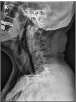
A posteroanterior chest x-ray and contrast-enhanced com- puted tomography (CT) of the chest showed large and diffuse subcutaneous emphysema around the neck and thorax wall, as well as pneumoretroperitoneum, pneumomediastinum, pul- monary interstitial emphysema, and pneumothorax (Fig. 1). An erect and supine abdominal x-rays also revealed pneumo- retroperitoneum (Fig. 2). Anteroposterior and both lateral neck x-rays revealed prevertebral air and extensive subcuta- neous emphysema around the neck (Fig. 3). Considering the dis- tribution of free air, perforation of the extraperitoneal portion of the gastrointestinal organ attached to the retroperitoneal cavity was suspected.
To rule out any possibilities of esophagus perforation, esophagography was performed. There was no evidence of contrast media leakage from the esophagus into the thoracic cavity. A colonoscopy was performed, with the quality of bow- el preparation being fair. A visible perforation was found at the rectosigmoid colon (Fig. 4A). Endoscopic clipping was performed, and the perforation site was closed successfully with seven endoscopic clips (Fig. 4B).
After successful primary closure, we decided to manage the patient conservatively because the patient showed sta- ble vital signs and was in good general condition without signs of peritoneal irritation. The patient was treated with
A B
Fig. 4. Colonocopic finding. (A) The perforation was found at the rectosigmoid colon. (B) Endoscopic clipping was performed to close the perforation.
Fig. 3. Lateral neck x-ray showing prevertebral air and subcuta- neous emphysema around the neck.
Fig. 2. Erect abdominal x-ray showing pneumoretroperitoneum.
bowel rest, intravenous nutrition support, systemic anti- biotics, oxygen, and symptomatic care. A closed thoracotomy was considered due to pneumothorax, but was not per- formed since the amount of air in the thorax was relatively small and she did not suffer from any symptoms of respira- tory distress.
Over the course of conservative care, symptoms improved, and the amount of free air gradually decreased according to simple x-rays. By the 8th day in hospital, abdominopelvic CT showed that pneumoretroperitoneum and pneumomediastinum were markedly decreased since the last CT scan, while pneu-
mothorax was eliminated. The patient’s voice also improved, and neck swelling and fever also subsided. On the 10th day in hospital, free air was no longer visible on simple x-rays. Oral intake was initiated and did not cause any problems. The pa- tient was then discharged with oral antibiotics. The patient was later followed-up at our outpatient clinic and was confirmed to be fully recovered without any further complications.
A B
DISCUSSION
Colorectal perforation is a serious complication of colono- scopy and can result in extended hospital stays, operations, peritonitis, sepsis, multiple organ failure, and even death.5 A retrospective review of cases between 1980 and 2006 showed that when iatrogenic colonic perforations were man- aged operatively, the morbidity and mortality rates were 35%
and 7%, respectively.6 Other studies have reported that the mortality rate can be as high as 25%.7 The rate of perforation during colonoscopy has been reported to be 0.1-0.3%.8 Therapeutic colonoscopy is known to have an increased risk of perforation compared with diagnostic-only colonoscopy.
The perforation rate has been reported to be about 0.16% for diagnostic colonoscopies and about 0.44% for therapeutic colonoscopies.9 However, a review conducted by Iqbal et al.
argued that a diagnostic colonoscopy does not necessarily car- ry a lower rate of perforation than therapeutic colonoscopy.6
Perforations can be intraperitoneal, extraperitoneal, or a combination of both, depending on the location of the perfo- ration site; however, it has been shown that intraperitoneal perforations are much more common.10 In the lower gastro- intestinal tract, extraperitoneal organs include ascending and descending colon, posterior walls of the sigmoid colon, and rectum.3 The sigmoid colon, including rectosigmoid, is known as the most frequent site of all types of colorectal per- foration, followed by the cecum.6
The risk factors of iatrogenic colonic perforation are elderly patients, diverticulosis, severe colitis, inflammatory bowel disease, malignancy, pelvic adhesions due to history of ab- dominal or pelvic surgery, radiation therapy or inflammation, and lack of experience of the physician.11-14 The patient in our case had none of the above risk factors.
In our case, perforation caused an intraluminal air leak that accumulated in the retroperitoneal space. Since the ret- roperitoneum, mediastinum and thorax are anatomically connected, extraluminal free air reached various compart- ments of the body and caused pneumomediastinum, pneu- mothorax and subcutaneous emphysema.15,16 Free air in one of these spaces can travel to nearby structures, including the fascial planes and large vessels.17 Large amount of air may rupture the mediastinal pleura, penetrating the pleural cavity.18 Alternatively, any free air in the peritoneal cavity can per- meate through small diaphragmatic fenestrations and enter
the pleural space.18 Pneumothorax resulting from colonic perforations is very rare. Indeed, a review by Zeno et al. found only nine cases of pneumothorax; of which only two occurred after diagnostic colonoscopy.11
Patients with perforations most commonly suffer from ab- dominal pain. Fever with leukocytosis and tachycardia may also develop. Physical examination may reveal a rigid abdo- men with tenderness, rebound tenderness, and muscle guarding. However, in extraperitoneal perforation cases, ab- dominal pain may not be the initial symptom. Tiwari et el. con- ducted a review of 32 extraperitoneal perforation cases and concluded that subcutaneous emphysema of the neck, face, or upper chest was the most common clinical finding, with a prevalence rate of 65%.17 Palpable crepitus is usually accom- panied with subcutaneous emphysema. Abdominal pain, on the other hand, was seen in only 34%, and dyspnea was seen in 25%.17 Nearly 10% of patients remained asymptomatic.17 Therefore, even if there is no abdominal pain, perforation should be considered if atypical symptoms occur after a colonoscopy. Rare but possible atypical complications in- clude pneumopericardium, periorbital swelling, pharyngeal swelling, and pneumoscrotum.19,20
Symptoms of perforation may appear after several hours.20 A review conducted by Tiwari et al. revealed that 52% of perfo- rations were detected immediately or within 1 hour, whereas 29% were found within 1-24 hoursand 19% found after 24 hours from the procedure.17 In our case, perforation was not detected immediately when it first happened at the private clinic because the patient did not show any symptoms during and immediately following the colonoscopy. Therefore, upon completion of the colonoscopy, esophagogastroduodeno- scopy was performed next as planned. After the esoph- agogastroduodenoscopy, neck swelling was then observed as the initial symptom, accompanied with a change in voice and mild chest discomfort. Because of these unusual symp- toms with a delayed onset, the patient was transferred to our hospital for further evaluation.
Endoscopic closure was done successfully in our case be- cause the perforation size remained relatively small, as the patient was transferred to our hospital in a timely manner. In ad- dition, bowel preparation was done properly so that fecal materi- als did not contaminate the peritoneal space. Furthermore, sys- temic antibiotics were quickly administered. All of these fac- tors contributed to a successful treatment of perforation
without the need for surgery.
In conclusion, we presented a rare case of rectosigmoid perforation occurring after a diagnostic colonoscopy that led to the extensive amount of extraperitoneal free air develop- ment with voice change and neck swelling as the initial symptoms. Atypical symptoms should not be overlooked by physicians as they can be indicative of perforation.
REFERENCES
1. ASGE Standards of Practice Committee, Fisher DA, Maple JT, et al. Complications of colonoscopy. Gastrointest Endosc 2011;74:
745-752.
2. Panteris V, Haringsma J, Kuipers EJ. Colonoscopy perforation rate, mechanisms and outcome: from diagnostic to therapeutic colonoscopy. Endoscopy 2009;41:941-951.
3. Singh JP, Steward MJ, Booth TC, Mukhtar H, Murray D. Evolution of imaging for abdominal perforation. Ann R Coll Surg Engl 2010;92:182-188.
4. Jung HC, Kim HJ, Ji SB, et al. Pneumoretroperitoneum, pneumo- mediastinum, subcutaneous emphysema after a rectal endo- scopic mucosal resection. Ann Coloproctol 2016;32:234-238.
5. Lohsiriwat V. Colonoscopic perforation: incidence, risk factors, management and outcome. World J Gastroenterol 2010;16:
425-430.
6. Iqbal CW, Cullinane DC, Schiller HJ, Sawyer MD, Zietlow SP, Farley DR. Surgical management and outcomes of 165 colonoscopic perforations from a single institution. Arch Surg 2008;143:701- 706; discussion 706-707.
7. Lüning TH, Keemers-Gels ME, Barendregt WB, Tan AC, Rosman C. Colonoscopic perforations: a review of 30,366 patients. Surg Endosc 2007;21:994-997.
8. Ko CW, Dominitz JA. Complications of colonoscopy: magnitude and management. Gastrointest Endosc Clin N Am 2010;20:659-671.
9. Kim HH, Park SJ, Lee SH, et al. Efficacy of endoscopic sub- mucosal resection with a ligation device for removing small rec- tal carcinoid tumor compared with endoscopic mucosal re- section: analysis of 100 cases. Dig Endosc 2012;24:159-163.
10. Ignjatović M, Jović J. Tension pneumothorax, pneumoretroperitoneum, and subcutaneous emphysema after colonoscopic polypectomy: a case report and review of the literature. Langenbecks Arch Surg 2009;394:185-189.
11. Zeno BR, Shan SA. Colonoscopy-associated pneumothorax: a case of tension pneumothorax and review of the literature. Am J Med Sci 2006;332:153-155.
12. Levy I, Gralnek IM. Complications of diagnostic colonoscopy, up- per endoscopy, and enteroscopy. Best Pract Res Clin Gastroenterol 2016;30:705-718.
13. Ho HC, Burchell S, Morris P, Yu M. Colon perforation, bilateral pneumothoraces, pneumopericardium, pneumomediastinum, and subcutaneous emphysema complicating endoscopic poly- pectomy: anatomic and management considerations. Am Surg 1996;62:770-774.
14. Webb T. Pneumothorax and pneumomediastinum during colonoscopy.
Anaesth Intensive Care 1998;26:302-304.
15. Ball CG, Kirkpatrick AW, Mackenzie S, et al. Tension pneumo- thorax secondary to colonic perforation during diagnostic colo- noscopy: report of a case. Surg Today 2006;36:478-480.
16. Maunder RJ, Pierson DJ, Hudson LD. Subcutaneous and media- stinal emphysema. Pathophysiology diagnosis, and management.
Arch Intern Med 1984;144:1447-1453.
17. Tiwari A, Sharma H, Qamar K, Sodeman T, Nawras A. Recognition of extraperitoneal colonic perforation following colonoscopy: a review of the literature. Case Rep Gastroenterol 2017;11:256- 264.
18. Marwan K, Farmer KC, Varley C, Chapple KS. Pneumothorax, pneumomediastinum, pneumoperitoneum, pneumoretroperitoneum and subcutaneous emphysema following diagnostic colonoscopy.
Ann R Coll Surg Engl 2007;89:W20-W21.
19. Kim BH, Yoon SJ, Lee JY, Moon JE, Chung IS. Subcutaneous em- physema, pneumomediastinum, pneumoretroperitoneum, and pneumoperitoneum secondary to colonic perforation during colonoscopy. Korean J Anesthesiol 2013;65(6 Suppl):S103-S104.
20. Anyfantakis D, Kastanakis M, Karona P, Papadomichelakis A, Bobolakis E. Subcutaneous facial and neck emphysema as first sign of intestinal perforation in a female patient after a routine colonoscopy. Eurasian J Med 2016;48:230-232.