大짧放射線醫學會홉‘ 第 22 卷 第6 YJÆ PÞ. 1083 - 1086, 1986 Journal of Korean Radiological Society, Vol.22, No.6, 1986
先天性 冠狀動眼樓*
양明大學校 醫科大學 放射線科學數室
吳흘熙·金 洪·숲石놈·徐修之
- Abstract-
Congenital Coronary Artery Fistula
Yeon Hee Oh, M.D., Hong Kim, M.D., 5eockil Zeon, M.D. and 500 Jhi Suh, M.D.
Department of Radi%gy, Schoo/ of Medicine, Keimyung University. Taegu, Korea
Congenital coronary artery fistula (CCAF) is communication of a coronary artery or its main branch with one of the atria or ventricles, the coronary sinus, the superior vena cava, or the pulmonary artery
In Korean peoples, only 4 cases of the CCAF were reported as rare as worldwide and authors want to report another case of CCAF, confirmed by operation.
10.years-old girl shows a fistula between sinus node artery of the right coronary artery and right atrium on root aor~ogram with left-to-right shunt and Qp/Qs=1.58, in which simple ligation of the origin of the sinus node artery from right coronary artery was performed‘
AII of the 5 Korean CCAF (4 were previously reported and 1 of authors) were originated from right coronary artery, and of which 4 were opening into Fight ventricle and 1 of authors were into right atrium
Associated cardiac anomaly was noted in only 1 case as single coronary artery Ages were from 9 months of age to 10 years old and no adult lefe case were found.
3 were female and 2 were male patients
績 論
先天性 冠狀動服짧 (Congenital Coronary Artery Fistula; CCAF)는 Krause (1 865)와 Brooks (1886) 에 의해 처음으로 報告된 。 l 래 文敵上 報告가 倫貴한 흉愚으로써 한국에서도 져우 4 例가 報告되었다.
CCAF는 冠狀動服파 心廳의 心室 또는 心房의 어느
* 이 논문은 1986 년도 계명대학교 동산의료원 임상연 쿠버로 이루어졌음.
이 논문은 1986 년 9 월 22 일에 접수하여 1986 년 12 월 8일에 채택되었음.
한 部位 또는 !Iitï動服사이에 짧를 形成하는 흉愚으로써 左右短絡을 냐타냐는 非줌色폼 先天性 心廳훌愚의 일종 이다. 최근에 兩面性 超흘波該斷의 發達로 手術前 양斷 이 可能해졌으나 역시 心職映畵進影術로써 手術前 確該 및 解용U學的 構造의 *4 明파 血力學的 檢훌가 필수척 이 다.
著者들은 양明大學校 醫科大學 放射線科學數室에서 최근에 先天性 冠狀動/lIiÆl 1 예 를 發見하고 文願考察과 함께 報告하는 바이다.
~ 例 報 告 깅 00, 여, 10 세
- 1083 -
人함}iX M없펙샤,~fr ‘녀‘ : 찌;22 상 짜 6 빠 1986
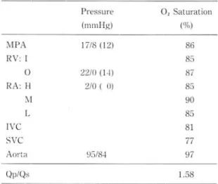
主訴 ; 반복적안 상기도 감염증 Table 1. Carcliac Catheterization Data 過去歷 및 家族歷, 득기샤항 없음.
現病歷, 약 2 년전부터 上氣道 %한었íìË이 頻發하였으 며, 고식적인 치료블 받아오던 가운데,최근 본원 小兒科 外來를 BJ-운하여 心雜흡을 發昆하고, 心!械 檢;안를 위 하 여 入院하였다
Pressure 0
,
Saturation理學的 檢효 所見 ; Jill젠↑'1:. 機械!’8 心쩌t 룹이 흉골 상 부위 우연을 α} 라서 청진되었으며,그 외에는 이상 소견 이 없었마.
心超흡波 所見; 左心房파 左心쫓으I }j잉大, 파bIDR 냐 상始 즙ß의 8E大 즉 心室*1챔缺.Jfl Jî에 양당한 간心室 容:fi'l t갖 쩌의 Tfr見이 냐타났냐.
心導子法 所見 ; (Table 1) 心房에 서의 左右표絡 所 見파 Qp/Qs 1.58 이 보였다.
心藏映畵造影frliÏ ;(Fig. l )上,iè의 여러가시 검사소간
MPA RV ! 0 RA: H
M L IVC SVC Ao
‘
ta Qp/Qs(1ll11l!-!g) ('끼)
17/8 (l2) 86
85
22/0 (1..j) 87
2/0 ( 0) 85
90 85 81 77
95/84 97
1.58
을 좀합하여 우션 心室이그隔1Iik fll 값을 여1 싱하고,끼「비1 쉐位 에서 右心끼써로 떠口되고 있었고, 手術中에 Sinus No- 의 左心室造影術을 실시 하여 右상狀파J!.派의 Sinus No- c1e Artery흔 일시 차단시켰으냐, 삼천도에서 아무런
de artery 가 불규걱적으로 심하게 llt大펀 컷융 확인하 변화가 없었으므로 이환부위의 기시부를 댄純 結삶하였 였고, 心室中隔缺댐은 없였다. 잇달아 t폐JWR 짧上픔11에서 나.
造影뺑j를 주사하여 上記으1 Sinus Node artery 가 긴 댄、者는 手 ~fcJ後 心써t 흡이 完全하게 消失되였고 특떨한 心房무로 開口하고 있는 것을 %認하고, CCAF릎 2 斷 合i:If:JJË 없이 12일만에 退院하였다. 手術後 2 년이 經 하였 다펴할 띠| 까시 아무런 異常 }꺼 見이 없으므로 外來 獅察을
手術所見 및 經過; 左心팅 파뻐의 표면에서 振패이 觸 4' 斷[하였다.
指되었고, 중등도의 右心房 ”잉大가 있있으며,;L; κ[:!Ä파1 IDR의 Sinus Node Artery 가 ll%大되어샤 까心꺼의 깎벽
Fig. 1. Root aortogral1l(A) ancl schellla(B) show irregular ancl tortuous c1ilatation of sinlls nocle artery ancl opening into the right atriul1l (*in B)
- 1084 -
- 吳進‘웰 외 · 先天↑'É 텀狀꾀IJÆ따행 -
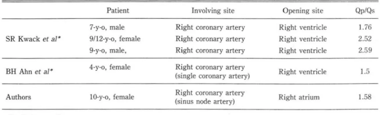
Table 2. Congenital Coronary Artery Fistula in Korean Literatures
Patient lnvolving
siteOpening site Qp/Qs
7.y-o
,male Right coronary artery Right ventricle
1.76 SR Kwack
et aI*9/12-y-o
,female Right coronary artery Right ventricle 2.52 9-y-o
,male
,Right coronary artery Right ventricle 2.59
BH Ahn
et aI*4-y-o
,female Right coronary artery
Right ventricle
1.5(single coronary artery)
Authors 10-y-o
,fema le Right coronary artery
Right atrium
1.58 (sinus node artery)
• . Reference 3)
•• : Reference 4)
考 察
先天性 冠狀動服짧는 冠狀動服 또는 그 分技가 心室 이 냐 心房의 어 느 부위 , 冠狀動服i1PJ, 上行大靜版,또는 뼈 動服과 交通하는 짧가 形成되는 先天性 心魔흉愚으로써
Coronary Arteriocameral Fistula , Congenital Cor-
onary Arteriovenous Fistula
등으로도 불리워 진다.이는
1855
년 Krausel-3) 와1866
년 Brooks1, 2 싸 처 음 으로 報告한 이래 천세계적으로 매우 드물게 보고되어 왔다. 한국인에서도 우미인과 마찬가지로 매우 드운 뾰 例로써 곽등 3) 파 안등에 4) 의해 4 例가 報告되었으며 著 者들의 뾰例와 함하연 5 例가 되어Table
2 와 같마.이는 %生初期의 心觸에 있는
intertrabecular space
가 發育過程에서 서서히 없어져야 히는데 生後까지 그 대로 남아서 생긴마고 한마 6)CCAF 의 起始部에 판한 Levin등5)의 보고는 구미 인 에서 右冠狀動版이
50
%, Md뼈nara등6의 보고에서는 右冠狀動服이 60%라고 하였으내, 적은 효例。 171는 하 지 만 한국인에 서는 5 例 모두가 右冠狀動服에서 起始하 였다. 이는 앞으로 더 않은 연우가 必훨할 것으로 사료 펀다. 또한 著者들의 증례와 같이 대부분의 CCAF는 單 -짧 6)를 形成하지만 多發性인 경우도 2 例가 報告되어있마 7)
CCAF 의 開口 부위는 動.IlIî擾의 起始部位에도 많은 聯|짧性이 있을 것으효 생각되지만,한국인에서는 右心室
4 例, 右心房 1 例로써 左心은 전혀 없었다.
또한 일반적으로 CCAF는 單一缺휩폼이라고 하지만
여 러 가지 先天性 또는 後天性 心職훌愚。l 同伴되 는 報 告1 , 6)도 있으며 單- 冠狀動.IlIî 4) 에서 發生한 증례가 한 국인에서 알려지고 있다.
이의 聽용소견은 기시부위와 開口부위에 따라서 각각 상이해지묘로 특벨한 감옐점은 없다고 하겠으냐,뼈’몹의 좌연에서 心雜륨이 聽該되는 경우가 않으므로 心室中隔 缺휩。l 냐 행ÏJ .IlIî管 開存'llE등과 강벨하여야 한마 5)
CCAF는 행孔의 크기에 따라서 전혀 무증상6)에서 JÜ 不全폼까지 여러 정도의 증상을 일£킬 수 있으며 1 , 2)合 f뺨에 따라서 여러 증/뿔 유발하게 될 수도 있으므로 특이한 임/픔상은 없으냐, 다만 左右短絡의 양에 따라 서 冠狀動服의 虛血現狀을 유발하여
Angina Pector-
iS2, 6)를 일으킬 수 있 다고 한마
흉부 x-선에서는 左右短絡의 양에 짜라서 Jlï1î혈판의 울혈소견。l 나타냐며 , 左心室 및 左心房의 HE大와 右心 의 開口部 하부의 Ðe:大를 볼 수 있 마고 한마J)
근간에 상당히 발달된 兩面超를波檢훌에서 어느 정도 장斷을 예측할 수 있으냐J)著者들의 1fË例와 마찬가지로 左心室 容積 負뼈를 나타내는 경우가 대부분으로써, 心 室中隔缺휩효과외 강별。 1 곤란하므로 역시 心職映畵造 影術을 실시 하여 確該과 머불어 解웹學的 구조 변화를 쐐明하여 야 한다 1, 2)
Shubrooks등 8) 이 14 세 낭아에서 CCAF 가 자연소 실되는 것을 報告하기도 했지만, Liberthson 등 2) 의 報 告에 의하연 오든 CCAF는 수술로써 橋正해 주어 야만 연령이 증가함으로써 생기는 증상의 發現이나 手術 合 션Hî을 줄일 수 있다고 한마. 著者들의 증례에서는 수 술로써 완전교정이 가능하였다.
- 1085 -
大혐放射~醫쩡會託 : 第 22卷 第6 號 1986
結 -ι:、iíf!ll
著者들은 最近에 혈明大學校 醫科大學 放射線科學 敎 室에서 매우 f;ffi월-한 先天性 冠狀動服짧 l 例를 經驗하 고 文敵考察파 함께 報告하는 바이 다.
REFERENCES
1. Neufeld HN, 5chneeweiss A: Coronary artery disease in infants and children. 31-58. Lea and Febigeι Philadelphia 1983
2. Liberthson RR, 5agar K, Berkoben jP, et al.: Congenital cor- onary arteriovenous fiswla. Circulation 59:849-851, 1979
3. Kwack 5R, Roh JR, 5uh KP et al.: Fistula between right cor- onary artery and right ventricle. The Korean journal of Thoracic and Cardiovascular Surgery 15:11ι117, 1982 4. Ahn BH, Lee DJ: Single coronary artery with coronary
arteriovenous fistula. The Korean journal of Thoracic and Cardiovascular surgery 15:366-369, 1982
5. Levin DC, Fellown KE, Abrams HL: Hemodynamically significant primary anomalies of the coronary arteries. Cir- culation 58:27-34, 1978
6. McNamara Jj, Gross RE: Congenital coronary artery fiswla Surg 65:59-69, 1969
7. Rose AG ι1ultiple coronary arterioventricular fiswlae. Cir culation 58:178-181, 1978
8. 5hubrooks 5J. Naggar CA: Spontaneous near closure of coronary artery fistula. Circulation 57:197-199, 1978.