대한내분비외과학회지:제 9 권 제 2 호
□ 증 례 □
Vol. 9, No. 2, June 2009
90
책임저자 : 장항석, 서울시 강남구 언주로 712
135-720, 연세대학교 강남세브란스병원 갑상선암 클리닉
Tel: 02-2019-3370, Fax: 02-3462-5994 E-mail: surghsc@yuhs.ac
게재승인일:2009년 6월 8일
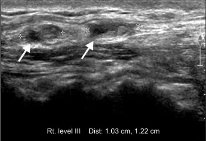
Fig. 1. Preoperative ultrasonographic feature of nodules at the right level III area. Nodules showing heterogenous echogenecity with irregular margin.
갑상선 수질암의 측경부 림프절 절제술 후에 발생한 외상성 신경종
연세대학교 의과대학 세브란스병원 갑상선암센터 외과학교실, 1병리학교실
김국진ㆍ홍순원1ㆍ김법우ㆍ강상욱ㆍ이용상ㆍ남기현ㆍ정웅윤ㆍ장항석ㆍ박정수
Traumatic Neuroma following Lateral Neck Lym- ph Node Dissection in Medullary Thyroid Cancer Patient
Kuk Jin Kim, M.D., Soon Won Hong, M.D.
1, Bub WooKim, M.D., Sang Wook Kang, M.D., Yong Sang Lee,
M.D., Kee-Hyun Nam, M.D., Woong Youn Chung, M.D.,Hang Seok Chang, M.D. and Cheong Soo Park, M.D.
A 57-year-old male patient was admitted for evaluation of an enlarged neck lymph node. Previously, the patient had undergone three operations for recurrent medullary thyroid cancer. In preoperative neck ultrasonography, several nod- ules were identified in right level 3, level 5 and central neck area, which were suspicious for recurrence of thyroid cancer. Selective neck dissection for nodules was per- formed. Pathologic reports for nodules in right level 3 area were consistent with traumatic neuroma. (Korean J Endo-
crine Surg 2009;9:90-92)
Key Words: Medullary thyroid cancer, Lymph node dis-
section, Traumatic neuroma중심 단어:갑상선 수질암, 림프절 절제술, 외상성 신 경종
Departments of Surgery and 1Pathology, Thyroid Cancer Center, Severance Hospital, Yonsei University College of Medicine, Seoul, Korea
증 례
57세 남자 환자가 경부 림프절 종대로 내원하였다. 환자 는 10년 전 타 병원에서 갑상선 수질암으로 갑상선 전절제 술 및 중앙 구획 림프절 절제술을 시행받았고, 그 후 두 차
례 재발하여 각각 우측 및 좌측 측경부 림프절 절제술을 시행받았다.
내원 시에 환자가 호소한 특이 증상은 없었으나, 이학적 검사에서 우측 측경부 3구역(Level III)에 약 1 cm 크기의 단단하지만 압통은 동반되지 않은 종괴가 만져졌다.
수술 전 시행한 경부 초음파 검사에서 우측 측경부 3구역 내부에 불균질한 음영을 보이며 경계가 불분명한 약 0.6 cm, 1.2 cm 크기의 종괴가 관찰되었고, 우측 측경부 5구역 (Level V)에도 유사한 소견의 약 1.3 cm 크기의 종괴가 보였 으며, 우측 중앙 구획 아래쪽에도 약 0.5 cm 크기의 경계가 불분명한 저 음영 종괴가 관찰되었다(Fig. 1). 환자가 관찰 된 병변에 대해 추가적인 검사 없이 수술적인 절제를 원하 여 세침 흡입 검사는 시행하지 않았다.
수술은 중앙 구획, 우측 측경부 3구역 및 5구역의 선택적 림프절 절제술이 시행되었다. 수술 소견에서 중앙 구획과 우측 측경부 5구역에서는 수술 전 초음파와 일치하는 위치 에 크기가 증가한 림프절들이 관찰되었다. 하지만, 우측 측 경부 3구역에서는 림프절과는 다른, 흰색의 단단하고 매끈 한 종괴들이 발견되었는데, 이전의 같은 부위의 림프절 절 제술 시행 후에 발생한 외상성 신경종(traumatic neuroma)으 로 추정되었다(Fig. 2). 최종 병리 결과에서는 중앙 구획 림 프절에서는 갑상선 수질암의 재발이 확인되었고, 우측 측경
김국진 외:갑상선 수질암의 측경부 림프절 절제술 후에 발생한 외상성 신경종
91
Fig. 2. Nodules at the right level 3 area identified during operation. Nodules were hard and had firm texture that different with metastatic lymph nodes.
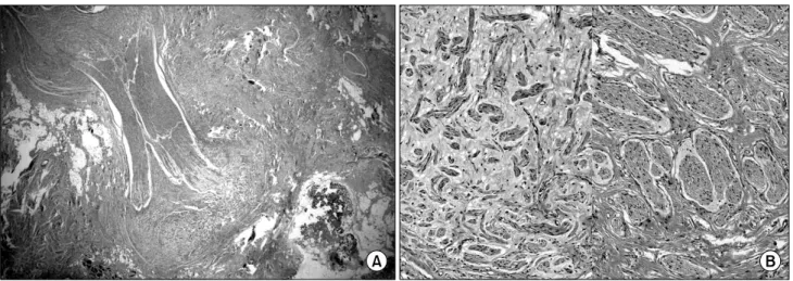
Fig. 3. Histology of the nodule at the right level 3 area. (A) Low power view shows ill defined fibrosis with well formed and haphazardly arranged nerve bundles (H&E, ×12), (B) The haphazardly arranged nerve bundles reveal small to medium sized nerve bundles with myxoid change (H&E, ×100).
부 3구역에서 절제한 종괴들에서는 신경 조직(nerve bundle) 들이 관찰되어 외상성 신경종에 합당한 소견이었다(Fig. 3).
외상성 신경종은 신경이 절단되었을 때 근위부 말단에서 일어나는 신경 조직의 과증식으로 인해 발생하는데, 두경 부 종양으로 인한 경부 림프절 절제술 후 약 1.1∼2.7% 정 도로 보고되고 있다.(1) 외상성 신경종은 림프절 재발로 오 인되어 임상적으로 혼란을 가져오기도 하는데,(2,3) 촉진 혹 은 세침 흡입 검사시 심한 통증을 호소하면 우선 이를 의심 해야 한다. 초음파에서는 불균질한 음영강도를 보이고 내 부에 평행한 줄무늬(parallel echogenic striae)가 관찰되는 경 우가 많다(Fig. 1). 세침 흡입 검사 결과에서는 티로글로불 린 농도가 낮게 측정되고, 말단 신경 조직의 파편들이 관찰 된다. 최종 진단은 조직 병리 검사 결과로 신경 조직을 확인
하면 된다.(4) 하지만 본 증례처럼 갑상선 암이 재발한 수차 례의 과거력이 있거나, 특징적인 촉진시의 통증이 없는 환 자들에서는 임상적으로 외상성 신경종과 재발한 림프절을 구분하기 어려운 경우가 많다. 따라서 각 환자에 대한 외과 의의 임상적인 경험과 영상 의학적, 세침 흡입 검사 소견들 을 종합하여 신중한 판단을 내리는 것이 환자의 불필요한 수술과 그에 따른 위험을 줄일 수 있는 방법일 것이다.
REFERENCES
1) Yabuuchi H, Kuroiwa T, Fukuya T, Tomita K, Hachitanda Y.
Traumatic neuroma and recurrent lymphadenopathy after neck dissection: comparison of radiologic features. Radiology 2004;
92
대한내분비외과학회지:제 9 권 제 2 호 2009
233:523-9.
2) Huang LF, Weissman JL, Fan C. Traumatic neuroma after neck dissection: CT characteristics in four cases. Am J Neur- oradiol 2000;21:1676-80.
3) Iida S, Shirasuna K, Kogo M, Matsuya T. Amputation neur-
oma following radical neck dissection--report of 3 cases. J Osaka Univ Dent Sch 1995;35:1-4.
4) Kwak JY, Kim EK, Kim MJ, Son E. Sonographic features of traumatic neuromas after neck dissection. J Clin Ultrasound 2009;37:189-93.