Copyright © 2013 Korean Neurological Association 61
Print ISSN 1738-6586 / On-line ISSN 2005-5013 http://dx.doi.org/10.3988/jcn.2013.9.1.61 CASE REPORT
J Clin Neurol 2013;9:61-64
Acute Myelitis in a Patient with Vogt-Koyanagi-Harada Disease:
Case Report and Review of the Literature
Shaojuan Gu,a Yu Liu,b Zhi Song,a Xiaohong Zi,a Hao Denga
aDepartment of Neurology and Center for Experimental Medicine, The Third Xiangya Hospital, Central South University, Changsha, China
bDepartment of Anesthesiology, Tumor Hospital Xiangya School of Medicine of Central South University, Changsha, China
Received October 18, 2011 Revised February 14, 2012 Accepted February 14, 2012 Correspondence Hao Deng, MD, PhD Department of Neurology and Center for Experimental Medicine, The Third Xiangya Hospital, Central South University, Changsha, 138 Tongzipo Road, Changsha, HN 410013, China Tel 86-731-8618372 Fax 86-731-8618339 E-mail [email protected]
BackgroundzzVogt-Koyanagi-Harada (VKH) disease is characterized by bilateral granuloma- tous uveitis with neurologic, auditory, and dermatologic manifestations. However, acute myeli- tis complicating VKH disease has rarely been reported.
Case ReportzzA 50-year-old Chinese Han woman presented with difficulty walking, numb- ness on the left side of the body, and difficulty with urination. The patient was diagnosed with incomplete VKH disease and received corticosteroid treatment prior to the neurological presen- tation. Acute myelitis was diagnosed based on both clinical and spinal-cord MRI findings.
ConclusionszzClinicians should consider acute myelitis as a rare possible neurological mani- festation in VKH disease patients, and early systemic administration of corticosteroids will suppress the acute inflammatory process and prevent recurrences. This report raises the possi- bility that VKH disease and acute myelitis share common pathogenic pathways
. J Clin Neurol 2013;9:61-64
Key Wordszz Vogt-Koyanagi-Harada disease, acute myelitis, pathogenesis.
Open Access
cc This is an Open Access article distributed under the terms of the Cre- ative Commons Attribution Non-Commercial License (http://creative- commons.org/licenses/by-nc/3.0) which permits unrestricted non-com- mercial use, distribution, and reproduction in any medium, provided the ori- ginal work is properly cited.
Introduction
Vogt-Koyanagi-Harada (VKH) disease is an idiopathic, multi- system autoimmune disorder characterized by its affects on pigmented tissues in the ocular, auditory, integumentary, and central nervous systems. The prevalence of VKH disease var- ies markedly, and the risk of developing at least one neurologi- cal manifestation exceeds 50%.1 Certain neurological manifes- tations-including aseptic meningitis, encephalitis, encephalo- myelitis, and cranial nerve neuropathy-are occasionally asso- ciated with this disorder, but acute myelitis has rarely been reported. Early systemic administration of corticosteroids will suppress the acute inflammatory process, and prevent recur- rences and the development of complications.
We present a case of VKH disease accompanied by acute myelitis, and review two previously published case reports in an attempt to elucidate the pathogenesis.
Case Report
A 50-year-old Chinese Han woman presented with sudden on- set of difficulty walking, numbness on the left side of the body, and difficulty with urination for 6 days. Twelve days prior to the presentation she had been diagnosed with incomplete VKH disease by an ophthalmologist based on blurred vision in both eyes, bilateral nontraumatic granulomatous iridocyclitis, retinal edema, and the presence of exudates. She had received cortico- steroid treatment (500-mg intravenous methylprednisone for 3 days followed by 300-mg intravenous methylprednisone for 3 days and a tapering course of 80-mg prednisone for 6 days).
Neurological manifestations (headache, tinnitus, difficulty cl- imbing stairs, numbness on the left side of the body, and dys- uria) emerged during the tapering of steroid treatment. She re- called a history of upper respiratory tract infection a month pre- viously but denied any history of vaccination. Physical exami- nations revealed normal vital signs. There was no lymphade- nopathy, or oral or genital ulcers. Her skin and hair showed no vitiligo, poliosis, or alopecia. Neurological examinations show- ed a normal mental status and cranial nerves except for a visu-
Acute Myelitis in a Patient with VKH Disease
62 J Clin Neurol 2013;9:61-64
al acuity of 20/200 bilaterally and papilledema of both eyes in a fundus examination. The strength of both upper and lower limbs was decreased. Her muscle tone was increased. Her deep tendon reflexes were hyperactive without clonus. The Babinski sign was present bilaterally, and her response to the finger-to-nose test was impaired. Her gait was slow and stiffly shuffling. Pinprick sensations were decreased in the left limbs, whereas position sense was preserved. No obvious sensory level was identified. Kernig’s sign was absent. Routine labora- tory evaluation showed all relevant values to be within normal limits. Screening for connective-tissue disease, including C re- active protein, rheumatic factor, antinuclear antibody, C-anti- neutrophil cytoplasmic autoantibody, anti-double-strand DNA, anti-Smith, anti-ribonucleoprotein, anti-Sjogren’s syndrome A, and anti-Sjogren’s syndrome B antibodies, produced normal or negative results. The erythrocyte sedimentation rate was 39 mm/hr. Cerebrospinal fluid (CSF) contained 128 WBCs/mm3 (normal <10/mm3) with 94% mononuclear cells and 4.83 g/L protein (normal 1.5-4.5 g/L). A CSF bacterial culture produced negative results. A nerve conduction study showed mild poly- neuropathy, and visual evoked potentials were normal. A chest X-ray was normal. A brain MRI scan was normal with no cal-
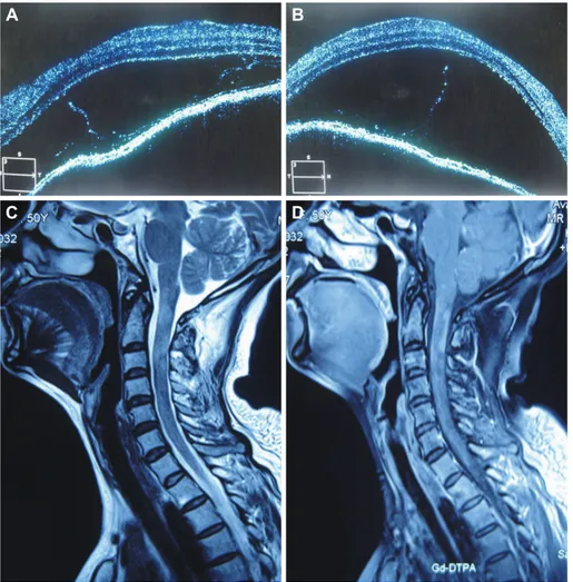
losal or significant periventricular lesions. Optical coherence tomography performed independently by two ophthalmologists revealed bilateral disc edema accompanied by serous retinal detachment (Fig. 1A and B). Spinal-cord MRI (Fig. 1C and D) revealed a hyperintense signal in the T2-weighted sequence with significant gadolinium enhancement between the C1-3 vertebrae in the cervical cord. The patient was diagnosed as aseptic meningitis with acute myelitis complicating VKH dis- ease, and was given pulse steroid therapy again (intravenous administration of 1000 mg of methylprednisolone for 3 days, 500-mg intravenous methylprednisolone for 10 days, 250-mg intravenous methylprednisolone for 10 days, and a tapering course of 60-mg prednisone over a 6-month period). After 4 months she was able to climb stairs without help. Her visual acuity recovered to 50/200 and her numbness and dysuria also improved significantly. At the 1-year follow-up she was back to her baseline overall condition and showed no recurrence of any visual or neurological symptoms.
Discussion
The neurological manifestations and spinal-cord MRI findings
Fig. 1. Optical coherence tomography demonstrating serous detachment of both eyes (A and B). T2-weighted sag- ittal view of the spinal cord showing a hyperintense signal of the cord at the level of the C1–3 vertebrae (C), and contrast-enhanced sagittal view show- ing gadolinium enhancement at the same level (D).
A
C
B
D
Gu S et al.
www.thejcn.com 63 of the patient were consistent with acute myelitis. The differ-
ential diagnosis for the etiology of acute myelitis primarily in- cluded Behçet disease, sarcoidosis, infections (e.g., syphilis, toxoplasmosis, and viruses), systematic diseases (e.g., lupus er- ythematosus and Sjogren’s syndrome), and neuromyelitis op- tica. Behçet disease, sarcoidosis, infections, and systematic dis- eases were excluded by the absence of disease-related clinical manifestations and negative serological tests. neuromyelitis op- tica was excluded based on previous ophthalmological exami- nations indicating that the visual impairment was caused by retinal edema and exudates, and the IgG index and VEP being normal.2
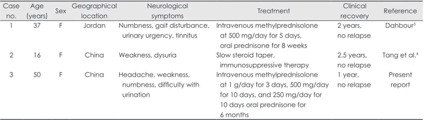
To our knowledge there are only two MRI-documented cas- es of myelitis in VHK disease in the literature,3,4 and our pa- tient (the third case) has the oldest onset age of 50 years (Table 1 and 2). Women were affected in all three cases (one was from Jordan and the other two were from China) with a wide range for the onset age (37, 16, and 50 years, respectively), which is consistent with reports that VKH disease occurs mostly in Na- tive Americans, East Indian, Asian, Middle Eastern, and His- panic populations, people aged between 20 and 50 years, and women.5,6 Spinal shock was not seen in the three cases, and is possibly less common in myelitis with VKH disease than in id- iopathic acute transverse myelitis. Spinal-cord MRI revealed cervical lesions in all three patients, while thoracic changes are more frequent in idiopathic acute transverse myelitis cases.
However, too few patients with VKH disease and acute myeli- tis have been reported to allow firm conclusions to be drawn
about potential differences in disease expression. In contrast to our patient, the other two cases did not present with headache and showed normal CSF cells or mild pleocytosis, which may be due to variations in the extraocular appearance, such as me- ningismus, tinnitus, vitiligo, and alopecia.7 Moreover, the oth- er two patients had already been off steroids before the neuro- logical presentation, which may be explained either as part of the normal course of the disease or adequate corticosteroid therapy not being introduced early enough.7 Although the clin- ical profile of VKH disease is well-established, little is known about its pathogenesis. Triggering of CD4+ T cells (Th1, T helper 17, and regulatory T cells) reactive to melanocyte-spe- cific proteins [e.g., tyrosinase, tyrosinase-related protein 1, and TRP-2] by an infectious agent is proposed to be involved in the pathogenesis.8 In addition, genetic factors, including HLA- DR4, HLA-DR1, and HLA-DRB1*0405, may also play an im- portant role.8-11 The absence of melanocytes in the spinal cord means that the precise mechanism by which VKH disease leads to acute myelitis is unclear. The effectiveness of steroid therapy in the three cases suggests underlying immunological pathogenic mechanisms, which might involve myelin basic protein.12 The history of upper respiratory tract infection of our patient suggests that infectious factors were involved in the pathogenesis, and the specific geographic distribution of these three cases suggests that genetic background also influences the development of VKH disease with acute myelitis. In sum- mary, new insights into immune responses and genetic abnor- malities will help to clarify the pathogenic mechanisms under- Table 1. Clinical data from the three reported VKH-disease patients accompanied with acute myelitis
Case no.
Age
(years) Sex Geographical location
Neurological
symptoms Treatment Clinical
recovery Reference 1 37 F Jordan Numbness, gait disturbance,
urinary urgency, tinnitus
Intravenous methylprednisolone at 500 mg/day for 5 days, oral prednisone for 8 weeks
2 years, no relapse
Dahbour3
2 16 F China Weakness, dysuria Slow steroid taper,
immunosuppressive therapy
2.5 years, no relapse
Tang et al.4
3 50 F China Headache, weakness,
numbness, difficulty with urination
Intravenous methylprednisolone at 1 g/day for 3 days, 500 mg/day for 10 days, and 250 mg/day for 10 days oral prednisone for 6 months
1 year, no relapse
Present report
F: female, VKH: Vogt-Koyanagi-Harada.
Table 2. Laboratory data for the three reported VKH-disease patients accompanied with acute myelitis Case
no.
ESR (mm/hr)
CSF WBC count (/mm3)
CSF protein (g/L)
CSF IgG
index Brain MRI Spinal-cord MRI Reference
1 22 Normal Normal None Bilateral small subcortical
enhanced lesions
Abnormal at C3 level Dahbour3
2 Normal 28 6.38 Normal Normal Abnormal at C6–T9 level Tang et al.4
3 39 128 4.83 Normal Normal Abnormal at C1–3 level Present
report ESR: erythrocyte sedimentation rate, VKH: Vogt-Koyanagi-Harada, WBC: white blood cell.
Acute Myelitis in a Patient with VKH Disease
64 J Clin Neurol 2013;9:61-64
lying VKH disease with acute myelitis.
Conflicts of Interest
The authors have no financial conflicts of interest.
Acknowledgements
This work was supported by the Fund of the “125” Project of The Third Xiangya Hospital, China (S.G.), the National Natural Science Foundation of China (30871351), and the Sheng Hua Scholars Program and Culture Foundation of National Outstanding Youth of Central South University, China (H.D.)
REFERENCES
1. Smith JR, Rosenbaum JT. Neurological concomitants of uveitis. Br J Ophthalmol 2004;88:1498-1499.
2. Wingerchuk DM, Lennon VA, Pittock SJ, Lucchinetti CF, Weinshenk- er BG. Revised diagnostic criteria for neuromyelitis optica. Neurology 2006;66:1485-1489.
3. Dahbour SS. MRI documented acute myelitis in a patient with Vogt- Koyanagi-Harada syndrome: first report. Clin Neurol Neurosurg 2009;
111:200-202.
4. Tang Y, Wang X, Ding Y, Li C, Jia J. Longitudinal lesion of the spinal cord in a patient with Vogt-Koyanagi-Harada disease. J Neurol Neuro-
surg Psychiatry 2010;81:941-942.
5. Moorthy RS, Inomata H, Rao NA. Vogt-Koyanagi-Harada syndrome.
Surv Ophthalmol 1995;39:265-292.
6. Fang W, Yang P. Vogt-koyanagi-harada syndrome. Curr Eye Res 2008;
33:517-523.
7. Rajendram R, Evans M, Rao NA. Vogt-Koyanagi-Harada disease. Int Ophthalmol Clin 2005;45:115-134.
8. Damico FM, Bezerra FT, Silva GC, Gasparin F, Yamamoto JH. New insights into Vogt-Koyanagi-Harada disease. Arq Bras Oftalmol 2009;
72:413-420.
9. Iqniebi A, Gaafar A, Sheereen A, Al-Suliman A, Mohamed G, Al-Hus- sein K, et al. HLA-DRB1 among patients with Vogt-Koyanagi-Harada disease in Saudi Arabia. Mol Vis 2009;15:1876-1880.
10. Du L, Kijlstra A, Yang P. Immune response genes in uveitis. Ocul Im- munol Inflamm 2009;17:249-256.
11. Tiercy JM, Rathinam SR, Gex-Fabry M, Baglivo E. A shared HLA- DRB1 epitope in the DR beta first domain is associated with Vogt- Koyanagi-Harada syndrome in Indian patients. Mol Vis 2010;16:353- 12. Damico FM, Cunha-Neto E, Goldberg AC, Iwai LK, Marin ML, Ham-358.
mer J, et al. T-cell recognition and cytokine profile induced by melano- cyte epitopes in patients with HLA-DRB1*0405-positive and -negative Vogt-Koyanagi-Harada uveitis. Invest Ophthalmol Vis Sci 2005;46:
2465-2471.