Introduction
Peripartum cardiomyopathy (PPCMP) is a cardiac condi- tion characterized by development of heart failure during the last month of pregnancy or during the first five months of post partum period without any other identifiable cause of heart failure.1) The hypercoagulable state in the pregnancy along with left ventricular (LV) systolic dysfunction predispos- es the patient to thromboembolic complications like intraven- tricular thrombi. Two dimensional (2D) and three dimension- al (3D) echocardiography are valuable tools for evaluation of intra-cardiac masses. We report an unusual case of a peduncu- lated highly mobile LV mass in a patient with PPCMP evalu- ated by 3D transthoracic echocardiography (TTE) responding to oral anticoagulation.
Case
A 30-year-old gravida 4 and para 4 female presented to our department for evaluation of recent onset dyspnea on exertion
along with orthopnea. She had delivered a healthy baby 3 weeks ago through normal vaginal delivery. There was no his- tory of pre-existing cardiac illness. On general examination, pulse rate was 110 per minute, blood pressure 120/70 mmHg, respiratory rate of 22 per minute and jugular venous pressure was raised. Cardiac auscultation did not reveal any significant finding and chest examination showed bilateral basilar fine crepitations. The blood investigations including complete blood count, renal function test and liver function tests were within normal range. Electrocardiogram showed sinus tachy- cardia and chest X-ray showed cardiomegaly along with fea- tures of raised pulmonary venous pressure. Evaluation by 2D TTE (Fig. 1, Supplementary movie 1) showed dilatation of all four cardiac chambers and global hypokinesia of the LV. The LV ejection fraction was 32%, LV end diastolic dimension in- dex was 3.05 cm/m2 and M mode LV fractional shortening was 15%. There was a highly mobile pedunculated mass (2.5
× 2.0 cm) attached to the interventricular septum protruding Rajiv Bharat Kharwar, MD, Sharad Chandra, DM, FACC,
Sudhanshu Kumar Dwivedi, MD, DM, and Ram Kirti Saran, DM, FACC
Department of Cardiology, King Georges’ Medical University, Lucknow, India
Peripartum cardiomyopathy is a cardiac condition characterized by development of heart failure during the last month of preg- nancy or during the first five months of post partum period without any other identifiable cause of heart failure. The hypercoagu- lable state in the pregnancy along with left ventricular (LV) systolic dysfunction predisposes the patient to thromboembolic com- plications like intraventricular thrombi. We report a case of a 30-year-old female with peripartum cardiomyopathy along with a highly mobile mass in the LV cavity on two dimensional echocardiography. Three dimensional transthoracic echocardiography clearly showed the pedicle of the mass attached to the interventricular septum along with internal echolucent areas within the mass. Due to denial of the patient to undergo surgery, she was started on oral anticoagulation, with complete dissolution of the mass within one month.
KEY WORDS: Three dimensional echocardiography · Pedunculated · Left ventricular thrombus · Peripartum cardiomyopathy.
• Received: February 10, 2014 • Revised: March 31, 2014 • Accepted: August 20, 2014
• Address for Correspondence: Rajiv Bharat Kharwar, Department of Cardiology, King Georges’ Medical University, Chowk, Lucknow 226003, Uttar Pradesh, India Tel: +91-522-2258949, Fax: +91-522-2258948, E-mail: [email protected]
• This is an Open Access article distributed under the terms of the Creative Commons Attribution Non-Commercial License (http://creativecommons.org/licenses/by-nc/3.0) which permits unrestricted non-commercial use, distribution, and reproduction in any medium, provided the original work is properly cited.
into the LV cavity. There was no history of diabetes, hyperten- sion, preeclampsia, eclampsia, family history of cardiomyopa- thy or any metabolic disorders that could lead to cardiomyop- athy. Hence the diagnosis of PPCMP with intra-cardiac thrombus was considered. 3D TTE evaluation (Fig. 2, Supple- mentary movie 2) was done to further delineate the anatomy of the LV mass. 3D TTE clearly delineated the attachment of the mass as well as the internal echolucent areas within the mass. Based on the clinical scenario and the 3D TTE finding the mass was suspected to be a thrombus. However, the possi- bility of cardiac tumour could not be excluded. Because of the mobility of the mass and uncertainty of its nature, the risk of embolization was considered to be high. The patient was ad- vised surgical removal of the mass, but she opted for oral anti- coagulation therapy. She was started on standard heart failure therapy along with warfarin (target international normalized ratio 2.0–3.0). Repeat echocardiography 1 month later (Fig.
3, Supplementary movie 3) showed complete dissolution of the LV mass along with an improvement in LV ejection frac-
tion (43%). There was no history of embolization in between.
The response to anticoagulation therapy suggested that the pendunculated LV mass was in fact a thrombus.
Discussion
The true incidence of PPCMP is unknown, but it is estimat- ed to be between 1:15000 to 1:1300 deliveries.2) PPCMP has been associated with several risk factors including increased age, gravidity or parity, African origin, toxaemia or hyperten- sion of pregnancy, use of tocolytics, twin pregnancy, obesity and low socioeconomic status.3) The exact etiology of PPCMP is not known and the patient are managed on the lines of stan- dard heart failure regimes. The hypercoagulable state during the pregnancy along with the poor LV systolic function pro- vides a fertile soil for the development of thromboembolic complications including intracardiac thrombi. Up to 53% of cases of PPCMP have been reported to have thromboembo- lism1)4) although majority have been reported at autopsy or af- ter the clinical occurrence of the embolic episode. Intra cardiac
Fig. 1. Two dimensional transthoracic echocardiography. A large single pedunculated mass (indicated by *) can be seen in the LV cavity in apical four chamber (A), apical two chamber (C), and parasternal short axis (B) views. There is dilatation of all the four cardiac chambers with poor left ventricular ejection fraction of 32% (D). LA: left atrium, LV: left ventricle, RA: right atrium, RV: right ventricle.
C A
D B
thrombus rarely presents as a pedunculated structure. Cardiac fibroma, cardiac hemangioma, blood cyst and aspergillus mu- ral endocarditits can all present as pedunculated mass in the LV cavity but the LV ejection fraction is usually preserved in these cases. The sensitivity of 2D echocardiography for detect- ing LV thrombi is 92% to 95% and the specificity is 86% to 88%.5) A retrospective study using surgical confirmation as the gold standard suggested that contrast-enhanced magnetic reso- nance imaging is superior to TTE and transesophageal echo- cardiography in both sensitivity and specificity.6) However the time required for the study and its high cost limits its routine clinical use. Real time 3D TTE is a newer technique for evalu- ation of intra cardiac masses. The 3D data set once acquired, can be cropped and sliced in many different ways and addi- tional information about mass location, shape, attaching inter- face and relationships with adjacent structures can be derived.
In our case, 3D TTE performed better than 2D TTE for exact delineation of 1) the irregular surface characteristics of the mass, 2) the narrow based attachment of the mass via a pedicle
to the interventricular septum, and 3) internal echolucency within the mass. The echolucency within the thrombi has been reported to indicate internal clot lysis and liquefaction.7)8) The irregular surface characteristics of the thrombi in our case indicated its recent origin and the internal echolucency pre- dicted its possible response to anticoagulation. A pedunculat- ed LV thrombus that is connected to septum or the LV wall, which is highly mobile and which moves throughout the car- diac cycle, is a risky situation as it has got a high potential for embolization despite adequate anticoagulation.9) Over the years, the primary therapeutic option for such a thrombi has included thrombectomy, anticoagulation and thrombolyis.
Still the definitive therapy is controversial and oral anticoagu- lation has variable success, with resolution rates between 13%
and 59%. Very few cases of PPCMP have been reported in the literature where the intra cardiac thrombi have rapidly re- sponded to anticoagulation with complete resolution of the thrombi.10)
Our patient had a pedunculated highly mobile LV mass in
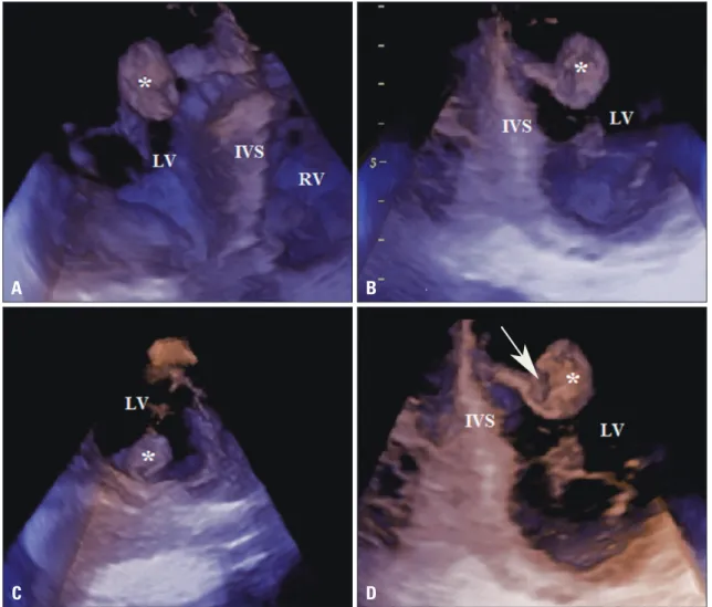
Fig. 2. Three dimensional (3D) transthoracic echocardiography. The mass within the LV cavity (indicated by *) can be viewed from different imaging planes (A-D) with the help of cropping the basic 3D data set. The site of attachment of the mass to the IVS via a pedicle, the irregular surface characteristics as well as the internal echo lucent areas within the mass (arrow in D) can be clearly delineated. IVS: interventricular septum, LV: left ventricle, RV: right ventricle.
C A
D B
the setting of PPCMP. Due to its narrow pedicle and high mobility, the risk of embolization was considered to be very high and patient was advised to undergo surgical removal of the mass. The information obtained from 3D TTE and its re- sponse to anticoagulation confirmed the nature of the mass to be a thrombus. Although surgical thrombectomy is the pre- ferred treatment strategy in such cases, our patient responded well to anticoagulation.
Supplementary movie legends
Movie 1. Two dimensional transthoracic echocardiography.
Apical four chamber view showing dilatation of all the four cardiac chambers along with poor LV systolic function. Note the presence of single, highly mobile mass in the LV cavity at- tached to the interventricular septum. LA: left atrium, LV: left ventricle, RA: right atrium, RV: right ventricle.
Movie 2. Three dimensional transthoracic echocardiogra- phy. The attachment of the mass to the interventricular sep- tum via a pedicle, the irregular surface characteristics, the ro- tation as well as the three dimensional structure of the mass can be clearly delineated. LV: left ventricle.
Movie 3. Two dimensional transthoracic echocardiography.
Apical 4 chamber view at the follow-up echocardiography af- ter one month of anticoagulation therapy showing complete resolution of the thrombus. Also note the improvent in the systolic LV function. LA: left atrium, LV: left ventricle, RA:
right atrium, RV: right ventricle.
References
1. Demakis JG, Rahimtoola SH. Peripartum cardiomyopathy. Circulation 1971;44:964-8.
2. Cunningham FG, Pritchard JA, Hankins GD, Anderson PL, Lucas MJ, Armstrong KF. Peripartum heart failure: idiopathic cardiomyopathy or compounding cardiovascular events? Obstet Gynecol 1986;67:157-68.
3. Bhattacharyya A, Basra SS, Sen P, Kar B. Peripartum cardiomyopathy:
a review. Tex Heart Inst J 2012;39:8-16.
4. Demakis JG, Rahimtoola SH, Sutton GC, Meadows WR, Szanto PB, Tobin JR, Gunnar RM. Natural course of peripartum cardiomyopa- thy. Circulation 1971;44:1053-61.
5. Stratton JR, Lighty GW Jr, Pearlman AS, Ritchie JL. Detection of left ventricular thrombus by two-dimensional echocardiography: sensitivity, specificity, and causes of uncertainty. Circulation 1982;66:156-66.
6. Srichai MB, Junor C, Rodriguez LL, Stillman AE, Grimm RA, Li- eber ML, Weaver JA, Smedira NG, White RD. Clinical, imaging, Fig. 3. Two dimensional transthoracic echocardiography. Follow-up echocardiography at 1 month after the onset of anticoagulation and heart failure therapy showed complete resolution of the intracardiac mass (A, B, and C) as well as improvement in the left ventricular ejection fraction (43%, D). LA: left atrium, LV: left ventricle, RA: right atrium, RV: right ventricle.
C A
D B