CASE REPORT
Copyright © 2011, the Korean Surgical Society J Korean Surg Soc 2011;81:287-290
http://dx.doi.org/10.4174/jkss.2011.81.4.287
JKSS
Journal of the Korean Surgical Society pISSN 2233-7903ㆍeISSN 2093-0488
Received September 10, 2010, Revised January 27, 2011, Accepted February 1, 2011 Correspondence to: Dae Hyun Yang
Department of Surgery, Hallym Sacred Heart Hospital, Hallym University College of Medicine, 896 Pyeongchon-dong, Dongan-gu, Anyang 431-070, Korea
Tel: +82-31-380-3772, Fax: +82-31-385-0157, E-mail: [email protected]
cc Journal of the Korean Surgical Society is an Open Access Journal. All articles are distributed under the terms of the Creative Commons Attribution Non-Commercial License (http://creativecommons.org/licenses/by-nc/3.0/) which permits unrestricted non-commercial use, distribution, and reproduction in any medium, provided the original work is properly cited.
Laparoscopic total extraperitoneal repair of lumbar hernia
Man Sup Lim, Hae Wan Lee, Chang Hee Yu
1, Dae Hyun Yang
Departments of Surgery and 1Urology, Hallym Sacred Heart Hospital, Hallym University College of Medicine, Anyang, Korea
Lumbar hernia is a rare surgical entity without a standard method of repair. With advancements in laparoscopic techniques, successful lumbar herniorrhaphy can be achieved by the creation of a completely extraperitoneal working space and secure fixation of a wide posterior mesh. We present a total extraperitoneal laparoendoscopic repair of lumbar hernia, which al- lowed for minimal invasiveness while providing excellent anatomical identification, easy mobilization of contents and wide secure mesh fixation. A total extraperitoneal method of lumbar hernia repair by laparoscopic approach is feasible and may be an ideal option.
Key Words: Lumbar hernia, Grynfeltt hernia, Laparoscopic repair, Extraperitoneal repair
INTRODUCTION
Lumbar hernia in the flank is an uncommon type of ven- tral hernia. Anatomically, lumbar hernia involves both the intraperitoneal and retroperitoneal areas bounded superi- orly by the 12th rib and inferiorly by the iliac crest. Its unique anatomical nature has made the diagnosis and proper repair of lumbar hernias a challenge. Since P.
Barbette first suggested the existence of this entity in 1672, its management has been controversial. With more accu- rate body imaging techniques such as computed tomog- raphy (CT) scan and magnetic resonance imaging and im- provements in laparoendoscopic techniques of hernia re- pairs with mesh there has been a lot of achievement in un- derstanding and surgical treatment of lumbar hernia.
This report is the first laparoscopic totally extraperi- toneal (TEP) approach to lumbar hernia in Korea.
CASE REPORT
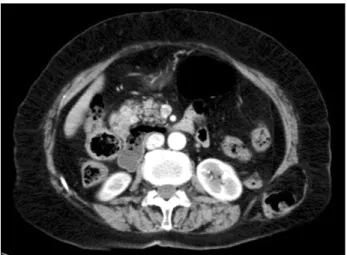
A 76 year old woman with a history of left flank bulge for 6 months presented with left-sided back pain in the same area for two months. She had no previous surgery or trauma in that area. On physical examination she had a 6 x 5 cm reducible bulge at the posterolateral part of left flank below the 12th rib. A CT scan showed herniation of retro- peritoneal fat and descending colon through the fascial defect at the left superior lumbar triangle (Fig. 1). We plan- ned the laparoscopic TEP approach for herniorrhaphy of
Man Sup Lim, et al.
288 thesurgery.or.kr
Fig. 1. A CT scan showed herniation of retroperitoneal fat and descending colon through the fascial defect at the left superior lumbar triangle.
Fig. 2. Position of trocars.
Fig. 3. Laparoscopic view of hernia defect.
Fig. 4. Design of polypropylene mesh (15 × 15 cm).
Fig. 5. Postoperative wounds.
this superior lumbar hernia, Grynfeltt hernia.
Under the general anesthesia the patient was placed in
a full right lateral decubitus position with a lumbar roll in place. A 2 cm transverse incision was made over the hernia defect of the already reduced hernia which was below the 12th rib along the mid axillary line. The retroperitoneal fat was detached first with blunt dissection by an index finger and then with smooth instrument to create space in the ret- roperitoneal cavity. An 11 mm trocar was inserted and the space was expanded by a balloon dissector (Tyco Health- care, Norwalk, CT, USA). Under the direct visualization with a 30o 10 mm scope, three 5 mm trocars were inserted anteriorly along the anterior axillary line approximately 5 cm apart (Fig. 2). Then, the 11 mm trocar was removed and the wound was plugged with gauze to maintain the pneumoretroperitoneum. With 30o 5 mm scope in the cen- tral port the retroperitoneal fat and structures were de- tached from the hernia defect and surrounding muscu- lar-bony structures using 2 working ports above and be-
Laparoscopic total extraperitoneal repair of lumbar hernia
thesurgery.or.kr 289
low the central port (Fig. 3). A 15 x 15 cm octagonal poly- propylene mesh (Fig. 4) was inserted through the re- inserted 11 mm trocar previously used for space making and placed on the lumbar wall to cover at least 4 cm mar- gin around the hernia defect. The mesh was then fixed with transfascial sutures of polypropylene at the center and the margins of each quadrant. To secure the mesh in place we used Tacker (Tyco Healthcare). The mesh was not fixed to the iliac crest or the 12th rib. Postoperative pain was controlled with injections of analgesic (non-steroidal anti-inflammatory drugs) until the postoperative second day. There was no seroma or other wound complication.
She was discharged on the 5th day after the operation (Fig.
5). There was no evidence of recurrence on follow up at 11 months.
DISCUSSION
Lumbar hernia has infrequently been reported with on- ly about 300 cases in the English literature, 62 cases in Japanese journals and 11 cases in Korean journals [1-3].
They are classified as either congenital or acquired. Acqui- red forms are further grouped as primary or sencondary types. The secondary lumbar hernias are attributed to pre- vious surgery, trauma or infection. Primary or sponta- neous lumbar hernias are usually in two anatomical loca- tions: in the inferior (Petit triangle) or in the superior (Grynfeltt or Grynfeltt-Lesshaft triangle). Inferior triangle is smaller and bordered by the iliac crest at the base, the ex- ternal oblique muscle laterally and the latissimus dorsi medially. Superior triangle is larger and more common lo- cation for a lumbar hernia and its borders are defined su- periorly by the 12th rib, medially by erector spinae, later- ally by the internal oblique muscle. Predisposing factors for primary acquired lumbar hernia are age, obesity, in- tense slimming, strenuous physical activity, and situations related to increased intra-abdominal pressure. The most common clinical sign is a bulge that increases with cough- ing or strenuous activity which tends to disappear when placed in the lateral decubitus position. In about 9% of cas- es the patients present with incarceration. Although the history, symptoms and physical signs suggest a lumbar
hernia, an abdominal CT scan is essential for diagnosis. CT scan is used to confirm the physical examination, evaluate abdominal wall muscles, identify the hernia contents, and assess the anatomical relation of lumbar area [4].
There are many options in management of a lumbar hernia including no repair, suture closure, muscle closure of diverse techniques, and various repair with prostheses.
Repairing the hernias is often difficult. The reasons for no standard treatment and difficulty in repair include diffi- culty in defining edges of the fascial defect, no adequate fascia and inherent weakness of the surrounding tissue, the bony boundaries, and the lack of collective experience of any one surgeon or group of surgeons to improve the operative technique [5].
Several surgeons have adapted the technique of laparo- scopic ventral herniorrhaphy to lumbar hernia repair. In 1996, Burick and Parascandola [6] reported the first lapa- roscopic repair for a traumatic superior lumbar hernia.
They explored the abdomen transperitoneally and re- paired the lumbar defect with polypropylene mesh by a technique similar to that of transabdominal preperitoneal inguinal herniorrhaphy. In 1997, Heniford et al. [7] re- ported laparoscopic transperitoneal repair using a 4 cm overlapping polytetrafluoroethylene mesh secured with transabdominal polypropylene sutures. In 1999 Wood- ward et al. [8] operated on a patient of recurrent lumbar hernia having a history of previous open mesh repair by the first laparoscopic retroperitoneal (extraperitoneal) approach. With extraperitoneal approach, Habib [9] in 2000 repaired a primary Grynfeltt hernia, and in 2001 Meinke [10] did a large inferior lumbar hernia by trans- fascial suture and Tacker fixation of 4 cm overlapping pol- ypropylene mesh. A flank incision permits easy dissection of extraperitoneal plane. With the aid of ballooning we can get generous operating space large enough to complete a repair with a large mesh which is secured to the abdominal wall with a transfascial suture and Tacker reinforcements.
Although there are favorable reports of extraperitoneal mesh repair using Prolene Hernia System (Ethicon Inc., Somerville, NJ, USA) via the anterior approach in Japan and Korea [2,3] the laparoscopic extraperitoneal approach could be an ideal option for lumbar hernia repair.
Laparoscopic approaches for lumbar hernia using TEP
Man Sup Lim, et al.
290 thesurgery.or.kr
technique of inguinal herniorrhaphy and laparoscopic ventral herniorrhaphy had been reported with promising results. Our experience of the laparoendoscopic TEP lum- bar hernia repair further supports the advantages of ex- cellent operative visualization as well as minimal invasi- veness.
CONFLICT OF INTEREST
No potential conflict of interest relevant to this article was reported.
REFERENCES
1. Moreno-Egea A, Baena EG, Calle MC, Martínez JA, Albasi- ni JL. Controversies in the current management of lumbar hernias. Arch Surg 2007;142:82-8.
2. Kurumiya Y, Kameoka N, Ogawa A, Hibino M, Chigira H.
A case of superior lumbar hernia- with review of 62 cases of lumbar hernia in Japan. Geka (Japanese) 2005;67:1761-5.
3. Chung M. Post-traumatic lumbar hernia repaired with PHS (Prolene Hernia System). J Korean Surg Soc 2009;77:
221-4.
4. Salameh JR. Primary and unusual abdominal wall hernias.
Surg Clin North Am 2008;88:45-60, viii.
5. Yavuz N, Ersoy YE, Demirkesen O, Tortum OB, Erguney S.
Laparoscopic incisional lumbar hernia repair. Hernia 2009;
13:281-6.
6. Burick AJ, Parascandola SA. Laparoscopic repair of a trau- matic lumbar hernia: a case report. J Laparoendosc Surg 1996;6:259-62.
7. Heniford BT, Iannitti DA, Gagner M. Laparoscopic inferior and superior lumbar hernia repair. Arch Surg 1997;132:
1141-4.
8. Woodward AM, Flint LM, Ferrara JJ. Laparoscopic retro- peritoneal repair of recurrent postoperative lumbar hernia.
J Laparoendosc Adv Surg Tech A 1999;9:181-6.
9. Habib E. Retroperitoneoscopic tension-free repair of lum- bar hernia. Hernia 2003;7:150-2.
10. Meinke AK. Totally extraperitoneal laparoendoscopic re- pair of lumbar hernia. Surg Endosc 2003;17:734-7.