J Korean Surg Soc 2013;84:273-280 http://dx.doi.org/10.4174/jkss.2013.84.5.273
ORIGINAL ARTICLE
JKSS JKSS JKSS
Journal of the Korean Surgical Society pISSN 2233-7903ㆍeISSN 2093-0488
Received August 16, 2012, Revised February 19, 2013, Accepted February 20, 2013 Correspondence to: Jeong Eon Lee
Division of Breast and Endocrine Surgery, Department of Surgery, Samsung Medical Center, Sungkyunkwan University School of Medicine, 81 Irwon-ro, Gangnam-gu, Seoul 135-710, Korea
Tel: +82-2-3410-0260, Fax: +82-2-3410-6982, E-mail: [email protected]
cc Journal of the Korean Surgical Society is an Open Access Journal. All articles are distributed under the terms of the Creative Commons Attribution Non-Commercial License (http://creativecommons.org/licenses/by-nc/3.0/) which permits unrestricted non-commercial use, distribution, and reproduction in any medium, provided the original work is properly cited.
Neoadjuvant human epidermal growth factor
receptor-2 targeted therapy in patients with locally advanced breast cancer
Dong Hui Cho, Se Kyung Lee
1, Sangmin Kim
1, Min-Young Choi
1, Seung Pil Jung
1, Jeonghui Lee
1, Jiyoung Kim
1, Min Young Koo
1, Soo Youn Bae
1, Jung-Han Kim
1, Jee Soo Kim
1, Kil Won Ho
1, Jeong Eon Lee
1, Seok Jin Nam
1, Jung-Hyun Yang
2Department of Surgery, Seoul Medical Center, Seoul, 1Division of Breast and Endocrine Surgery, Department of Surgery, Samsung Medical Center, Sungkyunkwan University School of Medicine, Seoul, 2Department of Surgery, Konkuk University Medical Center, Seoul, Korea
Purpose: We analyzed the responses of patients with locally advanced breast cancer to neoadjuvant chemotherapy (NAC) and NAC combined with neoadjuvant human epidermal growth factor receptor-2 (HER2) targeted therapy (NCHTT).
Methods: We retrospectively reviewed 59 patients with HER2 amplified locally advanced breast cancer among patients who were treated surgically after neoadjuvant therapy at Samsung Medical Center between 2005 and 2009. Thirty-one patients re- ceived conventional NAC and 28 patients received NCHTT. Pathologic responses were assessed according to response evalu- ation criteria in solid tumors (RECIST) guidelines. Results: Pathologic complete response (pCR) was achieved in 13 out of 28 patients treated with NCHTT and in 6 out of 31 patients treated with NAC alone (46.4% vs. 19.4%, respectively, P = 0.049).
Breast conserving surgery (BCS) was more frequently performed in the NCHTT group than in the NAC only group (71.4%
vs. 19.4%, P < 0.001). The 3-year recurrence-free survival (RFS) rate was 100% in the NCHTT group and 76.4% in the NAC group (P = 0.014). Together, NCHTT, type of operation (BCS vs. mastectomy) and pathologic nodal status were significant prognostic factors for RFS in univariate analysis. Conclusion: We found that NCHTT produced higher pCR rates than NAC alone in locally advanced breast cancer.
Key Words: Breast neoplasms, Neoadjuvant therapy, ErbB-2, Response
INTRODUCTION
Breast cancer is the second most common cancer and the sixth leading cause of cancer deaths in women in Korea.
Neoadjuvant therapy, surgery, and adjuvant therapy are
standard treatments for locally advanced breast cancer.
Typically, neoadjuvant therapy plays an important role in patients with locally advanced breast cancers and in treat- ing distant micrometastasis, downstaging tumors, im- proving operability, and sometimes in allowing breast
conserving surgery (BCS). The multidisciplinary manage- ment approach combined with neoadjuvant chemo- therapy (NAC) and the incorporation of anthracyclines and taxanes has resulted in a significant reduction in the risk of death and a subsequent reduction in the 5-year re- currence rate for patients with early and advanced stage breast cancers [1,2].
NAC was first introduced in the 1970s [3] and several clinical studies since then have corroborated the efficacy of NAC [4,5]. A majority of studies report that after NAC, the partial response rate was 60% to 80% and the complete re- sponse (CR) rate was 10% to 20% [5,6]. The primary goals of NAC are to increase the rate of BCS and to predict prog- nosis through the response of tumors to treatment [7,8].
On the other hand, studies have estimated that approx- imately 20% to 25% of invasive breast cancers exhibit over- expression and gene amplification of human epidermal growth factor receptor-2 (HER2, also known as ERBB2) [9,10]. This represents an adverse prognostic indicator as- sociated with aggressive histopathologic parameters and is correlated with decreased recurrence-free survival (RFS), overall survival (OS), and poor prognosis and outcome.
Trastuzumab, a monoclonal antibody directed against HER2, has become a standard treatment in HER2 positive breast cancer patients and has resulted in increased re- mission rates after neoadjuvant therapy [11]. Given the improvement in outcomes with the neoadjuvant use of trastuzumab with HER2 positive breast cancer patients, the optimal use of these compounds is still unclear. A num- ber of new treatment approaches directed against HER2 are currently being examined in clinical trials. The pur- pose of this retrospective study was to assess pathologic CR (pCR), RFS, and OS rates in patients treated with NAC combined with neoadjuvant HER2 targeted therapy (NCHTT) compared to patients treated with NAC only.
METHODS
From January 2005 to December 2009, 60 patients who were histologically confirmed to be HER2 positive (defined as either immunohistochemical 3+ or 2+ and
gene amplification determined by fluorescence in situ hy- bridization [FISH]) underwent NAC or NCHTT followed by surgery to treat breast cancer at Samsung Medical Center, Seoul, Korea. Histologic confirmation of invasive tumors was performed by fine needle aspiration biopsy or core needle biopsy. We excluded one patient because she had a remnant skin tumor that was detected 38 days post- operatively after modified radical mastectomy. We re- viewed clinicopathologic factors and treatment modalities (type of operation, use of hormonal therapy, and radiation therapy) in 59 patients. Before initiation of therapy, all pa- tients underwent staging evaluations that included com- plete histories, physical examinations, complete blood counts, chemical profiles, chest radiographs, ultrasounds or computed tomography scans of the liver, and bone scans. Mammography of both breasts was performed and additional breast and axillary assessment was conducted by ultrasound.
The pathologic tumor stage was assessed according to the American Joint Committee on Cancer 7 staging system [12]. The histologic grade was determined according to the Bloom-Richardson classification. The Allred score was used for estrogen receptor and progesterone receptor pos- itivities, and HER2 was scored as 0–3. Patients with an im- munohistochemistry score of 3+ or 2+ and gene amplifi- cation determined by FISH were considered positive.
Responses to neoadjuvant therapy were categorized by pCR. The pCR was defined as no detectable invasive or noninvasive residual cancer cell in the breast or axillary lymph nodes by histopathology.
NAC regimens consisted of adriamycin with cyclo- phosphamide, adriamycin with docetaxel, adriamycin with cyclophosphamide plus docetaxel. All patients were treated with 3–8 cycles according to their regimen proto- cols in 3-week intervals. In the NAC only group, 31 pa- tients received anthracycline-based chemotherapy. In the NCHTT group, trastuzumab, pertuzumab or lapatinib were used for HER2 targeted therapy concurrently with a chemotherapeutic agent. Neoadjuvant trastuzumab was administered as a loading dose of 4 mg/kg intravenously over 90 minutes on the first day and then subsequently given weekly at a dose of 2 mg/kg over 30 minutes, con- comitantly with the above chemotherapy. Statistical anal-
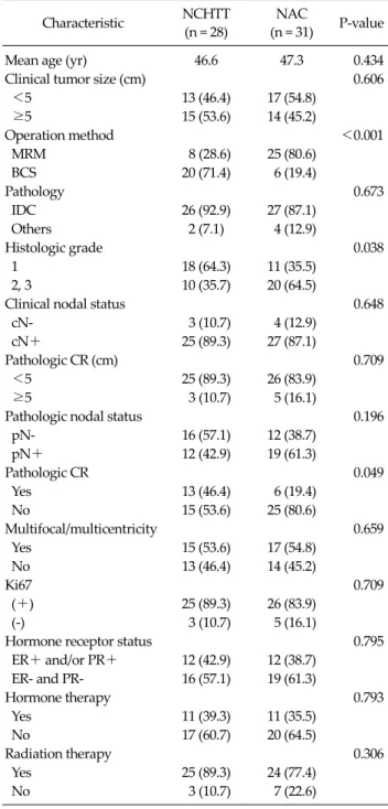
Table 1.Clinicopathologic characteristics of patients with neo- adjuvant HER2 targeted therapy and without HER2 targeted therapy
Characteristic NCHTT (n = 28)
NAC
(n = 31) P-value
Mean age (yr) 46.6 47.3 0.434
Clinical tumor size (cm) 0.606
<5 13 (46.4) 17 (54.8)
≥5 15 (53.6) 14 (45.2)
Operation method <0.001
MRM 8 (28.6) 25 (80.6)
BCS 20 (71.4) 6 (19.4)
Pathology 0.673
IDC 26 (92.9) 27 (87.1)
Others 2 (7.1) 4 (12.9)
Histologic grade 0.038
1 18 (64.3) 11 (35.5)
2, 3 10 (35.7) 20 (64.5)
Clinical nodal status 0.648
cN- 3 (10.7) 4 (12.9)
cN+ 25 (89.3) 27 (87.1)
Pathologic CR (cm) 0.709
<5 25 (89.3) 26 (83.9)
≥5 3 (10.7) 5 (16.1)
Pathologic nodal status 0.196
pN- 16 (57.1) 12 (38.7)
pN+ 12 (42.9) 19 (61.3)
Pathologic CR 0.049
Yes 13 (46.4) 6 (19.4)
No 15 (53.6) 25 (80.6)
Multifocal/multicentricity 0.659
Yes 15 (53.6) 17 (54.8)
No 13 (46.4) 14 (45.2)
Ki67 0.709
(+) 25 (89.3) 26 (83.9)
(-) 3 (10.7) 5 (16.1)
Hormone receptor status 0.795
ER+ and/or PR+ 12 (42.9) 12 (38.7) ER- and PR- 16 (57.1) 19 (61.3)
Hormone therapy 0.793
Yes 11 (39.3) 11 (35.5)
No 17 (60.7) 20 (64.5)
Radiation therapy 0.306
Yes 25 (89.3) 24 (77.4)
No 3 (10.7) 7 (22.6)
Values are presented as number (%).
HER2, human epidermal growth factor receptor-2; NCHTT, neoadjuvant chemotherapy combined with neoadjuvant HER2 targeted therapy; NAC, neoadjuvant chemotherapy; MRM, modified radical mastectomy; BCS, breast conserving surgery;
IDC, invasive ductal carcinoma; CR, complete response; ER, estrogen receptor; PR, progesterone receptor.
yses were performed using the IBM SPSS ver. 18.0 (IBM Co., Armonk, NY, USA). Student’s t-test, Pearson’s chi-square test or Fisher’s exact test were used to compare the clinicopathological characteristics between the NCHTT group and the NAC only group in HER2 positive cases. The Kaplan-Meier method and Logistic regression analysis were used to determine RFS and OS rates.
RESULTS
Clinicopathological characteristics and treatment mo- dalities were analyzed for 59 patients who underwent NAC with or without NCHTT due to invasive breast carcinoma. Twenty-four patients had estrogen and/or pro- gesterone receptor positive disease. Fifty-three patients had invasive ductal carcinoma, five patients had micro- papillary carcinoma, and one patient had invasive papil- lary carcinoma. The mean breast tumor size was 5.3 cm.
After adjuvant therapy, 33 patients (55.9%) underwent mastectomies, whereas 26 patients (44.1%) had BCS.
Nineteen patients had stage III disease. Sentinel lymph node biopsy was performed on only one patient. An aver- age of 19 lymph nodes were dissected in patients who un- derwent axillary lymph node dissection. The mean fol- low-up period was 33.3 months (range, 5.7 to 70.0 months).
Table 1 summarizes clinicopathologic characteristics of 31 patients treated with NAC only and 28 patients treated with NCHTT. The mean age of patients treated with NCHTT was 46.6 years, and the mean age of patients treat- ed with NAC only was 47.3 years (P = 0.434). Thirteen pa- tients achieved pCR in the NCHTT group and 6 patients achieved pCR in the NAC only group (46.4% vs. 19.4%, P
= 0.049). BCS was more frequently performed in the NCHTT group than in the NAC only group (71.4% vs.
19.4%, P < 0.001). All 28 patients treated with NCHTT showed normal cardiac function before and after chemo- therapy (Table 2). There were no significant differences in tumor size, nodal status, receptor status or hormone ther- apy use between the two groups.
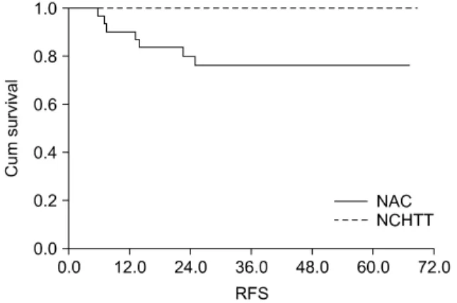
During the median follow-up of 28.7 months (range, 5.7 to 70.0 months), seven patients experienced disease re-
Table 2. Adverse events
Event No. of patients
NCHTT (n = 28) NAC (n = 31)
Neutropenic fever 8 5
LFT increase 2 1
Diarrhea 5 5
Stomatitis 12 5
Cardiac problem 0 0
NCHTT, neoadjuvant chemotherapy combined with neoadjuvant human epidermal growth factor receptor 2 targeted therapy; NAC, neoadjuvantchemotherapy; LFT, liver function test.
Fig. 2. Overall survival (OS) curve for advanced breast cancer patients treated by neoadjuvant chemotherapy combined with neoadjuvant human epidermal growth factor receptor 2 targeted therapy (NCHTT) and neoadjuvant chemotherapy (NAC).
Fig. 1. Recurrence-free survival (RFS) curve for advanced breast cancer patients treated by neoadjuvant chemotherapy combined with neoadjuvant human epidermal growth factor receptor 2 targeted therapy (NCHTT) and neoadjuvant chemotherapy (NAC).
currence; interestingly, all were from the NAC only group.
The 3-year RFS rate was 100% in the NCHTT group and 76.4% in the NAC only group (P = 0.014) (Fig. 1). During follow-up, only three patients died and the 3-year OS rate was 100% in the NCHTT group and 89.3% in the NAC only group (P = 0.149) (Fig. 2).
Table 3 shows the results of the RFS and OS-related uni- variate analysis of all patients in this study. NCHTT, type of operation (BCS vs. mastectomy), and pathologic nodal status were significant prognostic factors for RFS. For death and recurrence, multivariate analysis could not be applied to this study because of low incidence (3 deaths, 7 recurrences).
DISCUSSION
The results of this study indicate that NCHTT induced higher pCR rates than NAC only in patients with locally advanced breast cancer. BCS was more frequently per- formed in the NCHTT group than in the NCT only group and 3-year RFS rate was significantly higher in the NCHTT group. In comparison with the results from western coun- tries, we could observe a similar benefit from NCHTT.
NAC has become a standard therapy to treat patients with locally advanced breast cancer [8,13]. The primary goals of NAC are to increase the rate of BCS and to predict prognosis through the response of the tumor to the treat- ment [8,14]. NAC can reduce tumor size, which increases the rate of BCS and in some cases has the effect of prolong- ing OS. In addition, it improves the rates of operability in previously inoperable patients, can treat early stages of micrometastasis before the presence of chemo-resistant cell lines, reduces the risk of recurrence by preventing seeding during surgery and can predict further ther- apeutic efficacy of chemotherapeutic agents. Conversely, due to its downstaging effects, it can lead to therapeutic confusion due to the diminishment of traditional prog- nostic factors such as tumor size, axillary metastatic lymph node numbers, and histologic grading which are needed to assess patients for systemic treatment and over- treatment who may benefit by surgical intervention alone.
Although there are no clinical practice guidelines for
Table 3. Univariate analysis of the recurrence-free survivals (RFS) and overall survival (OS)
Variable 3-Year RFS (%) P-value 3-Year OS (%) P-value
Age (yr) NS NS
<50 88.6 94.3
≥50 87.5 95.8
Clinical tumor size (cm) NS NS
<5 90.0 96.7
≥5 86.2 93.1
NCHTT 0.014 NS
Yes 100 100
No 77.4 90.3
Operation method 0.018 NS
BCS 100 100
MRM 78.8 90.9
Pathology NS NS
IDC 83.8 100
Others 88.7 94.3
Histologic grade NS NS
1, 2 93.1 96.6
3 83.3 93.3
Clinical nodal status NS NS
cN- 92.7 96.7
cN+ 84.0 92.3
Pathologic tumor size (cm) NS NS
<5 88.2 94.1
≥5 87.5 100
Pathologic nodal status 0.009 NS
pN (-) 100 100
pN (+) 77.4 90.3
Response after NT NS NS
CR 94.7 94.7
Non-CR 85.0 95
Response after NT NS NS
CR+PR 88.7 94.3
SD+PD 83.3 100
p53 NS NS
(+) 95.3 96.8
(-) 85.2 92.6
Ki67 NS NS
(+) 87.5 94.6
(-) 100 100
Hormone receptor status NS NS
ER+ and/or PR+ 95.8 100
ER- and PR- 82.9 91.4
Hormone therapy NS NS
Yes 95.5 100
No 83.8 91.9
Radiation therapy NS NS
Yes 89.8 98.0
No 80.0 80.0
NS, not significant; NCHTT, neoadjuvant chemotherapy combined with neoadjuvant human epidermal growth factor receptor 2 targeted therapy; NAC, neoadjuvant chemotherapy; BCS, breast conserving surgery; MRM, modified radical mastectomy; IDC, invasive ductal carcinoma; CR, complete response; PR, partial response; SD, stable disease; PD, persistent disease; ER, estrogen receptor; PR, progesterone receptor.
NAC, it is more frequently used as a preliminary treat- ment for patients undergoing surgery than as pretreat- ment for locally advanced breast cancer.
The HER is the cell-surface receptor for members of the epidermal growth factor family (EGF-family) of ex- tracellular protein ligands and a potent mediator of nor- mal cell growth and development [15]. This family of re- ceptors consists of four closely related type 1 trans- membrane tyrosine kinase (TK) receptors: HER1 (epider- mal growth factor receptor; EGFR), HER2, HER3 and HER4. Each receptor comprises an extracellular domain where ligand binding occurs, an α-helical transmem- brane segment and an intracellular protein TK domain.
Receptor dimerization is essential for HER function and for the signaling activity of all HER receptors. Dimeriza- tion can occur between two different HER receptors (heterodimerization) or between two molecules of the same receptor (homodimerization). However, HER re- ceptors normally exist as inactive monomers with the mol- ecules folded to prevent dimerization [16]. The HER2:
HER3 heterodimer is considered the most potent HER re- ceptor pair with respect to strength of interaction, li- gand-induced tyrosine phosphorylation and downstream signaling, and functions as an oncogenic unit.
The humanized monoclonal antibody trastuzumab (Herceptin) was developed as a therapy targeted against HER2, which is over expressed in roughly one fourth of patients with invasive breast cancer. Readily available markers of overexpression and/or gene amplification of HER2 in tumor tissue predict the activity of this agent, and exclude those who will not benefit from this therapy.
Trastzumab binds to the extracellular domain of HER2.
Several mechanisms of action underlie the antitumor ef- fects of trastuzumab. Trastuzumab blocks HER2-activated cell signaling, thereby reducing cell proliferation and re- storing a capacity for apoptosis by inhibiting the PI3K/Akt pathway [17,18], which increases cellular sensitivity to chemotherapy and radiotherapy [19]. It has also been shown to prevent the formation of p95HER2 (a truncated, ac- tive form of HER2), which may lead to inhibition of tumor development [17]. Finally, trastuzumab has been shown to inhibit HER2 regulated angiogenesis and leads to anti- body-dependent cell-mediated cytotoxicity and triggers
the activation of natural killer cell-mediated apoptosis [17,20]. Trastuzumab is now the standard therapy used in the adjuvant setting.
Pertuzumab is a humanized monoclonal antibody that binds to an epitope in domain II, the dimerization domain of the HER2 receptor extracellular domain, which is a re- gion of HER2 distinct from the domain IV binding site of trastuzumab [21]. Pertuzumab inhibits HER2 dimeriza- tion by preventing HER2 from pairing with other HER re- ceptors, including HER3 [22]. Lapatinib is a dual inhibitor of HER1 and HER2 TKs to be used in clinical practice. It has been shown to inhibit the intracellular domain phos- phorylation of both HER1 and HER2 in a reversible man- ner with a long dissociation time of receptor-drug com- plex estimated as ≥300 minutes [23].
The role of trastuzumab in combination with chemo- therapy has been tested in the neoadjuvant setting. A phase III trial conducted at the M.D. Anderson Cancer Center evaluated the addition of trastuzumab to an an- thracycline based regimen. In this study, HER2 positive stage II–IIA patients were randomly assigned to receive chemotherapy with paclitaxel followed by FEC (5-fluo- rouracil, epirubicin and cyclophosphamide) or the same chemotherapy regimen plus weekly trastuzumab. The ad- dition of trastuzumab to chemotherapy resulted in more than a doubling in pCR rates compared to chemotherapy alone (65.2% vs. 26%, P = 0.016) [11]. The phase III NeOAdjuvant Herceptin (NOAH) trial evaluated the ad- dition of trastuzumab to an anthracycline- and tax- ane-based chemotherapy for HER2- positive patients with locally advanced or inflammatory breast cancer. The event-free survival rate at three years was significantly better in the chemotherapy plus trastuzumab group com- pared to chemotherapy alone: 71% versus 56%, re- spectively (HR, 0.59; P = 0.013); the pCR rate was also sig- nificantly higher in the chemotherapy plus trastuzumab group than the chemotherapy only group: (38% vs. 19%, respectively P = 0.001) [24]. In the phase III GeparQuattro trial, 1,509 patients with operable or locally advanced tu- mors were randomized to receive NAC with four cycles of epirubicin/cyclophosphamide followed by four cycles of docetaxel, with or without capecitabine [25]. The 445 HER2 positive patients enrolled also received trastuzu-
mab 6 mg/kg (with a loading dose of 8 mg/kg) every 3 weeks during all chemotherapy cycles. The pCR rate in the HER2 positive subset was 31.7% with no relevant early toxicity observed.
In conclusion, we found that patients treated with NCHTT experienced higher pCR rates, more breast con- serving rates and lower RFS rates compared to patients treated with NAC only. Unfortunately, we have few cases with HER2 amplified locally advanced breast cancer who were treated surgically after NAC. For various survival benefits, additional studies with a larger patient enroll- ment are required to identify survival differences between NCHTT and NAC alone with the same chemotherapy regimen.
CONFLICTS OF INTEREST
No potential conflict of interest relevant to this article was reported.
REFERENCES
1. Early Breast Cancer Trialists' Collaborative Group (EBCTCG).
Effects of chemotherapy and hormonal therapy for early breast cancer on recurrence and 15-year survival: an over- view of the randomised trials. Lancet 2005;365:1687-717.
2. Jemal A, Siegel R, Ward E, Murray T, Xu J, Smigal C, et al.
Cancer statistics, 2006. CA Cancer J Clin 2006;56:106-30.
3. De Lena M, Zucali R, Viganotti G, Valagussa P, Bonadonna G. Combined chemotherapy-radiotherapy approach in lo- cally advanced (T3b-T4) breast cancer. Cancer Chemother Pharmacol 1978;1:53-9.
4. Rubens RD, Sexton S, Tong D, Winter PJ, Knight RK, Hayward JL. Combined chemotherapy and radiotherapy for locally advanced breast cancer. Eur J Cancer 1980;
16:351-6.
5. Kantarjian HM, Hortobagyi GN, Smith TL, Blumenschein GR, Montague E, Buzdar AU, et al. The management of lo- cally advanced breast cancer: a combined modality approach. Eur J Cancer Clin Oncol 1984;20:1353-61.
6. Lippman ME, Sorace RA, Bagley CS, Danforth DW Jr, Lichter A, Wesley MN. Treatment of locally advanced breast cancer using primary induction chemotherapy with hormonal synchronization followed by radiation therapy with or without debulking surgery. NCI Monogr 1986;(1):
153-9.
7. Fisher B, Bryant J, Wolmark N, Mamounas E, Brown A,
Fisher ER, et al. Effect of preoperative chemotherapy on the outcome of women with operable breast cancer. J Clin Oncol 1998;16:2672-85.
8. Kim JW, Jung SK, Eum T, Koo BY, Kang HJ, Kim LS.
Pathologic findings of residual tumor according to the re- sponse rate after neoadjuvant chemotherapy for breast cancer. J Korean Surg Soc 2008;75:1-8.
9. Press MF, Bernstein L, Thomas PA, Meisner LF, Zhou JY, Ma Y, et al. HER-2/neu gene amplification characterized by fluorescence in situ hybridization: poor prognosis in node-negative breast carcinomas. J Clin Oncol 1997;15:
2894-904.
10. Moy B, Goss PE. Lapatinib: current status and future direc- tions in breast cancer. Oncologist 2006;11:1047-57.
11. Buzdar AU, Ibrahim NK, Francis D, Booser DJ, Thomas ES, Theriault RL, et al. Significantly higher pathologic com- plete remission rate after neoadjuvant therapy with trastu- zumab, paclitaxel, and epirubicin chemotherapy: results of a randomized trial in human epidermal growth factor re- ceptor 2-positive operable breast cancer. J Clin Oncol 2005;23:3676-85.
12. Edge SB, Compton CC. The American Joint Committee on Cancer: the 7th edition of the AJCC cancer staging manual and the future of TNM. Ann Surg Oncol 2010;17:1471-4.
13. Rastogi P, Anderson SJ, Bear HD, Geyer CE, Kahlenberg MS, Robidoux A, et al. Preoperative chemotherapy: up- dates of National Surgical Adjuvant Breast and Bowel Project Protocols B-18 and B-27. J Clin Oncol 2008;26:
778-85.
14. Kuerer HM, Newman LA, Smith TL, Ames FC, Hunt KK, Dhingra K, et al. Clinical course of breast cancer patients with complete pathologic primary tumor and axillary lymph node response to doxorubicin-based neoadjuvant chemotherapy. J Clin Oncol 1999;17:460-9.
15. Baselga J, Swain SM. Novel anticancer targets: revisiting ERBB2 and discovering ERBB3. Nat Rev Cancer 2009;
9:463-75.
16. Burgess AW, Cho HS, Eigenbrot C, Ferguson KM, Garrett TP, Leahy DJ, et al. An open-and-shut case? Recent insights into the activation of EGF/ErbB receptors. Mol Cell 2003;12:541-52.
17. Nahta R, Esteva FJ. Herceptin: mechanisms of action and resistance. Cancer Lett 2006;232:123-38.
18. Longva KE, Pedersen NM, Haslekas C, Stang E, Madshus IH. Herceptin-induced inhibition of ErbB2 signaling in- volves reduced phosphorylation of Akt but not endocytic down-regulation of ErbB2. Int J Cancer 2005;116:359-67.
19. Pegram MD, Konecny GE, O'Callaghan C, Beryt M, Pietras R, Slamon DJ. Rational combinations of trastuzumab with chemotherapeutic drugs used in the treatment of breast cancer. J Natl Cancer Inst 2004;96:739-49.
20. Arnould L, Gelly M, Penault-Llorca F, Benoit L, Bonnetain F, Migeon C, et al. Trastuzumab-based treatment of HER2-positive breast cancer: an antibody-dependent cel- lular cytotoxicity mechanism? Br J Cancer 2006;94:259-67.
21. Franklin MC, Carey KD, Vajdos FF, Leahy DJ, de Vos AM, Sliwkowski MX. Insights into ErbB signaling from the
structure of the ErbB2-pertuzumab complex. Cancer Cell 2004;5:317-28.
22. Mendoza N, Phillips GL, Silva J, Schwall R, Wickrama- singhe D. Inhibition of ligand-mediated HER2 activation in androgen-independent prostate cancer. Cancer Res 2002;
62:5485-8.
23. Bouchalova K, Cizkova M, Cwiertka K, Trojanec R, Friedecky D, Hajduch M. Lapatinib in breast cancer - the predictive significance of HER1 (EGFR), HER2, PTEN and PIK3CA genes and lapatinib plasma level assessment.
Biomed Pap Med Fac Univ Palacky Olomouc Czech Repub 2010;154:281-8.
24. Gianni L, Eiermann W, Semiglazov V, Manikhas A, Lluch A, Tjulandin S, et al. Neoadjuvant chemotherapy with tras- tuzumab followed by adjuvant trastuzumab versus neo- adjuvant chemotherapy alone, in patients with HER2-posi- tive locally advanced breast cancer (the NOAH trial): a randomised controlled superiority trial with a parallel HER2-negative cohort. Lancet 2010;375:377-84.
25. Untch M, Rezai M, Loibl S, Fasching PA, Huober J, Tesch H, et al. Neoadjuvant treatment with trastuzumab in HER2-positive breast cancer: results from the Gepar- Quattro study. J Clin Oncol 2010;28:2024-31.