submit.radiology.or.kr J Korean Soc Radiol 2012;66(2):155-158
155 INTRODUCTION
Plexiform schwannoma is a benign neoplasm of the peripheral nerves. It is generally localized in the skin and subcutaneous tis- sues of the head, neck, arms, and chest. Visceral localization of plexiform schwannoma is very uncommon (1). In particular, gastric involvement of plexiform schwannoma is extremely rare.
Neurofibromatosis type 2 (NF2) is a dominantly inherited tumor-prone disorder characterized by the development of multiple schwannoma and meningiomas (2). The clinical fea- tures of NF2 typically include nervous system tumors and ocu- lar abnormalities (3).
We present a case in an 18-year-old man affected by NF2 with a gastric plexiform schwannoma, which was confirmed by endoscopic biopsy. To our knowledge, the case we present is the first report of a patient affected by NF2 with extremely rare case of gastric schwannoma.
CASE REPORT
An 18-year-old male presented with right lower quadrant ab- dominal pain. He was previously diagnosed bilateral vestibular schwannoma, left cerebellar extraaxial nodule, multiple intra- and extradural nodules of the spine and calcification of the left choroid plexus. He also had a mass on his eyelid that was con- firmed to be a neuroma after excisional biopsy. NF2 because bi- lateral vestibular schwannomas on MRI or CT are the hallmark and definitely diagnostic for NF2 (3).
For evaluation of his abdominal pain, the patient underwent abdominal CT scan with 5-mm section thicknesses using a multidetector CT scanner. Abdominal CT revealed fluid-filled dilatation of the appendix with appendiceal wall thickening and periappendiceal infiltration, suggestive of acute appendici- tis. Additionally, a 2.3 cm oval-shaped, hypoattenuated mass- like lesion abutting the stomach body was noted (Fig. 1A). The
Case Report
pISSN 1738-2637
J Korean Soc Radiol 2012;66(2):155-158
Received August 11, 2011; Accepted November 8, 2011 Corresponding author: Hyun Sun Cho, MD
Department of Radiology, Sanggye Paik Hospital, Inje University College of Medicine, 1342 Dongil-ro, Nowon-gu, Seoul 139-707, Korea.
Tel. 82-2-950-1182 Fax. 82-2-950-1220 E-mail: [email protected], [email protected] Copyrights © 2012 The Korean Society of Radiology
Plexiform schwannoma is a relatively rare benign subepithelial tumor arising from the peripheral nerve sheath, and associated with Neurofibromatosis type 2 (NF2). There are a few reports of plexiform schwannomas arising from the gastro- intestinal tract, and to our knowledge, there is no report of it arising from the stomach in a patient with NF2. Here we present the first case of a plexiform schwannoma of the stomach in an NF2 patient a submucosal tumor on radiologic finding.
Index terms
Plexiform Schwannoma Neurofibromatosis Type 2 Stomach
Plexiform Schwannoma of the Stomach in Neurofibromatosis Type 2: A Case Report
1제2형 신경섬유종증 환자에서 발생한 위의 얼기형신경초종: 1예 보고1
Dong Heon Yeom, MD
1, Hyun Sun Cho, MD
1, Hyun-Jung Kim, MD
2,
Woo Ho Cho, MD
1, Jae Hyung Kim, MD
1, Myeong Ja Jeong, MD
1, Soung Hee Kim, MD
1, Ji Young Kim, MD
1, Soo Hyun Kim, MD
1, Mi Jin Kang, MD
1, Jihae Lee, MD
1,
Han Bee Lee, MD
1Departments of 1Radiology, 2Pathology, Sanggye Paik Hospital, Inje University College of Medicine, Seoul, Korea
Plexiform Schwannoma of the Stomach in Neurofibromatosis Type 2
submit.radiology.or.kr
J Korean Soc Radiol 2012;66(2):155-158
156
body (Fig. 1D). EUS was also able to show a well-marginated hypoechoic mass with hyperechoic strands arising from the third layer of the gastric wall, which suggested the possibility of a neurogenic tumor (Fig. 1E).
For final diagnosis, endoscopic biopsy was performed. As the histopathologic aspect of the mass, the spindle cells were rela- tively bland looking with pointed ends and palisading arrange- ment (Verrocay bodies) (Fig. 1F). Low power view showed sev- eral fragments of a multinodular growing neoplasm, separated by fibrous capsule (Fig. 1G). These findings were consistent with attenuation of the mass was 25 HU on unenhanced CT (Fig.
1B), and 40 HU on contrast-enhanced CT. The margin of the mass was well-demarcated, and a thin peripheral wall connect- ed with adjacent stomach mucosa was suspected (Fig. 1C).
There was no calcification within the mass. Therefore, we ini- tially diagnosed the patient with a submucosal tumor of the stomach.
Sequential endoscopy and endoscopic ultrasound (EUS) was performed for characterization of the mass. Endoscopy revealed a submucosal tumor at the lesser curvature of the gastric lower
E D A
F B
G C
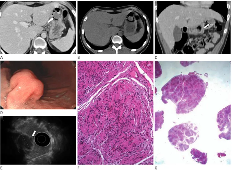
Fig. 1. Plexiform schwannoma of the stomach associated with NF2 in an 18-year-old male.
A, B. Axial MDCT scan shows a well-defined, ovoid-shaped, homogeneous soft tissue density lesion surrounded by mucosa in the gastric body (arrow) (A). The attenuation of the mass is 25 HU on unenhanced CT (B), and 40 HU on contrast-enhanced CT.
C. Contrast-enhanced coronal reformatted CT scan definitively shows that overlying mucosa is connected with adjacent stomach mucosa, indic- ative of a submucosal tumor (arrow).
D. Endoscopy shows a submucosal tumor at the lesser curvature of the gastric lower body.
E. EUS shows a well-marginated hypoechoic mass with hyperechoic strands arising from the third layer of gastric wall (arrow).
F. Photomicrograph (H&E staining × 200 magnification) shows that the spindle cells are relatively bland looking with pointed ends and palisad- ing arrangement (Verrocay bodies), compatible with schwannoma.
G. Photomicrograph (H&E staining × 10 magnification) shows several fragments of multinodular growing neoplasm separated by fibrous capsule.
Note.-EUS = endoscopic ultrasound, MDCT = multidetector computed tomography, NF2 = neurofibromatosis type 2
Dong Heon Yeom, et al
submit.radiology.or.kr J Korean Soc Radiol 2012;66(2):155-158
157
vealed as plexiform schwannoma of the gastrointestinal (GI) tract (1, 4-10). The location of the lesions were three in the esophagus, one in the small bowel, one in the ascending colon, two in the sigmoid colon, and one in the rectosigmoid colon, respectively. Among these cases, one case in the esophagus of a pediatric patient and one case in the sigmoid colon of a second patient were associated with NF2. Therefore, to our knowledge, this is the first report that demonstrated stomach localization of a plexiform schwannoma associated with NF2.
In the GI tract, the differential diagnosis of plexiform schwanno- ma includes gastrointestinal stromal tumor with plexiform growth pattern. In this situation, a negative immunohisto- chemical stain for CD 117, along with strong and diffuse stain- ing for S-100 protein, would be helpful in confirming the diag- nosis for a plexiform schwannoma (6). Also, neurogenic tumors of the GI tract that must be distinguished from plexi- form schwannoma include conventional schwannoma, neuro- fibroma, malignant peripheral nerve sheath tumor, ganglioneu- roma, and ganglioneuromatosis (7).
The distinction among plexiform schwannoma, plexiform neurofibroma, and malignant peripheral nerve sheath tumor (MPNST) is crucial for the correct clinical management of the patient (1). While plexiform schwannoma is just a benign form, plexiform neurofibroma presents a 2% to 5% risk of malignant transformation, and MPNST is, by definition, a malignant neo- plasm (1). The strong connection between NF1 and plexiform neurofibroma, and between NF2 and plexiform schwannoma is established in literature (1). This notion could be helpful in diagnosing the nature of a tumor.
The diagnosis of GI tract schwannoma is difficult preopera- tively, as these lesions appear as subepithelial tumors. To our knowledge, there are no typical endosonographic features of GI tract schwannoma (9). Because of their plexiform pattern of growth, it is relatively common for benign schwannomas to traverse several layers of bowel wall and even extend into the surrounding adipose tissue (9). Definitive treatment requires complete surgical resection.
In summary, we have reported with a case of plexiform schwannoma of the stomach in an NF2 male patient. If a sub- epithelial mass of the stomach or GI tract is noted in a patient with NF2, although it is rare, the possibility of plexiform schwannoma should be considered.
a plexiform schwannoma pathologically.
DISCUSSION
Plexiform schwannoma is a widely documented variant of schwannoma (1) and a benign peripheral nerve sheath tumor composed exclusively of schwann cells arranged in a plexiform pattern (4). These lesions tend to present in early adulthood, lack- ing obvious sex predilection (5). Almost all plexiform schwanno- mas have been reported as dermal or subcutaneous tumors, and most commonly appeared as multinodular, well-circum- scribed tumors (3-5).
NF2 has an autosomal dominant pathology. It is a separate entity from neurofibromatosis type 1 (NF1), and its clinical characteristics include 1) schwannoma of acoustical nerves, 2) central nervous system tumors (meningioma, astrocytoma, ependymoma), 3) juvenile subcapsular cataract, and some- times 4) café au lait spots (recurring less frequently than in NF1) (1).
Several reports demonstrated an association between plexi- form schwannoma and neurofibromatosis type (4). Solitary plexiform schwannomas are generally considered to be unasso- ciated with NF1 (5), and rare associations with NF2 have been described (6). According to previous reports, patients with plexiform schwannoma associated with NF2 had acoustic neu- rinomas, but the association between these two diseases is still controversial, including the hereditary conditions (1).
Most plexiform schwannoma cases associated with NF2 showed multiple, dermal, and subcutaneous locations (6). Visceral loca- tion of plexiform schwannoma is extremely rare. The first case of a solitary plexiform schwannoma in the visceral organ was de- scribed in 1997 by Hirose et al. (7). Agaram et al. (6) assert that plexiform schwannoma with visceral localization occur more often in females than in males and have a high risk of recur- rence but do not show malignant features or metastatic spread- ing. In our case, the patient was male and there was no evi- dence of metastatic spreading, but we could not know about recurrence and malignant transformation because follow up was not done. More generally, it is not yet clear whether plexi- form schwannoma in unusual localizations have the same be- havior as skin and subcutaneous lesions (1).
According to our literature search, only eight cases were re-
Plexiform Schwannoma of the Stomach in Neurofibromatosis Type 2
submit.radiology.or.kr
J Korean Soc Radiol 2012;66(2):155-158
158
6. Agaram NP, Prakash S, Antonescu CR. Deep-seated plexi- form schwannoma: a pathologic study of 16 cases and com- parative analysis with the superficial variety. Am J Surg Pathol 2005;29:1042-1048
7. Hirose T, Scheithauer BW, Sano T. Giant plexiform schwanno- ma: a report of two cases with soft tissue and visceral in- volvement. Mod Pathol 1997;10:1075-1081
8. Coron R, Boucard H, Richards R. Case report: sigmoid schwannoma as the lead point for intussusception in an adult patient with neurofibromatosis. Department of Medicine Faculty of Papers 8-9-2006
9. Jacobson BC, Hirsch MS, Lee JH, Van Dam J, Shoji B, Far- raye FA. Multiple asymptomatic plexiform schwannomas of the sigmoid colon: a case report and review. Gastroin- test Endosc 2001;53:801-804
REFERENCES
1. Retrosi G, Nanni L, Ricci R, Manzoni C, Pintus C. Plexiform schwannoma of the esophagus in a child with neurofibro- matosis type 2. J Pediatr Surg 2009;44:1458-1461
2. Evans DG. Neurofibromatosis type 2 (NF2): a clinical and molecular review. Orphanet J Rare Dis 2009;4:16
3. Miyakawa T, Kamada N, Kobayashi T, Hirano K, Fujii K, Sa- sahara Y, et al. Neurofibromatosis type 2 in an infant with multiple plexiform schwannomas as first symptom. J Der- matol 2007;34:60-64
4. Iida A, Imamura Y, Katayama K, Hirose K, Yamaguchi A.
Plexiform schwannoma of the small intestine: report of a case. Surg Today 2003;33:940-943
5. Cokelaere K, Sciot R, Geboes K. Esophageal Plexiform Schwannoma. Int J Surg Pathol 2000;8:353-357
제2형 신경섬유종증 환자에서 발생한 위의 얼기형신경초종: 1예 보고1
염동헌
1· 조현선
1· 김현정
2· 조우호
1· 김재형
1· 정명자
1· 김성희
1· 김지영
1· 김수현
1· 강미진
1· 이지혜
1· 이한비
1얼기형신경초종은 말초신경초에서 기원하는 드문 양성 종양으로 대개 상피하종괴로 발현되며 제2형 신경섬유종증과 연 관성이 있는 것으로 알려져 있다. 위장관에 발생한 얼기형신경초종에 대한 증례는 매우 적으며, 지금까지 제2형 신경섬유 종증 환자에서 위에 생긴 증례는 보고된 바가 없다. 본 증례에서 얼기형신경초종은 제2형 신경섬유종증 환자의 위에 점막 하종괴로 발현하였으며, 지금까지 제2형 신경섬유종증환자에서 위에 발생한 얼기형신경초종을 보고한 예가 없기에 이의 영상의학적 소견을 기술하고자 한다.
인제대학교 의과대학 상계백병원 1영상의학과학교실, 2병리과학교실