J Korean Soc Radiol 2017;77(1):14-18 https://doi.org/10.3348/jksr.2017.77.1.14
INTRODUCTION
Paraganglioma is a family of tumors originating from the neu- ral crest cells. Tumor occuring within the adrenal medulla are called pheochromocytoma, and those developing in extra-adre- nal locations (5–10% of cases) are referred to as paraganglioma (1, 2). Paraganglioma secrete catecholamines and cause malig- nant hypertension or symptoms such as headaches, palpitations, and diaphoresis. They can be located at any anatomical site along the paravertebral and para-aortic areas, from the head to the pel- vic floor. Extra-adrenal paraganglioma that develops within the abdomen are distributed along the paravertebral and para-aortic axis, in particular, along the organ of Zuckerkandl (close to the origin of the inferior mesenteric artery). However, several studies have reported the occurrence of extra-adrenal paraganglioma in
relatively rare locations, such as the urinary bladder, gallbladder, and hepatoduodenal ligament (3-5). Paraganglioma located in the mesentery or pararectal space is extremely rare, and only a few reports of such masses have been published (1, 2, 6-9).
We report here an unusual case of malignant, extra-adrenal paragangliomas, presenting as mesenteric and pararectal masses with lymph node metastasis. This paper includes their imaging features and histopathology characteristics.
CASE REPORT
A 70-year-old man visited our institution for evaluation and management of abdominal masses incidentally detected at a lo- cal clinic. The patient had no specific symptoms or relevant past medical history. All laboratory findings, including plasma cate-
Extra-Adrenal Malignant Paragangliomas Presenting as Mesenteric and Pararectal Masses: A Case Report
장간막과 직장 주변부에 발생한 악성 부신경절종: 증례 보고
Sunhye Lee, MD
1, Jongmee Lee, MD
2*, Baek Hui Kim, MD
3, Kyeong Ah Kim, MD
2, Cheol Min Park, MD
21Department of Radiology, Korea University Anam Hospital, Korea University College of Medicine, Seoul, Korea
Departments of 2Radiology, 3Pathology, Korea University Guro Hospital, Korea University College of Medicine, Seoul, Korea
Extra-adrenal paraganglioma is a rare tumor arising from the neural crest cells. Most tumors that develop in the abdomen arise from paraganglia along the paravertebral and para-aortic areas, in particular the organ of Zuckerkandl, which is close to the ori- gin of the inferior mesenteric artery. However, extra-adrenal paraganglioma also oc- curs in relatively rare places such as the urinary bladder, gallbladder, hepatoduodenal ligament, and gastrointestinal tract. Here, we report imaging findings of extra-adrenal paragangliomas presenting as mesenteric and pararectal masses with lymph node metastasis.
Index terms Paraganglioma Mesentery Neoplasms
Computed Tomography Magnetic Resonance Imaging
Received November 22, 2016 Revised January 4, 2017 Accepted January 22, 2017
*Corresponding author: Jongmee Lee, MD
Department of Radiology, Korea University Guro Hospital, Korea University College of Medicine, 148 Gurodong-ro, Guro-gu, Seoul 08308, Korea.
Tel. 82-2-2626-1338 Fax. 82-2-863-9282 E-mail: [email protected]
This is an Open Access article distributed under the terms of the Creative Commons Attribution Non-Commercial License (http://creativecommons.org/licenses/by-nc/4.0) which permits unrestricted non-commercial use, distri- bution, and reproduction in any medium, provided the original work is properly cited.
cholamine levels, were within the normal range. The patient had undergone computed tomography (CT) scans at an outside hos- pital to evaluate the intra-abdominal masses using a 16-slice multidetector CT scanner (SOMATOM Emotion 16; Siemens, Erlangen, Germany). Contrast-enhanced axial CT images re- vealed a multilobulated, solid mass, approximately 15 cm in
size, showing heterogeneous and strong enhancement, with ad- jacent engorged vessels in the small bowel mesentery (Fig. 1A).
A second round mass at the right side of the rectum, 2.8-cm in size, well defined, and with a similar enhancement pattern, was observed on the CT scan (Fig. 1B). The patient further under- went magnetic resonance (MR) imaging to assess the exact an-
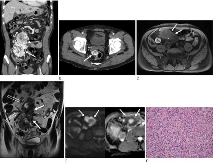
Fig. 1. Mesenteric and pararectal paragangliomas in a 70-year-old man.
A. The portal venous phase coronal computed tomography (CT) scan shows a 15 cm-sized, heterogeneous, hypervascular soft tissue mass, with a lobulated contour (arrows) in the small bowel mesentery of the abdomen.
B. The contrast enhanced portal venous phase axial CT image shows another well-defined, hypervascular mass, approximately 2.8 cm in size, ad- jacent to the right rectal wall (arrow).
C. The T1-weighted axial magnetic resonance (MR) image without a contrast agent shows the heterogeneous signal intensity of the mesenteric mass (arrows), which is similar to that of the pararectal mass (not shown).
D. The T2-weighted coronal MR image reveals a large, lobulated soft tissue mass (arrows) with heterogeneous high signal intensity, internal sig- nal void structures, and engorged supplying vessels (blank arrows) in the small bowel mesentery. Heterogeneous high signal intensity with inter- nal signal void was observed in the pararectal mass (not shown).
E. The high signal intensity of the mesenteric mass (arrows) is shown on the diffusion-weighted MR image with a b-value of 800 s/mm2 (left).
Some portions of the mesenteric mass show a low apparent diffusion coefficient value (arrows), indicating the presence of diffusion restriction (right).
F. Microscopic features of the paraganglioma. Some tumor cells show a nested pattern, and others show a diffuse pattern. The tumor cells exhibit eosinophilic cytoplasm and ‘salt and pepper’ patterned chromatin with moderate nuclear pleomorphism (hematoxylin-eosin stain; original mag- nification, × 200), consistent with the findings of a paraganglioma.
A B C
D E F
atomic location, using a 3T unit (MAGNETOM Skyra, Siemens, Erlangen, Germany) with a standard 18-channel body matrix coil and table-mounted 32-channel spine matrix coil. For the enhancement studies, gadoxetic acid as a contrast agent (Gd- EOB-DTPA, Primovist; Bayer Healthcare, Berlin, Germany) was administered intravenously. MR imaging revealed well-de- fined solid masses in the small bowel mesentery and pararectal area, showing heterogeneous hypo- and hyper-signal intensities on both T1- (Fig. 1C) and T2-weighted images (Fig. 1D). On dynamic enhancement, the tumors showed avid, heterogeneous enhancement with poor central enhancement that matched the high signal intensity on the T2-weighted image. On the diffu- sion-weighted MR image with a b-value of 800 s/mm2, the mes- enteric mass showed high signal intensity, and the peripheral portion of the mass had a low value on the apparent diffusion coefficient (ADC) map, indicating the presence of restricted dif- fusion (Fig. 1E). No evidence of restricted diffusion in the para- rectal mass was seen.
Differential diagnoses based on these imaging findings includ- ed a gastrointestinal stromal tumor, neuroendocrine tumor, paraganglioma, and hypervascular metastasis. A subsequent exploratory laparotomy revealed a large mass with hemorrhage, originating from the small bowel mesentery, and another mass in the right pararectal area. Since the pararectal mass had in- vaded the rectal wall, a low anterior resection was performed.
The histopathological examination revealed cellular neoplasms composed of uniform small round cells separated by fibrovascu- lar connective tissue, showing a characteristic zellballen pattern (Fig. 1F). The mitotic count was low (< 1/10 high power field).
Immunohistochemically, the tumor cells were positive for syn- aptophysin and chromogranin. The proportion of Ki-67 posi- tive cells was 10%.
On the basis of the histopathologic findings, the masses in the small bowel mesentery and pararectal area were confirmed as paragangliomas. Metastasis was identified in 1 out of the 18 re- sected mesenteric lymph nodes, and the masses were finally di- agnosed as malignant paragangliomas. The postoperative course was uneventful, and the patient was discharged on postoperative day 8.
DISCUSSION
Paraganglioma is a rare tumor derived from neural crest cells that develop into the sympathetic and parasympathetic paragan- glia. When it secretes catecholamines, it is called a functioning paraganglioma. The most common clinical symptoms associated with catecholamine secretion include headaches, palpitations, and profuse sweating. From a diagnostic viewpoint, a functional tu- mor is easier to diagnose. However, similar to our case, most ex- tra-adrenal paragangliomas are nonfunctional (9). Our patient was devoid of any clinical symptoms or abnormal laboratory find- ings. Therefore, the clinical presentation and biochemical markers were non-informative in making the diagnosis. In these situations, imaging studies play an important role in characterizing the tu- mors and determining their location.
Extra-adrenal paraganglioma are usually observed as wellde- fined, round- or oval-shaped masses of soft-tissue density on un- enhanced CT scans. Punctate parenchymal calcification is ob- served in about 15% of such tumors. As the tumor progresses, it often appears cystic with central necrosis. Intense contrast en- hancement is observed due to the hypervascular nature of para- gangliomas. On MR imaging, extra-adrenal paraganglioma is usually hypointense or isointense on T1-weighted images, and hyperintense on T2-weighted images. Heterogeneous signal in- tensity is often seen, owing to the presence of hemorrhage. In ad- dition, a speckled appearance with multiple flow voids is typical.
Imaging findings of extra-adrenal paraganglioma on diffusion- weighted MR imaging and ADC map have been described in very few reports (3). Extra-adrenal paraganglioma shows high signal intensity on diffusion-weighted MR imaging with high b- values. It displays a low value on the ADC map, indicative of re- stricted diffusion. In our patient, the mesenteric and pararectal paragangliomas showed high signal intensity on diffusion- weighted MR imaging with a high b-value gradient. However, re- stricted diffusion was seen only in the peripheral portions of the mesenteric paraganglioma.
Despite positive laboratory test results, if a lesion is not identi- fied on radiologic studies, radionuclide imaging with 123I-me- taiodobenzylguanidine or 18F-fluorodeoxyglucose positron emis- sion tomography is recommended to identify the occult lesion.
Radionuclide imaging is a more practical way of examining the entire body, and is valuable in localizing a functional extra-adre-
nal paraganglioma and its associated metastases.
The gross morphology of a paraganglioma is usually sharply circumscribed with expanding borders; on observing the cross section, this tumor has resiliently firm and grey-white surface, with areas of hemorrhage and cystic degeneration within the tumor. Microscopically, the most characteristic pattern is an or- ganoid or trabecular arrangement (zellballen) of neoplastic chief cells, separated by a rich microvasculature. Intracytoplasmic granules, similar to those seen in healthy paraganglia, can be seen in some tumors. Although prominent nuclear hyperchromasia and a remarkable degree of nuclear pleomorphism may be pres- ent, these are not reliable features in diagnosing malignancy.
Benign and malignant paragangliomas have an identical histo- logic appearance, and the criteria for malignancy on the basis of histopathology are not well defined. Therefore, local invasion or distant metastasis are important features to be considered in the diagnosis of a malignant paraganglioma. Approximately 2–36% of paragangliomas are diagnosed as malignant, based on extensive local invasion or metastasis (10). In general, a para- ganglioma is more likely to be malignant than a pheochromo- cytoma.
Although most paragangliomas are solitary and arise sporadi- cally, they can be multicentric and can occur synchronously or metachronously; the incidence rate of multicentricity is approxi- mately 10% (10). Our case showed 2 extra-adrenal paraganglio- mas in the small bowel mesentery and the pararectal area. The smaller paraganglioma in the pararectal area may have been a synchronous tumor or metastases from the mesenteric paragan- glioma. Either way, the mesenteric paraganglioma was identified as malignant due to the presence of mesenteric lymph node me- tastasis.
The differential diagnosis for such hypervascular masses in- cludes solitary fibrous tumors, gastrointestinal stromal tumors, and hypervascular metastases of carcinoma and melanoma.
Considering the hypervascular nature of the tumors in this case, extra-adrenal paraganglioma was included in the differential di- agnosis. However, its distant location from the para-aortic area and the rarity of mesenteric and pararectal paragangliomas, made a preoperative diagnosis difficult.
We conclude by summarizing that extra-adrenal paraganglio- ma in the abdomen is rarely found in the mesenteric or pararec- tal location. It can manifest as a heterogeneous, hypervascular solid mass that should be differentiated from a mesenchymal tu- mor or metastases. Although rare, extra-adrenal paraganglioma should be included in the preoperative differential diagnosis of a solid hypervascular intra-abdominal mass, regardless of the tu- mor location.
REFERENCES
1. Fujita T, Kamiya K, Takahashi Y, Miyazaki S, Iino I, Kikuchi H, et al. Mesenteric paraganglioma: report of a case. World J Gastrointest Surg 2013;5:62-67
2. Ozkan Z, San Ozdemir C, Yasar G, Altas O, Koc M, Gul Y, et al. An unusual mesenteric tumor ‘paraganglioma’: a case report. Iran Red Crescent Med J 2014;16:e16837
3. Wang H, Ye H, Guo A, Wei Z, Zhang X, Zhong Y, et al. Blad- der paraganglioma in adults: MR appearance in four pa- tients. Eur J Radiol 2011;80:e217-e220
4. Baker C, Bhagwat P, Wan A. Mesenteric paraganglioma with gallbladder paraganglion nest. J Surg Case Rep 2012;2012:8 5. Furukawa K, Ishida H, Komatsuda T, Yagisawa H, Yamada M,
Miyauchi T, et al. Malignant paraganglioma draining into the main portal vein. Abdom Imaging 2005;30:758-760 6. Chetrit M, Dubé P, Royal V, Leblanc G, Sideris L. Malignant
paraganglioma of the mesentery: a case report and review of literature. World J Surg Oncol 2012;10:46
7. Kudoh A, Tokuhisa Y, Morita K, Hiraki S, Fukuda S, Eguchi N, et al. Mesenteric paraganglioma: report of a case. Surg To- day 2005;35:594-597
8. Moslemi MK, Abolhasani M, Vafaeimanesh J. Malignant ab- dominal paraganglioma presenting as a giant intra-perito- neal mass. Int J Surg Case Rep 2012;3:537-540
9. Bhatt S, Vanderlinde S, Farag R, Dogra VS. Pararectal para- ganglioma. Br J Radiol 2007;80:e253-e256
10. Lee KY, Oh YW, Noh HJ, Lee YJ, Yong HS, Kang EY, et al. Ex- traadrenal paragangliomas of the body: imaging features.
AJR Am J Roentgenol 2006;187:492-504
장간막과 직장 주변부에 발생한 악성 부신경절종: 증례 보고
이선혜
1· 이종미
2* · 김백희
3· 김경아
2· 박철민
2부신경절종은 신경능선세포에서 기원하는 부신경절의 실질세포에서 발생하는 종양으로 드물게 부신 외에서 발생한다. 부 신 외 형태로 복강내에서 발생할 때는 주로 하장간막동맥 기시부의 대동맥 주위에 분포하는 부신경절인 Zuckerkandl 기 관에서 발생하나 드물게 방광이나 담낭 등 다른 장기에 발생한 보고들도 있다. 저자들은 매우 드물게 장간막과 직장 주변 에 동시에 발생한 악성 부신경절종에 대해 문헌고찰과 함께 보고하고자 한다.
1고려대학교 의과대학 안암병원 영상의학과, 고려대학교 의과대학 구로병원 2영상의학과, 3병리과