INTRODUCTION
Atopic dermatitis (AD) is the most common chronic inflam- matory skin disease in pediatric populations. The prevalence of this condition has been increasing worldwide over the past few decades, particularly in countries, such as South Korea, where a Western diet and lifestyle are more common.1,2 Although genet- ic susceptibility remains the most important factor for the de- velopment of AD, genetics alone cannot account for the abrupt increase in disease prevalence. The etiology of AD should there- fore be considered a multifactorial process consisting of genet- ic, epigenetic, developmental, and environmental factors.
Proper skin care, avoidance of known environmental triggers, and topical corticosteroid therapy have long been the standard of care for the management and treatment of AD. However, poor compliance, frequent disease recurrence, and complications as a result of chronic corticosteroid use have led to calls for new therapeutic alternatives.5
Microbial exposure early in life has been shown to confer pro- tection against AD.3,4 This observation has led to significant in-
Efficacy of Probiotic Therapy on Atopic Dermatitis in Children:
A Randomized, Double-blind, Placebo-controlled Trial
Hyeon-Jong Yang, Taek Ki Min, Hae Won Lee, Bok Yang Pyun*
Pediatric Allergy and Respiratory Center, Department of Pediatrics, Soonchunhyang University Hospital, Soonchunhyang University College of Medicine, Seoul, Korea
terest in the use of probiotics as an alternative strategy for the prevention of atopic diseases in susceptible individuals.6,7 Pro- biotics are defined as live organisms that can confer beneficial effects on the health of the host. Probiotic therapies have been proven to be effective in treating a variety of medical conditions, including antibiotic-associated diarrhea, AD, and other chronic inflammatory conditions. The therapeutic potential of these treatments appear to be mediated through a number of mech- anisms of action, including modulation of the immune response, competitive inhibition of invading flora in the gut, modification of pathogenic toxins and host products, and enhancement of epithelial barrier function.8,9
Allergy Asthma Immunol Res. 2014 May;6(3):208-215.
http://dx.doi.org/10.4168/aair.2014.6.3.208 pISSN 2092-7355 • eISSN 2092-7363
Purpose: To evaluate a therapeutic efficacy of probiotics mixture (probiotics) in the treatment of children with mild-to-moderate atopic dermatitis (AD). Methods: Randomized, double-blind, placebo-controlled, parallel trial with a washout period of 2 weeks and an intervention period for 6 weeks, conducted from November 2010 to October 2011. One hundred children with mild to moderate AD (2-9 years old) were randomly allocated to the probiotics (Lactobacilluss casei, Lactobacillus rhamnosus, Lactobacillus plantarum, and Bifidobacterium lactis) or placebo groups. The assessment of efficacy was based on the change in eczema area severity index (EASI), visual analogue scale for pruritus (VASP), fecal cell counts of each strains (log10[cell counts/g stool]), and serum cytokine levels (Interleukin-4 [IL-4]; IL-10; Tumor necrosis factor alpha, [TNF-α]) in weeks 0 and 6. Results: De- mographics and baseline characteristics at the week 0 were not significantly different between the 2 groups. The significant increments in fecal-cell counts were observed in the probiotcs group at week 6 (P=0.00), while the cytokine levels between the 2 groups were not significantly different in week 6 (IL-4, P=0.50; IL-10, P=0.58; TNF-α, P=0.82). The probiotics significantly improved clinical severity after 6 weeks’ intervention of probiotics;
however, the placebo group also showed significant improvement (EASI; P=0.00, VASP; P=0.00). Conclusions: Our findings showed that probiot- ics successfully colonized in the intestine after 6 weeks’ intervention; nevertheless, we could not find an additional therapeutic or immunomodulato- ry effects on the treatment of AD. Further long-term studies will be necessary to clarify the therapeutic efficacy of probiotics.
Key Words: Atopic dermatitis; cytokines; placebo-controlled trial; probiotics; Randomized Controlled Trial
This is an Open Access article distributed under the terms of the Creative Commons Attribution Non-Commercial License (http://creativecommons.org/licenses/by-nc/3.0/) which permits unrestricted non-commercial use, distribution, and reproduction in any medium, provided the original work is properly cited.
Correspondence to: Bok Yang Pyun, MD, PhD, Department of Pediatrics, Pediatric Allergy and Respiratory Center, Soonchunhyang University College of Medicine, Soonchunhyang University Hospital, 22 Daesagwan-gil,
Yongsan-gu, Seoul 140-743, Korea.
Tel: +82-2-709-9339; Fax: +82-2-794-5471; E-mail: [email protected] Received: April 26, 2013; Revised: June 13, 2013; Accepted: July 18, 2013
•There are no financial or other issues that might lead to conflict of interest.
Although a handful of species have been considered effective for the prevention of AD, the efficacy of probiotics in the man- agement of AD is unknown.10 Humans regularly eat foods con- taining probiotic bacteria, such as yogurt or other fermented foods.11 However, because most studies use only single strains of probiotic bacteria, little is known about the efficacy of probi- otic mixtures, particularly whether mixing strains result in syn- ergistic or inhibitory effects on the treatment of atopic disease.
We therefore sought to evaluate the therapeutic efficacy of a de- fined probiotic mixture (Lactobacillus casei, L. rhamnosus, L.
plantarum, and Bifidobacterium lactis), and to determine the effects of this mixture on serum cytokine levels in children with mild-to-moderate AD.
MATERIALS AND METHODS Patients and study setting
This study was performed between November 2010 and Octo- ber 2011. One hundred children (2 to 9 years of age) with mild- to-moderate AD were recruited from the Pediatric Allergy and Respiratory Center of Soonchunhyang University Hospital, a tertiary medical center located in Seoul, Korea. Diagnosis of AD was performed using the diagnostic criteria set forth by Hanifin and Rajka in 1980.12 The study protocol was approved by the Soonchunhyang University Hospital Research Ethics Commit- tee prior to initiation of the study. Written informed consent was obtained from each patient and his or her parents at the time of recruitment and before the study-related interview was per- formed.
Study design
This study was designed as double-blind, placebo-controlled, randomized parallel trial with a 2-week washout period prior to intervention.
Enrollment criteria
The severity of AD was assessed using the SCORAD index13 at the time of enrollment. Children with mild-to-moderate AD (SCORAD score ≤40) were included.
Exclusion criteria
The exclusion criteria used in this study were children with (1) severe AD (SCORAD score >40), (2) exposure to commercial probiotic products during the previous 4 weeks, (3) acute gas- trointestinal infections, (4) chronic underlying disease or base- line factors predisposing to the infection (e.g., neurologic dis- ease, metabolic disease, chronic respiratory disease, congenital anomaly of the heart, gastrointestinal system, or lung), (5) known or suspected immunodeficiency, (6) prematurity, and (7) re- ceiving antibiotic, systemic corticosteroid, immunosuppressive, or Chinese herbal therapies within 4 weeks prior to enrollment.
Sample size
Sample size was calculated assuming a 30% reduction in Ec- zema Area and Severity Index (EASI) score in the probiotics group and a 15% reduction in the placebo group at week 6, ap- plied with 5% significance levels and 90% power. On the basis of our pilot data, ~40 children are required for each group. Ulti- mately, a sample size of 100 children was deemed necessary with reference to an expected drop-out rate of 20%.
Wash-out period
At the time of enrollment, parents were trained in appropriate bathing and skin care practices, and received instructions re- garding the application of topical emollients. Use of topical cor- ticosteroids (TCSs), topical calcineurin inhibitors (TCIs), oral antihistamines, or any commercial probiotic-containing prod- ucts was stopped 2 weeks prior to initiation of the study (week 0).
Intervention and assignments
The probiotics mixture consisted of L. casei, L. rhamnosus, L.
plantarum, and B. lactis in glucose anhydrous crystalline pow- der derived from cornstarch. The study preparation was manu- factured by CellBiotec (Seoul, Korea). A single dose of the prep- aration contained 1×10⁹ colony forming units (CFU) of each bacterial strain; the placebo consisted of pure glucose anhydrous crystalline powder. The probiotics mixture and placebo controls were identical in color, taste, smell, packing, and manner of ad- ministration. All formulations were dispensed by a pharmacist not associated with the study; both investigators and study sub- jects were blinded to the identity of the intervention. Both prep- arations were administered in warm water or juice immediately after meals, twice a day (2×10⁹ CFU in each strain) for 6 weeks.
During the study period, neither TCSs nor TCIs were allowed for either group.
Randomization
Randomization software14 was used to randomly allocate chil- dren to receive either the probiotics mixture (probiotics group, n=50) or placebo control (placebo group, n=50) for 6 weeks.
Drop-out criteria
The drop-out criteria included patients with poor compliance in either the probiotics or placebo groups (<80% of study pro- tocol), severe flare-ups requiring oral steroid or oral antibiotic therapy, and infectious diseases (i.e., upper respiratory infection, lower respiratory infection, acute gastroenteritis, urinary tract infection) which made the interpretation of outcomes difficult.
Outcome measurement
Measurements were taken at the beginning (week 0) and end (week 6) of the study. Efficacy of probiotics was assessed on the basis of clinical improvement as determined by EASI and visual analog scale for pruritus (VASP) scores, as the primary outcome.
As secondary outcomes we compared fecal CFUs for each pro- biotic strain (L. casei, L. rhamnosus, L. plantarum, and B. lactis), and serum cytokine levels (interleukin-4, IL-4; interleukin 10, IL-10; tumor necrosis factor alpha, TNF-α) in weeks 0 and 6.
Atopic sensitization
Serum total immunoglobulin E (IgE) and specific IgE for food allergens (cow’s milk, egg white, soy, peanuts, and cod) and ae- ro-allergens (Dermatophagoides farinae, D. pteronyssinus, dog hair, and Aspergillus fumigatus) were measured using Immu- noCAP (Phadia AB, Uppsala, Sweden) in week 0. Allergen sen- sitization was defined as specific IgE levels ≥0.35 kU/L, which is the positive cutoff value recommended by the manufacturer.
Fecal samples and real-time quantitative PCR (RT-qPCR) Each fecal sample was collected at the subject’s home in a sterile container, frozen, and then transported to the laborato- ry, where it was stored at -80°C prior to analysis. RT-qPCR was performed using a LightCycler 480 system (Roche, Germany).
Primers were synthesized commercially by Bioneer (Daejeon, Korea), and the specificity of each primer was verified using DNA isolated from closely and distantly related bacteria as a template.
Quantitative PCR was performed in a 96-well plate in a final volume of 20 µL. Reaction mixtures consisted of 1-µL fecal DNA, 0.5-µL primers (10 pmol each), 10-µL SYBR Green I master mix (Roche, Germany), and 8-µL H2O. PCR amplification was car- ried out under the following conditions: pre-incubation at 94°C for 4 min, followed by 55 cycles of amplification (denaturation at 94°C for 15 seconds, primer annealing at 55°C for 15 seconds, and elongation at 72°C for 20 seconds). The melting curve was analyzed by heating the reaction from 50 to 90°C with a temper- ature transition time of 5°C/sec.
Fecal cell count
Standard curves were constructed to convert the cross point (Cp) value obtained from the qPCR analysis to cell counts. The total number of cells in 1 mL of pure culture was determined using a hemocytometer (Marienfeld, Germany). Genomic DNA was prepared from these suspensions, as described above, quan- tified using an e-spect spectrophotometer (Malcom, Tokyo, Ja- pan), and 10-fold serially diluted. Cp values for each of the dilu- tions were plotted against the corresponding cell number. Bac- terial cell numbers in 1-g feces were calculated using the stan- dard curve.
Statistical analysis
The primary goal for data analysis was estimating the clinical improvement of the probiotics group relative to the placebo group. The secondary goal was to compare differences in fecal cell composition and serum cytokines levels between the two groups. We performed the Chi-square test or Fisher’s exact test for nominal variables. Comparisons of continuous variables
within groups were performed using a paired t-test for normal- ly distributed data, or Wilcoxon’s matched-pairs signed-ranks test if the data were not normally distributed. Comparisons of continuous variables between groups were performed using independent-sample Student’s t-test for normally distributed data, or the Mann-Whitney U-test for non-normally distributed data. All calculated P values were 2-tailed; P values <0.05 were considered statistically significant. All analyses were performed using SPSS, version 15.0 (SPSS Inc., Chicago, IL, USA).
RESULTS
A total of 141 children with AD were enrolled in the study and assessed for eligibility. Forty-one were excluded due to severe AD (n=21), underlying chronic diseases (n=6), acute gastroen- teritis (n=2), preterm delivery (n=2), or recent antibiotic use (n=10). The remaining 100 children were randomized and evenly assigned to either the probiotics or placebo groups. Thir- teen subjects in the probiotics group and 16 subjects in the pla- cebo group failed to complete the study due to patient with- drawal, missed visit, respiratory infection requiring oral antibi- otics use, or violation of study protocol. A total of 37 subjects in the probiotics group and 34 subjects in the placebo group com- pleted the study. An overview of study design is shown in Fig. 1.
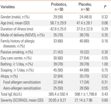
No statistically significant differences in baseline demograph- ics and clinical characteristics were observed between the 2 groups (Table 1).
Fecal cell counts and clinical severity
Clinical severity was measured using standard EASI and VASP methods; no differences were observed between the 2 groups in weeks 0 or 6. Fecal cell counts for all probiotic strains were similar in treatment and control groups at week 0, with the ex- ception of L. rhamnosus which was higher in the placebo group (probiotics group, 4.03±0.93; placebo group, 4.82±1.25; P<0.01).
By week 6, fecal cell counts for all probiotic strains were signifi- cantly higher in the probiotics group than in the placebo group (B. lactis, P≤0.001; L. casei, P≤0.001; L. rhamnosus, P≤0.001; L.
plantarum, P≤0.001) (Table 2).
Changes in fecal cell counts and clinical severity within each group
Clinical severity was significantly improved in the probiotics group in week 6 relative to baseline (week 0); however, signifi- cant improvement was also seen in the placebo group. Fecal cell counts for all probiotic strains were increased substantially in the probiotics group (P<0.01). Within this group, the greatest increase was seen in L. rhamnosus (514-fold); ~100-fold increas- es were also observed in both B. lactis and L. casei relative to baseline. L. casei was also increased in the placebo group in week 6 (P=0.03; Table 3); no significant differences were seen in any of the other species tested.
Comparison of ∆ESAI, ∆VASP, and ∆fecal cell counts between groups
The degree of improvement (∆ESAI and ∆VASP) was similar in both groups (∆ESAI, probiotics group, -2.00 vs placebo group, -2.75, P=0.28; ∆VASP, probiotics group, -1.00 vs placebo group, -1.00, P=0.32). Fecal cell counts for all stains were significantly increased in the probiotics group relative to placebo controls (P≤0.001; Table 4).
Serum cytokine levels
No differences in serum cytokine levels between the 2 groups were detected in either week 0 or 6 (Table 2). However, the IL- 10 level was significantly decreased in both groups in week 6 relative to baseline (probiotics group, 1.19 to 0.40 pg/mL, P=0.03;
placebo group, 1.39 to 0.40, P=0.04), and TNF-α was significant- ly increased in both groups in week 6 compared to week 0 (pro- biotics group, 6.69 to 10.65 pg/mL, P<0.01; placebo, 7.44 to 10.28, P<0.01; Table 3 and Fig. 2).
DISCUSSION
In this study, we conducted a randomized, double-blind, pla- cebo-controlled parallel trial to evaluate the therapeutic effect of a probiotic mixture on children with AD. Outcomes were de- termined on the basis of clinical improvement, the number CFUs for each of the probiotic strains detectable in feces, and serum cytokine levels after completion of the 6-week trial.
Upon completion of the study, our main findings were as fol- lows: (1) fecal cell counts were increased in the probiotics group compared to placebo controls, (2) modest clinical improvement was observed in both groups regardless of probiotic therapy, and (3) administration of probiotics for 6 weeks did not change serum cytokine levels (e.g., IL-4, IL-10, TNF-α).
Certain aspects of the Western lifestyle, such as diet and low rates of breast feeding, along with improved hygiene, are con- sidered important factors for the development of atopic dis- ease.4,15 Certain environmental factors, such as vaginal deliv- ery16 or exposure to more agricultural lifestyles,3 are associated
Excluded, n=41 SCORAD>40, N=21 Other chronic disease, n=6 AGE, n=2
Preterm infants, n=2 Recent antibiotics use, n=10
Assessed for eligibility
n=141
Enrollment
Weeks-2 Week 0 Weeks 6
SCORAD Mild to moderate
EASIVAS (pruritus) IL-4, 10, TNF-α Fecal cell counts
EASIVAS (pruritus) IL-4, 10, TNF-α Fecal cell counts Wash-out period for 2 weeks
Randomization n=100
Probiotics mixture n=50
Placebo n=50
Analyzed, n=37
Analyzed, n=34 Drop out, n=13
Refused, n=1 Missed visit, n=2 Antibiotics use, n=2 Lost to follow up, n=7 Protocol-violation, n=1
Drop out, n=16 Refused, n=3 Missed visit, n=0 Antibiotics use, n=1 Lost to follow up, n=11 Protocol-violation, n=1
Fig. 1. Overview of the study design.
Table 1. Demographics and baseline characteristics of the study population in week 0
Variables Probiotics,
n=50 Placebo,
n=50 P
Gender (male), n (%) 29 (58) 24 (48.0) 0.32
Age (mo), mean (SD) 58.7±29.9 47.4±28.1 0.06
Duration of illness (mo) 42.8±25.0 37.0±22.9 0.29 Mode of delivery (NSVD), n (%) 35 (70) 38 (76) 0.35 Family history of allergic
diseases, n (%)
33 (66) 40 (80) 0.16
Passive smoking, n (%) 21 (42) 16 (32) 0.30
Day care center, n (%) 30 (60) 27 (54) 0.55
Bathing ≥1/day, n (%) 39 (78) 39 (78) 1.00
Soaping ≥1/day, n (%) 45 (90) 44 (88) 0.75
Atopy, n (%) 32 (64) 35 (70) 0.52
Food allergen sensitization 22 (44) 17 (34) 0.31 Aero-allergen sensitization 25 (50) 28 (56) 0.55
Total IgE (kU/L) 385.4±592.4 598.1±1,798.8 0.43
Severity (SCORAD), mean (SD) 20.05±9.27 21.14±7.96 0.60
with protection from atopic and allergic disease, strongly sup- porting the so-called ‘hygiene hypothesis.’ Growing support for this hypothesis has led to a number of studies examining the use of probiotics for the prevention and treatment of atopic dis- ease; however, results of these trials remain inconclusive. Both positive17,18 and negative19,20 results have been seen in trials evaluating the efficacy of probiotics in children with AD. Van der Aa and colleagues10 argued that these conflicting results stem from the underlying heterogeneity between individual studies (e.g., timing of supplementation, different strains or dosage, different age or ethnicity of participants, severity of AD, study duration, and sample size) and that substantial improve- ment is often seen in placebo groups due to the natural tenden-
cy of AD to improve over time. Patients in our study showed similar improvements over time regardless of intervention, in- dicating that conventional therapies, including proper skin care and TCS, remain an important part of any treatment for AD.
The normal intestinal microbiota consists of over 400 species and is important in the development and maintenance of in- testinal immune function, intestinal barrier function, and ab- sorption of nutrients.21 Recent publications suggest that atopic diseases are strongly associated with the composition of the in- testinal microbiota. Species, such as Lactobacillus and Bifido- bacterium, predominated in the intestinal flora of healthy indi- viduals, while species such as Clostridium or Staphylococcus were more commonly associated with atopic diseases.16,22 Table 2. Fecal cell counts, clinical severity, and serum cytokine levels in weeks 0 and 6
Variables Week 0 Week 6
Probiotics, n=37 Placebo, n=34 P Probiotics, n=37 Placebo, n=34 P
Fecal cell count, log10 [cell/g, stool]
B. lactis 6.75±1.22 6.99±1.33 0.48 8.81±1.02 7.05±1.32 <0.01*
L. casei 5.29±0.83 5.49±1.13 0.44 7.24±0.90 6.07±1.17 <0.01*
L. rhamnosus 4.03±0.93 4.82±1.25 <0.01* 6.74±1.08 4.29±1.30 <0.01*
L. plantarum 5.84±0.77 5.94±0.73 0.61 7.24±1.21 5.75±0.98 <0.01*
Clinical severity, median (IQR)
EASI 7.2 (4.8-10.3) 8.3 (4.7-10.6) 0.46 4.7 (2.2-6.8) 4.5 (2.6-9.0) 0.95
VASP 4.0 (3.0-6.0) 4.0 (4.0-5.3) 0.95 3.0 (2.0-5.0) 2.5 (2.0-4.3) 0.68
Serum cytokine level (pg/mL), median (IQR)
IL-4 0.27 (0.27-0.30) 0.27 (0.27-0.27) 0.30 0.27 (0.27-0.50) 0.27 (0.17-0.34) 0.53
IL-10 1.19 (0.81-1.98) 1.39 (0.65-1.92) 0.84 0.40 (0.40-1.55) 0.40 (0.40-1.10) 0.58
TNF-α 6.69 (5.48-7.90) 7.44 (5.64-8.21) 0.47 10.65 (9.31-11.71) 10.28 (9.39-11.37) 0.82
B. lactis, Bifidobacterium lactis; L. casei, Lactobacillus casei; L. rhamnosus, Lactobacillus rhamnosus; L. plantarum, Lactobacillus plantarum.
*Independent-sample Student’s t-test, P<0.01.
Table 3. Fecal cell counts, clinical severity, and serum cytokine levels in weeks 0 and 6
Variables Probiotics group, n=37 Placebo group, n=34
Week 0 Week 6 P Week 0 Week 6 P
Clinical severity, median (IQR)
EASI 7.2 (4.8-10.3) 4.7(2.2-6.8) <0.01* 8.3 (4.7-10.6) 4.5 (2.6-9.0) <0.01*
VASP 4.0 (3.0-6.0) 3.0(2.0-5.0) <0.01* 4.0 (4.0-5.3) 2.5 (2.0-4.3) <0.01*
Fecal cell count, log10 [cells/g, stool]
B. lactis 6.75±1.22 8.81±1.02 <0.01‡ 6.99±1.33 7.05±1.32 0.80
L. casei 5.29±0.83 7.24±0.90 <0.01‡ 5.49±1.13 6.07±1.17 0.03†
L. rhamnosus 4.03±0.93 6.74±1.08 <0.01‡ 4.82±1.25 4.29±1.30 0.81
L. plantarum 5.84±0.77 7.24±1.21 <0.01‡ 5.94±0.73 5.75±0.98 0.31
Serum cytokine level (pg/mL), median (IQR)
IL-4 0.27 (0.27-0.30) 0.27 (0.27-0.50) 0.73 0.27 (0.27-0.27) 0.27(0.17-0.34) 0.16
IL-10 1.19 (0.81-1.98) 0.40 (0.40-1.55) 0.03† 1.39 (0.65-1.92) 0.40 (0.40-1.10) 0.04†
TNF-α 6.69 (5.48-7.90) 10.65 (9.31-11.71) <0.01‡ 7.44 (5.64-8.21) 10.28 (9.39-11.37) <0.01‡ B. lactis, Bifidobacterium lactis; L. casei, Lactobacillus casei; L. rhamnosus, Lactobacillus rhamnosus; L. plantarum, Lactobacillus plantarum.
*Wilcoxon’s matched-pairs signed-ranks test, P<0.01; †Paired t-test, P<0.05; ‡Paired t-test, P<0.01.
To date, the clinical efficacy of probiotics on the treatment and prevention of AD has been inconsistent, though probiotics continue to be viewed as an effective strategy for the manage- ment of this disease.23 While the primary source of this incon- sistency may be differences in study populations and research protocols, it may also be related to differences in dietary habits associated with different ethnicities. Humans can consume a wide range of probiotics bacteria in fermented and other foods;
however, most studies examined a single strain of probiotic bacteria (e.g., Lactobacillus or Bifidobacterium). The narrow fo- cus of these studies fails to reflect the real-world exposure to multiple probiotic species, potentially limiting the efficacy of these therapies. Further research is needed to determine the ef-
fects of specific probiotic species on the growth of other bacte- rial strains, and whether a combination of species is more ef- fective than a single strain.
Our findings showed that fecal cell counts for L. rhamnosus were higher in the placebo group that in the probiotics group at baseline, and increments of those for L. casei were observed in both group in week 6 despite the absence of probiotic supple- mentation in placebo group. Two possible mechanisms might explain the higher, fecal L. rhamnosus counts in the placebo group, either a lag effect revealing exposure to probiotics prior to enrollment in the study or alternatively exposure to probiot- ics from an unknown source. The former mechanism is unlike- ly to explain the increase in L. rhamnosus in the placebo group, for some reasons. First, we excluded patients with previous ad- ministration of commercial probiotics at the stage of enroll- ment, reducing the likelihood of recent exposure. Probiotic bacteria have been reported to persist at least 6 months after the cessation of the supplementation,7 supporting this hypoth- esis. Second, fecal L. casei counts were increased after 6 weeks, rather than at baseline, showing an active increase in this pop- ulation over the course of the study. Third, the mode of delivery was equivalent in both groups.
Exposure to probiotic bacteria during the course of the study is therefore a more likely explanation for the increase in L. casei.
Humans consume a wide array of fermented foods containing multiple probiotic strains, including Lactobacillus species in Kimchi,24 Baldi cheese,25 and Alheira.26 Accidental exposure to these or other fermented foods may therefore interfere with the interpretation of results. In our study, both the probiotics and control groups were randomly assigned, and were not instruct- ed to avoid fermented foods. Unreported exposure to these foods may have affected fecal cell counts, though exposure would be Probiotics groups Placebo groups Probiotics groups Placebo groups Probiotics groups Placebo groups
IL-4 (pg/mL)
0.6
0.5
0.4
0.3
0.2
0.1 Week 0 Week 6 Week 0 Week 6 P=0.73
P=0.30 P=0.50
P=0.16
IL-10 (pg/mL)
2.4 2.1 1.8 1.5 1.2 0.9 0.6 0.3
0 Week 0 Week 6 Week 0 Week 6 P=0.03
P=0.84 P=0.58
P=0.04
TNF-
α (pg/mL) 14 12 10 8 6 4 2
P=0.00 P=0.47
P=0.82
P=0.00 Week 0 Week 6 Week 0 Week 6
A B C
Fig. 2. Serum cytokine levels in the probiotics and placebo groups in weeks 0 and 6. (A) Serum IL-4 levels were similar in both groups in weeks 0 (P=0.30) and 6 (P=
0.53). No significant difference in serum IL-4 levels was observed in weeks 0 and 6 in either the probiotics (P=0.73) or control (P=0.16) groups. (B) Serum IL-10 lev- els were similar in both groups in weeks 0 (P=0.84) and 6 (P=0.58). IL-10 levels were significantly decreased in 6 weeks, relative to baseline in the probiotics (P=
0.03) and control (P=0.04) groups. (C) Serum TNF-α levels were similar in both groups in weeks 0 (P=0.47) and 6 (P=0.82). TNF-α levels were significantly increased in 6 weeks relative to baseline in the probiotics (P<0.01) and control (P<0.01) groups.
Table 4. Comparison of ∆ESAI, ∆VASP, and ∆ fecal cell counts between the 2 groups
Scale Probiotics, n=37 Placebo, n=34 P
Clinical severity, median (IQR)
∆ESAI -2.00 (0.05-4.25) -2.75 (0.58-5.48) 0.28
∆VASP -1.00 (0.00-2.00) -1.00 (0.00-3.00) 0.32
∆Fecal cell count, log10 [cells/g, stool], mean (SD)
B. lactis 2.05 (1.63) 0.06 (1.25) <0.01*
L. casei 1.95 (1.09) 0.58 (1.29) <0.01*
L. rhamnosus 2.71 (1.44) 0.74 (1.61) <0.01*
L. plantarum 1.40 (0.90) 0.20 (0.99) <0.01*
EASI, Eczema Area and Severity Index; IQR, interquartile range; VASP, Visual Analogue Scale for Pruritus; ∆EASI: the change in EASI between weeks 0 and 6;
∆VASP: the change in VASP between weeks 0 and 6; ∆Fecal cell count: the change in stool cell counts between weeks 0 and 6. B. lactis, Bifidobacterium lactis; L. casei, Lactobacillus casei; L. rhamnosus, Lactobacillus rhamnosus; L.
plantarum, Lactobacillus plantarum.
*Independent-sample Student’s t-test, P<0.01.
expected to occur randomly in both the treatment and control groups.
If probiotic bacteria successfully colonize the intestine, chang- es in the intestinal flora would ensue, which may modulate the immune response, both locally and systemically. It is now wide- ly accepted that probiotics down-regulate production of Th2 cytokines (e.g., IL-4, IL-13) and up-regulate production of either Th1 cytokines (e.g., IL-12, IFN-γ) or regulatory T cells (e.g., IL-10, TGF-β) in vitro.10 Numerous clinical trials in atopic diseases, particularly AD, have examined this effect in vivo, though as with other outcomes the results remain inconclusive. Our find- ings did not support the role of probiotics in modulating im- mune responses, despite evidence of intestinal colonization. A recent meta-analysis of the immunomodulatory effects of pro- biotics in vivo failed to identify a consistent, reproducible effect on immune responses, probably due to inconsistency in study design, including in probiotic strains and methods of stimulat- ing cytokine production.10,23
Probiotics appear to affect immune function by modulating production of pro- and anti-inflammatory cytokines; these ef- fects appear to be strain-specific. An in vitro study of multiple Lactobacillus species, except L. rhamnosus, showed a tendency for these species to induce proinflammatory cytokines, such as TNF-α, while Bifidobacterium species generally induced anti- inflammatory cytokines, such as IL-10.27 Our findings showed a consistent increase in TNF-α and a decrease in IL-10 in 6 weeks, regardless of the intervention used. The observed increase in TNF-α is consistent with those of previous reports; however, we could not explain the decrease in IL-10. This effect may stem from exposure to fermented foods, or interactions between in- dividual strains, though further investigations are needed.
The major strengths of our study included use of a placebo that was identical in color, taste, smell, packing, and manner of administration as the probiotic treatment, assessment of fecal cell counts before and after intervention, which was examined alongside clinical and serological data for both groups, and the equivalence of the treatment and control groups after random- ization.
There were also some limitations to our study. First, no differ- ences in clinical improvement were seen between the 2 groups.
This result suggests that unexpected interference (e.g., ferment- ed foods) may have occurred. Failure to eliminate dietary fer- mented foods in both groups may have affected increased fecal counts; restriction of fermented foods should therefore be tak- en into account when designing clinical trials using probiotics, even in a double-blind, randomized trial like this study. Second, the drop-out rate was higher than anticipated, with 13 patients (26%) in the probiotics group and 16 (32%) in the placebo group eliminated from the study. This was higher than the expected 20% drop-out rate, leading to a decrease in the overall power of this study. Clinical improvement was observed in both groups;
however loss to follow-up was more prominent in the placebo
group than in the probiotics group (11 vs 7 patients). Finally, the short study period further limited our conclusions. We con- ducted a randomized, parallel trial using a 6-week intervention period to reduce the influence of outside factors on the inter- pretation of data and to allow us to restrict the use of TCS. How- ever, this short study period may be insufficient to assess clini- cal efficacy, despite proof of successful fecal implantation.
In summary, this study was not designed to evaluate the su- periority or inferiority of the probiotic mixture but to prove that all strains could successfully colonize the intestine after 6 weeks and modulate the intestinal immune response. Our findings in- dicate that our probiotics mixture successfully colonized the in- testine after 6 weeks and did not suppress the growth of other strains. However, we failed to demonstrate any immunomodu- latory effects of this probiotic mixture. Further investigations using a prolonged observation period will be necessary to bet- ter understand the therapeutic effect of probiotics on AD.
ACKNOWLEDGMENTS
This work was supported by CellBiotec (Seoul, Korea). We would like to thank the children and their parents for their par- ticipation in this study.
REFERENCES
1. Williams H, Robertson C, Stewart A, Aït-Khaled N, Anabwani G, Anderson R, Asher I, Beasley R, Björkstén B, Burr M, Clayton T, Crane J, Ellwood P, Keil U, Lai C, Mallol J, Martinez F, Mitchell E, Montefort S, Pearce N, Shah J, Sibbald B, Strachan D, von Mutius E, Weiland SK. Worldwide variations in the prevalence of symptoms of atopic eczema in the International Study of Asthma and Allergies in Childhood. J Allergy Clin Immunol 1999;103:125-38.
2. Oh JW, Kim KE, Pyun BY, Lee HR, Choung JT, Hong SJ, Park KS, Lee SY, Song SW, Kim CH, Ahn KM, Nam SY, Shon MH, Kim WK, Lee MH, Kwon BC, Choi SY, Lee SY, Lee HB, Lee SI, Lee JS. Nationwide study for epidemiological change of atopic dermatitis in school aged children between 1995 and 2000 and kindergarten aged chil- dren in 2003 in Korea. Pediatr Allergy Respir Dis 2003;13:227-37.
3. Lluis A, Schaub B. Lesson from the farm environment. Curr Opin Allergy Clin Immunol 2012;12:158-63.
4. Martino DJ, Prescott SL. Silent mysteries: epigenetic paradigms could hold the key to conquering the epidemic of allergy and im- mune disease. Allergy 2010;65:7-15.
5. Misery L. Therapeutic perspectives in atopic dermatitis. Clin Rev Allergy Immunol 2011;41:267-71.
6. Ghadimi D, Fölster-Holst R, de Vrese M, Winkler P, Heller KJ, Schrezenmeir J. Effects of probiotic bacteria and their genomic DNA on TH1/TH2-cytokine production by peripheral blood mononuclear cells (PBMCs) of healthy and allergic subjects. Im- munobiology 2008;213:677-92.
7. Rose MA, Stieglitz F, Köksal A, Schubert R, Schulze J, Zielen S. Effi- cacy of probiotic Lactobacillus GG on allergic sensitization and asthma in infants at risk. Clin Exp Allergy 2010;40:1398-405.
8. Oelschlaeger TA. Mechanisms of probiotic actions - A review. Int J
Med Microbiol 2010;300:57-62.
9. Chapman CM, Gibson GR, Rowland I. Health benefits of probiot- ics: are mixtures more effective than single strains? Eur J Nutr 2011;
50:1-17.
10. van der Aa LB, Heymans HS, van Aalderen WM, Sprikkelman AB.
Probiotics and prebiotics in atopic dermatitis: review of the theo- retical background and clinical evidence. Pediatr Allergy Immunol 2010;21:e355-67.
11. Stanton C, Ross RP, Fitzgerald GF, Van Sinderen D. Fermented functional foods based on probiotics and their biogenic metabo- lites. Curr Opin Biotechnol 2005;16:198-203.
12. Hanifin JM, Rajka G. Diagnostic features of atopic dermatitis. Acta Derm Venereol Suppl (Stockh) 1980;(92):S44-7.
13. Consensus Report of the European Task Force on Atopic Dermati- tis. Severity scoring of atopic dermatitis: the SCORAD index. Der- matology 1993;186:23-31.
14. Saghaei M. Random allocation software for parallel group random- ized trials. BMC Med Res Methodol 2004;4:26.
15. Kalliomäki M, Antoine JM, Herz U, Rijkers GT, Wells JM, Mercenier A. Guidance for substantiating the evidence for beneficial effects of probiotics: prevention and management of allergic diseases by probiotics. J Nutr 2010;140:713S-721S.
16. van Nimwegen FA, Penders J, Stobberingh EE, Postma DS, Koppel- man GH, Kerkhof M, Reijmerink NE, Dompeling E, van den Brandt PA, Ferreira I, Mommers M, Thijs C. Mode and place of delivery, gastrointestinal microbiota, and their influence on asthma and at- opy. J Allergy Clin Immunol 2011;128:948-55.e1-3.
17. Gerasimov SV, Vasjuta VV, Myhovych OO, Bondarchuk LI. Probiot- ic supplement reduces atopic dermatitis in preschool children: a randomized, double-blind, placebo-controlled, clinical trial. Am J Clin Dermatol 2010;11:351-61.
18. Woo SI, Kim JY, Lee YJ, Kim NS, Hahn YS. Effect of Lactobacillus
sakei supplementation in children with atopic eczema-dermatitis syndrome. Ann Allergy Asthma Immunol 2010;104:343-8.
19. Kuitunen M, Kukkonen K, Juntunen-Backman K, Korpela R, Pous- sa T, Tuure T, Haahtela T, Savilahti E. Probiotics prevent IgE-associ- ated allergy until age 5 years in cesarean-delivered children but not in the total cohort. J Allergy Clin Immunol 2009;123:335-41.
20. Kopp MV, Hennemuth I, Heinzmann A, Urbanek R. Randomized, double-blind, placebo-controlled trial of probiotics for primary prevention: no clinical effects of Lactobacillus GG supplementation.
Pediatrics 2008;121:e850-6.
21. Vanderhoof JA. Probiotics in allergy management. J Pediatr Gas- troenterol Nutr 2008;47 Suppl 2:S38-40.
22. Candela M, Rampelli S, Turroni S, Severgnini M, Consolandi C, De Bellis G, Masetti R, Ricci G, Pession A, Brigidi P. Unbalance of intes- tinal microbiota in atopic children. BMC Microbiol 2012;12:95.
23. Lee J, Seto D, Bielory L. Meta-analysis of clinical trials of probiotics for prevention and treatment of pediatric atopic dermatitis. J Aller- gy Clin Immunol 2008;121:116-21.e11.
24. Won TJ, Kim B, Lim YT, Song DS, Park SY, Park ES, Lee DI, Hwang KW. Oral administration of Lactobacillus strains from Kimchi in- hibits atopic dermatitis in NC/Nga mice. J Appl Microbiol 2011;
110:1195-202.
25. Albesharat R, Ehrmann MA, Korakli M, Yazaji S, Vogel RF. Pheno- typic and genotypic analyses of lactic acid bacteria in local ferment- ed food, breast milk and faeces of mothers and their babies. Syst Appl Microbiol 2011;34:148-55.
26. Albano H, van Reenen CA, Todorov SD, Cruz D, Fraga L, Hogg T, Dicks LM, Teixeira P. Phenotypic and genetic heterogeneity of lac- tic acid bacteria isolated from “Alheira”, a traditional fermented sausage produced in Portugal. Meat Sci 2009;82:389-98.
27. Dong H, Rowland I, Yaqoob P. Comparative effects of six probiotic strains on immune function in vitro. Br J Nutr 2012;108:459-70.