INTRODUCTION
Gorham’s disease, or vanishing bone disease, is a rare disorder of unknown etiology characterized by a benign proliferation of vascular structures originating in the bone with progressive bony destruction and often extension into the surrounding soft tissues (1). The degree of complications ranges from mild to severe, even death, depending on the site of bony involve- ment. Gorham’s disease has a propensity for involvement of the maxilla, shoulder girdle, ribs, spine, and pelvis (1).
Chylothorax and vertebral disease are uncommon but usu- ally fatal in Gorham’s disease. This report describes a case of Gorham’s disease presenting with vertebral body destruction and chylothorax, which was treated with radiotherapy and pleurodesis.
CASE REPORT
A 25-yr-old woman was transferred to our hospital under the impression of lymphangioma with thoracic vertebral body invasion with a pig-tail tube placed in the thoracic cavity for the relief of dyspnea. Physical examination revealed that the range of motion of spine diminished due to provoked pain on bending, and percussion dullness was noted at the right lower
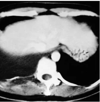
chest wall. Plain radiographs of chest and T-spine taken at a local hospital revealed right pleural effusion and lytic destruc- tion of right pedicles of 9th through 12th thoracic vertebral bodies including proximal part of ribs (Fig. 1). Chylous effu- sion was confirmed by increased triglyceride level (481 mg/dL) of pleural fluid aspirate. The pleural fluid drainage persisted at a rate of more than 500 mL/day. The drained fluid was mainly bloody initially and turned into reddish turbid cream-like fluid without foul odor. Analysis of the pleural fluid showed a sterile lymphocyte-dominant exudate. Computed tomographic (CT) scanning of the chest and the abdomen checked before insertion of a pig-tail tube showed right pleural effusion, a large low-attenuated, irregular-shaped mass lesion with peripheral rim enhancement, and destruction of posterior arch of right 9th and 12th ribs and T10 through L2 vertebral transverse process- es (Fig. 2). Magnetic resonance imaging (MRI) showed high- signal intensity on T1-weighted image and iso-signal inten- sity on T2-weighted image for the involved right side of the T11 and T12 vertebral bodies and pedicles (Fig. 3). The MRI findings suggested that vascular structures such as heman- gioma or lymphangioma replaced the vertebral body, and induced osteolysis. Alkaline phosphatase level was normal.
Bone scan showed no abnormal increase in uptake of 99mTc- diphosphonate. Open biopsy was performed in prone position to make histologic diagnosis. On opening the thoracic cavity,
Won Sup Lee*, �, Sung Han Kim*, Inho Kim*, Hark Kyun Kim*, Keun Seok Lee*, Sang Yoon Lee*, Dae Seog Heo*, Bong-Soon Jang�, Yung-Jue Bang*, Noe Kyeong Kim*
Department of Internal Medicine*, and Orthopedic Surgery�, Cancer Research Institute, Seoul National University College of Medicine, Seoul; �Department of Internal Medicine, College of Medicine Gyeong-Sang National University, Chinju, Korea
Received : 24 October 2001 Accepted : 24 January 2002
Address for correspondence Dae Seog Heo, M.D.
Department of Internal Medicine and Cancer Research Institute, Seoul National University College of Medicine, 28 Yongun-dong, Chongno-gu, Seoul 110-744, Korea
Tel : +82-2-760-2587, Fax : +82-2-742-6689 E-mail : [email protected]
826 J Korean Med Sci 2002; 17: 826-9
ISSN 1011-8934
Copyright � The Korean Academy of Medical Sciences
Chylothorax in Gorham’ s Disease
A 25-yr-old woman presented with a right pleural effusion. Destruction of 9th through 12th ribs, adjacent vertebral bodies, and transverse processes was noted on plain radiograph and a large low-attenuated, irregular shaped mass lesion with peripheral rim enhancement, destroying vertebral body and transverse process, was revealed on the computed tomographic scan. Magnetic resonance imaging showed high signal on T1- weighted image and iso- and low signal on T2-weighted image for the mass lesion replacing the vertebral bony cortex and marrow space. An open rib biopsy revealed the histopathological changes of Gorham’s disease (essential osteolysis), even though only bloody fluid filling the empty space and rib and ver- tebral transverse process destruction were grossly observed on operation. Even though there was no definite response to radiotherapy and pleurodesis, the patient showed stable condition up to 20 months after diagnosis.
Key Words : Gorham’s Disease; Osteolysis, Radiotherapy; Pleurodesis; Chylothorax
Chylothorax in Gorham’s Disease 827
a collection of bloody fluid filling the empty space, which might have resulted from osteolysis, was noticed in accordance with the mass lesion with peripheral rim-enhancement seen on CT scanning. Excisional biopsy specimens of remaining vertebral body and rib showed bony destruction and extensive vascular proliferation, mainly of capillary type (Fig. 4). There was no histological evidence of malignancy. The histological
findings were compatible with those of Gorham’s disease. Tho- racoscopy revealed no lymphatic duct or vascular dysplasia from which the chyle could leak out. Radioisotope lymphan- giography was carried out to find the possible focus of lym- phatic leak, but failed to reveal it in thoracic cavity.
Insertion of a chest tube was performed for pleurodesis after removal of a pig-tail tube. Radiation therapy (30.6 Gy in 17
Fig. 1.Plain radiograph showing the absence of pedicle of the 9th through 12th thoracic vertebral bodies and proximal part of ribs.
Fig. 2.Computed tomographic scan showing pleural effusion, large low-attenuated, irregular-shaped mass lesions with periph- eral rim enhancement.
Fig. 3.Magnetic resonance imaging (MRI) showing a iso- to high-signal intensity lesion replacing vertebral pedicle on T1-weighted image (A) which is enhanced .after gadollinium-based media injection (B).
A B
828 W.S. Lee, S.H. Kim, I. Kim, et al.
fractions) was given. Pleurodesis was attempted using OK-432 (picibanil) when the tube drainage diminished to the rate of 100 mL/day. Chest radiographs were serially followed up.
However, reaccumulation of pleural effusion developed on chest radiograph 2 months after the completion of radiother- apy. CT scan of chest and abdomen also revealed a moderate amount of pleural effusion as well as a progression of osteolysis that extended to the irradiated L3 transverse process. Even though there was no definite response to radiotherapy and pleurodesis, the patient showed stable condition up to 20 months after diagnosis.
DISCUSSION
Gorham’s disease is a very rare disorder that is characterized by local osseous invasion by angiomatous vascular mass with- out skip area, eventually causing lysis of the affected bone. To date, despite the varying MRI appearances of Gorham disease, T1-weighted images were reported to be useful in diagnosis (2). In the present case, MRI finding that showed high sig- nal on T1-weighted image also suggested that the vertebral lesions were angiomatous in nature (capillary type). At the time of operation, the vertebral lesions on MRI were found to be cavities filled with bloody fluid. Bone scan is also considered useful in excluding other vascular malignancies although it cannot demonstrate the lesions. This is consistent with a histologic feature of Gorham’s disease, which hardly shows evidence of osteoblasts or new bone formation.
As in the present case, vertebral disease in Gorham’s disease
may be complicated with severe chylothorax (3). Chylothorax results from leakage of chyle from penetration of the lymphat- ic dysplasia (4), or leakage of chyle from thoracic duct due to its invasion or blockage (3, 5, 6). Of the non-traumatic causes of chylothorax, the most common cause is the malignant lym- phoma. Whenever chylothorax of unknown etiology occurs, underlying malignant lymphoma should be suspected (7).
However, the Gorham’s disease should be included in dif- ferential diagnosis of chylothorax, especially when it is asso- ciated with osteolytic lesion. There have been about 30 cases of chylothorax complicating Gorham’s disease in the English literature (4, 8, 9).
For the prompt control of chylothorax, the available evidence suggests that early surgical intervention to ligate the thoracic duct reduce mortality related with malnutrition and sepsis, even though there are only a small number of cases reported (4, 9, 10). Ligation of thoracic duct and excision of the patho- logic lymphagiomatous tissue is indicated (4). However, we failed to demonstrate ductal structures to ligate or a lymphan- giomatous lesion to excise to control the chylothorax on tho- racoscopy as well as radioisotope lymphangiography. Therefore, we performed pleurodesis as an alternative treatment.
Even after thoracic duct ligation, radiotherapy is still needed to prevent the progression of vertebral osteolysis and to remove lymphatic vessels that replace the vertebral bone and marrow space. And there has been a report (11) that radiotherapy can be effective in arresting the process of proliferation of endothe- lial vascular channels which induces osteolysis.
Considering that the patient described by McNeil et al. (11), whose disease process was arrested after radiotherpy, a total dose of 40 Gy, the treatment failure of the present case could be associated with the lower radiation dose. However, there are some cases that show the contrary. One is the patient described by Stove and Reichelt who showed only a short-term control of osteolysis, though the patient had received a total dose of 40 Gy (12). In other cases, spontaneous resolution of osteolysis were described (13, 14), while it frequently occurred at more peripheral sites.
The review of reported cases of Gorham’s disease with chy- lothorax and the present case suggests the higher dose of radiation might be useful for disease control. More experience on the outcomes of the total dose of 40 Gy is still needed to give some guidelines on the management of Gorham’s disease.
REFERENCES
1. Choma ND, Biscotti CV, Bauer TW, Mehta AC, Licata AA. Gorham’s syndrome: a case report and review of the literature. Am J Med 1987;
83: 1151-6.
2. Chung C, Yu JS, Resnick D, Vaughan LM, Haghighi P. Gorham syndrome of the thorax and cervical spine: CT and MRI findings.
Skeletal Radiol 1997; 26: 55-9.
3. Fairfax AJ, Mc Nabb WR, Spiro SG. Chylothorax: a review of 18 Fig. 4.Photograph of a biopsy specimen of the 12th vertebral pedi-
cle showing replacement of medullary bone and early destruction of cortical bone by abnormal vascular structure (H&E, ×200).
Chylothorax in Gorham’s Disease 829
cases. Thorax 1986; 41: 880-5.
4. Chavanis N, Chaffanjon P, Frey G, Vottero G, Brichon P-Y. Chy- lothorax complicating Gorham’s Disease. Ann Thorac Surg 2001;
72: 937-9.
5. Sassoon CS, Light RW. Chylothorax and pseudochylothorax. Clin Chest Med 1985; 6: 163-71.
6. Goorwitch J. Traumatic chylothorax and thoracic duct ligation. J Thorac Surg 1955; 29: 467-79.
7. Hillerdal G. Chylothorax and pseudochylothorax. Eur Respir J 1997;
10: 1157-62.
8. Riantawan P, Tansupasawasdikul S, Subhannachart P. Bilateral chylothorax complicating massive osteolysis (Gorham’s syndrome).
Thorax 1996; 51: 1277-8.
9. Feigl D, Seidel L, Marmor A. Gorham’s disease of the clavicle with bilateral pleural effusion. Chest 1981; 79: 242-4.
10. Tie ML, Poland GA, Rosenow EC III. Chylothorax in Gorham’s syndrome: a common complication of a rare disease. Chest 1994;
105: 208-13.
11. McNeil KD, Fong KM, Walker QJ, Jessup P, Zimmerman PV.
Gorham’s syndrome: a usually fatal cause of pleural effusion treated successfully with radiotherapy. Thorax 1996; 51: 1275-6.
12. Stove J, Reichelt A. Massive osteolysis of the pelvis, femur and sacral bone with a Gorham-Stout syndrome. Arch Orthop Trauma Surg 1995; 114: 207-10.
13. Patrick JH. Massive osteolysis complicated by chylothorax successful- ly treated by pleurodesis. J Bone Joint Surg (Br) 1976; 58: 347-9.
14. Vanetti A, Picard JD, Fandre M, Roche J, Chollet M, Logeais Y, Mathey J. A propos d’une observation de chylothorax en apparence spontane de l’enfant, associe a des lesions osseuse de type osteoly- tique. Ann Chir Thorac Cardiovasc 1968; 7: 99-104.