INTRODUCTION
Early detection and correction of the risk factors for diabet- ic nephropathy (DN) might slow the development or pro- gression of DN. The urinary albumin excretion rate (AER) is the mainstay in early detection of DN (1). In addition, several factors such as hypertension, hyperglycemia, genetic predisposition, retinopathy, age, dyslipidemia, family histo- ry, smoking history, and the amount and origin of dietary protein were also suggested as predictors of DN risk in indi- vidual patients (1, 2).
Cardiac autonomic neuropathy is an early and common com- plication of diabetes mellitus (DM) (3). Previous studies have reported that cardiac autonomic neuropathy might be a risk factor for DN and associated with deterioration of renal func- tion in people with DM (4-16). However, there are not enough longitudinal data to show that cardiac autonomic neuropathy could be an early predictor of deterioration of renal function in normoalbuminuric, normotensive diabetics, especially in patients with type 2 DM. The aim of our study, which was performed retrospectively, was to evaluate whether cardiac autonomic neuropathy, as an early complication of DM, is asso- ciated with deterioration in glomerular filtration rate (GFR) in normoalbuminuric, normotensive patients with type 2 DM.
MATERIALS AND METHODS Patients
This retrospective longitudinal study was performed in accordance with the principles of the 1983 Declaration of Helsinki. From January 1996 to June 1997, consecutive patients with type 2 DM, who had undertaken the cardiac autonomic function test as one of the initial screening tests of diabetic complications at the Samsung Medical Center, Seoul, Republic of Korea, were eligible for inclusion if they 1) had systolic blood pressure of less than 130 mmHg and diastolic blood pressure of less than 85 mmHg; 2) had a urinary AER of less than 20 μg/min; 3) had an initial serum creatinine level noted at the time of the cardiac autonomic function test and a follow-up serum creatinine level record- ed at least 9 yr from that time; 4) had no evidence of cardiac or renal disease at the time of the cardiac autonomic func- tion test; and 5) had no history of antihypertensive medica- tion at the time of the autonomic function test. Type 2 DM was diagnosed if they 1) had no episodes of ketoacidosis; 2) diagnosed DM after the age of 40 yr; 3) treated by diet alone, or in combination with oral hypoglycemic agents or fasting serum C-peptide values greater than 1.0 ng/mL in patients
S69
Yong Kyun Kim*, Jung Eun Lee, Yoon Goo Kim, Dae Joong Kim, Ha-Young Oh, Chul Woo Yang*, Kwang-Won Kim, and Wooseong Huh
Department of Internal Medicine*, College of Medicine, The Catholic University of Korea, Seoul;
Department of Internal Medicine, Samsung Medical Center, Sungkyunkwan University School of Medicine, Seoul, Korea
Address for correspondence Wooseong Huh, M.D.
Division of Nephrology, Department of Medicine, Samsung Medical Center, 50 Irwon-dong, Gangnam-gu, Seoul 135-710, Korea Tel : +82.2-3410-3449, Fax : +82.2-3410-0064 E-mail : [email protected] DOI: 10.3346/jkms.2009.24.S1.S69
Cardiac Autonomic Neuropathy as a Predictor of Deterioration of the Renal Function in Normoalbuminuric, Normotensive Patients with Type 2 Diabetes Mellitus
Our study was performed to determine whether cardiac autonomic neuropathy can predict deterioration of the renal function in normoalbuminuric, normotensive peo- ple with type 2 diabetes mellitus (DM). One hundred and fifty-six normoalbuminuric, normotensive people with type 2 DM were included in our retrospective longitudi- nal study. We categorized normal patterns, early patterns, and definite or severe patterns according to the results of the cardiac autonomic function test. Of 156 patients included, 54 had normal patterns, 75 had early patterns, 25 had definite or severe patterns, and 2 had atypical patterns. During a median follow-up of nine years, glomerular filtration rates (GFR) remained stable in the normal and early pattern groups (mean changes, 4.50% and 0.77%, respectively) but declined in those with definite or severe patterns (mean change, -10.28%; p=0.047). An abnor- mal heart response to the deep breathing test of the cardiac autonomic function tests was an independent predictor of GFR decline. Our data suggest that cardiac autonomic neuropathy, especially with a definite or severe pattern, might be asso- ciated with a subsequent deterioration in renal function in normoalbuminuric, nor- motensive people with type 2 DM.
Key Words : Autonomic Neuropathy; Glomerular Filtration Rate; Diabetes Mellitus, Type 2
Received : 27 May 2008 Accepted : 12 January 2009
administered with insulin.
When multiple follow-up creatinine levels were available, we used the most recent value, and excluded values that were associated with an acute illness or hospitalization. One hun- dred and 56 patients met the inclusion criteria.
Cardiac autonomic function tests
We conducted five tests for each patient to evaluate car- diac autonomic function. Three tests to assess parasympa- thetic neuropathy were heart-rate response to deep breath- ing, standing up and the Valsalva manoeuvre. Two tests to assess sympathetic neuropathy were blood-pressure response to sustained handgrip and standing up. The methods of each test are described below (17).
Heart-rate response to deep breathing
Six maximal expirations and inspirations were performed within one minute in the supine position, during which heart rate was continuously recorded by electrocardiography (ECG), and the R-R intervals were recorded. The E/I ratio was cal- culated as the mean of the longest R-R interval during expi- ration (E) divided by the mean of the shortest R-R interval during inspiration (I). ‘Abnormal’ was defined as a ratio of less than 1.00; ‘borderline’ as a ratio of 1.01-1.09; and ‘normal’
as a ratio of more than 1.11.
Heart-rate response to standing up
The patient lay quietly and stood up unaided. The heart rate was recorded continuously by ECG. The ‘30:15 ratio’, which is the ratio of the longest R-R interval (around the 30th beat after starting to stand up) to the shortest R-R inter- val (around the 15th beat), was then calculated. ‘Abnormal’
was defined as a ratio of less than 1.00; ‘borderline’ as a ratio of 1.01-1.03; and ‘normal’ as a ratio of more than 1.04.
Heart-rate response to the Valsalva manoeuvre
The patient sat quietly and then blew into a mouthpiece at a pressure of 40 mmHg for 15 sec. Heart rate was record- ed continuously using ECG during and after the manoeuvre.
The ratio of the longest R-R interval shortly after the manoeu- vre to the shortest R-R interval during the manoeuvre was then calculated. ‘Abnormal’ was defined as a ratio of less than 1.10; ‘borderline’ as a ratio of 1.11-1.20; and ‘normal’
as a ratio of more than 1.21.
Blood-pressure response to sustained handgrip
The patient squeezed a handgrip dynamometer as hard as possible for a few seconds and then maintained steady pres- sure at 30% of the maximum pressure for three to four min- utes. Blood pressure was measured each minute, and the dif- ference between the diastolic blood pressure before starting and that just before the release of the handgrip was measured.
‘Abnormal’ was defined as a difference of less than 10 mmHg;
‘borderline’ as a difference of 11-15 mmHg; and ‘normal’ as a difference of more than 16 mmHg.
Blood-pressure response to standing up
Blood pressure was measured while the patient was lying down and again one minute after standing up, and the dif- ference in systolic blood pressure was noted. ‘Abnormal’ was defined as a difference of more than 30 mmHg; ‘borderline’
as a difference of 11-29 mmHg; and ‘normal’ as a difference of less than 10 mmHg.
Classification of test result
We categorized the results of these five tests as: ‘normal pattern’, all tests normal or one borderline; ‘early pattern’, one of the three heart-rate tests abnormal or two borderline;
‘definite or severe pattern’, two or more of the three heart- rate tests abnormal (definite pattern), or definite pattern plus one or both of the blood-pressure results abnormal, or both borderline (severe pattern); ‘atypical pattern’, any other com- bination of abnormal tests.
Clinical information and laboratory data
Clinical information was assessed from written and electron- ic medical records, which included medical history, current medications and laboratory data. The data collected includ- ed age, sex, urinary AER, serum creatinine, systolic blood pressure, diastolic blood pressure, retinopathy, glycosylated hemoglobin level (HbA1c), total cholesterol level, high-den- sity lipoprotein (HDL) cholesterol level, low-density lipopro- tein (LDL) cholesterol level, triglycerides level, homeostasis model for insulin resistance (HOMA-IR), body fat mass (%), use of statin, use of aspirin, duration of diabetes and estimated GFR. Urinary AER was assessed on 24 hr urine collection.
Normoalbuminuria was defined as less than 20 μg/min in 2 out of 3 consecutive tests taken within 2-3 months. Blood pressure was measured twice, 5 min apart, using a random zero sphygmomanometer with the patient seated after 10 min of rest. Based upon the average of readings at each of two or more visits, hypertension was defined as a systolic blood pressure of more than 140 mmHg and/or a diastolic blood pressure of more than 90 mmHg for three months and/or the commencement of antihypertensive therapy. The presence of retinopathy was assessed by fundus photography, which was interpreted by ophthalmologist. HOMA-IR (%), index of insulin resistance, was calculated by the formula, fasting glucose/18×insulin/22.5, with insulin expressed in μU/mL and fasting glucose in mg/dL (18). Body fat mass (%) was measured in a bioimpedance analysis (Inbody 2.0, Biospace, Seoul, Korea). Estimated GFR was calculated using the Modification of Diet in Renal Disease four-variable equa- tion at the time of the cardiac autonomic function test (19).
During the follow-up period, we also calculated estimated
glomerular filtration rate (eGFR) using the chemistry profile at the last visit.
Statistical analysis
Demographic, clinical, and biochemical data are expressed as means±SD. We used a paired t test to compare differences in continuous variables within the groups. One-way ANOVA test or Kruskal-Wallis test was used to compare differences in continuous variables, and the chi-square test or Fisher’s exact test to compare categorical variables among groups. The asso- ciation of the change of eGFR with demographic, clinical or biochemical variables were determined using univariate lin- ear regression analysis. Multivariate linear regression analy- ses were conducted to detect independent predictors of eGFR decline. Kaplan-Meier life-table analysis was used to calculate the cumulative incidence of hypertension during follow-up period. A p value of <0.05 was considered statistically sig- nificant. All statistical analyses were performed with SPSS 12.0 for Windows (SPSS, Chicago, IL, U.S.A.).
RESULTS
Of 156 patients, 54 had normal patterns on the autonom- ic function test, 75 had early patterns, 25 had definite or
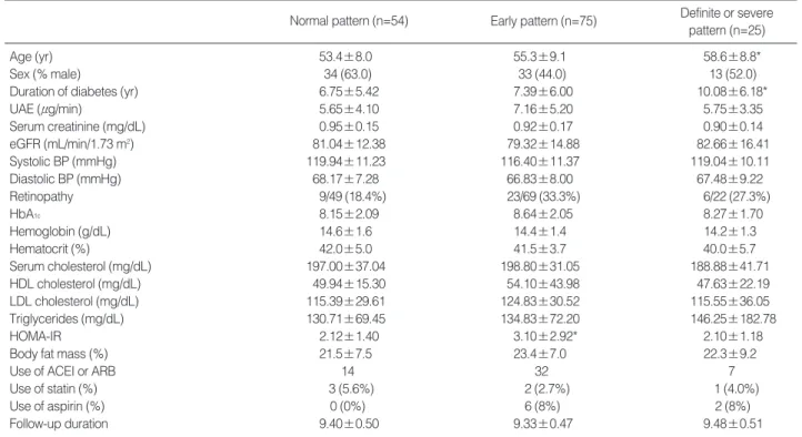
severe patterns, and 2 had atypical patterns. The prevalence of cardiac autonomic neuropathy was 65.4% in normoalbu- minuric, normotensive stage. Forty-one patients had concor- dant abnormal heart-rate response to deep breathing, 59 con- cordant abnormal heart-rate response to standing up, 29 con- cordant abnormal heart-rate response to the Valsalva manoeu- vre, 6 concordant abnormal blood-pressure response to sus- tained handgrip and no patient concordant abnormal blood- pressure response to standing up. Two patients with atypi- cal patterns were eliminated from the analysis because their number was too small to obtain statistically valid results. The characteristics of the patients, based on their patterns of auto- nomic function are given in Table 1. There were no signifi- cant differences in sex, serum creatinine, eGFR, urinary AER, systolic and diastolic blood pressure, HbA1c, retinopathy, lipid profiles and body fat mass at baseline among the three groups. There were also no significant differences in numbers of patients with treatment with statin and aspirin among the groups. However, the definite or severe pattern group was older and had DM longer than the other groups. HOMA- IR values in the early pattern group were significantly high- er than those in the normal pattern group.
Median follow-up duration was 9 yr. During the follow- up period, there was no significant difference between the baseline eGFR and the last follow-up eGFR in the normal pattern and early pattern groups. However, in the definite
Normal pattern (n=54) Early pattern (n=75) Definite or severe pattern (n=25)
Age (yr) 53.4±8.0 55.3±9.1 58.6±8.8*
Sex (% male) 34 (63.0) 33 (44.0) 13 (52.0)
Duration of diabetes (yr) 6.75±5.42 7.39±6.00 10.08±6.18*
UAE (μg/min) 5.65±4.10 7.16±5.20 5.75±3.35
Serum creatinine (mg/dL) 0.95±0.15 0.92±0.17 0.90±0.14
eGFR (mL/min/1.73 m2) 81.04±12.38 79.32±14.88 82.66±16.41
Systolic BP (mmHg) 119.94±11.23 116.40±11.37 119.04±10.11
Diastolic BP (mmHg) 68.17±7.28 66.83±8.00 67.48±9.22
Retinopathy 9/49 (18.4%) 23/69 (33.3%) 6/22 (27.3%)
HbA1c 8.15±2.09 8.64±2.05 8.27±1.70
Hemoglobin (g/dL) 14.6±1.6 14.4±1.4 14.2±1.3
Hematocrit (%) 42.0±5.0 41.5±3.7 40.0±5.7
Serum cholesterol (mg/dL) 197.00±37.04 198.80±31.05 188.88±41.71
HDL cholesterol (mg/dL) 49.94±15.30 54.10±43.98 47.63±22.19
LDL cholesterol (mg/dL) 115.39±29.61 124.83±30.52 115.55±36.05
Triglycerides (mg/dL) 130.71±69.45 134.83±72.20 146.25±182.78
HOMA-IR 2.12±1.40 3.10±2.92* 2.10±1.18
Body fat mass (%) 21.5±7.5 23.4±7.0 22.3±9.2
Use of ACEI or ARB 14 32 7
Use of statin (%) 3 (5.6%) 2 (2.7%) 1 (4.0%)
Use of aspirin (%) 0 (0%) 6 (8%) 2 (8%)
Follow-up duration 9.40±0.50 9.33±0.47 9.48±0.51
Table 1. Clinical and laboratory features at baseline and cardiac autonomic function
*p<0.05 compared with those whose autonomic neuropathy was normal pattern.
UAE, urinary albumin excretion; eGFR, estimated glomerular filtration rate; BP, blood pressure; HbA1c, glycosylated haemoglobin; HDL, high-density lipoprotein; LDL, low-density lipoprotein; HOMA-IR, homeostasis model for insulin resistance; ACEI, angiotensin converting enzyme inhibitor; ARB, angiotensin receptor blocker.
or severe pattern group, eGFR decreased significantly. In an intergroup comparison, the definite or severe pattern group had a greater change in eGFR than that of the normal pat- tern group or early pattern group (Table 2).
Predictors of a decline in renal function
Table 3 shows the univariate analyses for relationship of the change of eGFR with clinical parameters at baseline of the study. The decline in eGFR was associated with an abnor- mal heart-rate response to the deep breathing test, age, eGFR, total serum cholesterol level and diabetic retinopathy. Sixty- two patients progressed to hypertension during the follow-
Group differences, normal pattern vs. early pattern (p=0.354).
*Group differences, normal pattern vs. definite or severe pattern (p=
0.007); �Group differences, early pattern vs. definite or severe pattern (p=0.035).
eGFR, estimated glomerular filtration rate.
Autonomic neuropathy
% GFR change from
baseline p eGFR (mL/min/1.73 m2)
Baseline Follow-up
Normal pattern 81.04±12.38 84.42±17.63 4.50±17.78 0.092 Early pattern 79.32±14.88 79.32±21.10 0.77±24.30 1.000 Definite or 82.66±16.41 72.25±20.92 -10.28±25.77*,�
0.047 severe pattern
Table 2. Cardiac autonomic neuropathy and glomerular filtra- tion rate changes
Cumulative incidence (%)
100 90 80 70 60 50 40 30 20 10
0.00 1 2 3 4 5 6 7 8 9 10 11
Years
Normal pattern Early pattern
Definite or severe pattern
Fig. 1. Cumulative incidence of the development of hypertension in 154 initially normoalbuminuric, normotensive people with type 2 diabetes over a nine-year follow-up.
ACEI, angiotensin converting enzyme inhibitor; ARB, angiotensin recep- tor blocker; UAE, urinary albumin excretion; eGFR, estimated glomerular filtration rate at baseline; HbA1c, glycosylated haemoglobin; HOMA-IR, homeostasis model for insulin resistance.
Categorical variables
p value
% eGFR change from baseline Variable
present
Variable absent
Abnormal heart-rate response -14.0±30.2 5.4±17.0 <0.001 to deep breathing test (n=41)
Abnormal heart-rate response 1.2±20.8 -0.3±24.2 0.686 to standing up (n=59)
Abnormal heart-rate response -3.9±26.6 1.3±22.0 0.282 to Valsalva manoeuvre (n=29)
Abnormal blood-pressure -13.0±18.3 0.8±23.0 0.150 response to sustained
handgrip (n=6)
Male sex (n=80) -0.3±20.2 0.9±25.6 0.746 Retinopathy (n=38) -12.1±25.2 5.7±18.8 <0.001 Use of ACEI or ARB -9.3±27.8 -0.7±26.3 0.392 Use of statin (n=6) 8.1±16.0 -0.0±23.1 0.394 Use of aspirin (n=8) -14.5±34.5 1.1±22.0 0.061 New-onset hypertension (n=62) -8.0±27.5 5.9±17.2 0.001
Age -0.244 0.002
UAE -0.097 0.234
eGFR -0.225 0.005
Systolic blood pressure -0.093 0.251
Diastolic blood pressure 0.096 0.236
HbA1c -0.114 0.160
Total serum cholesterol -0.225 0.005
HOMA-IR -0.114 0.204
Body fat mass 0.038 0.651
Duration of diabetes -0.098 0.224
Table 3. Univariate analyses for relationship of the change of eGFR with clinical parameters at baseline of study
Continuous variable Correlation coefficient p value
UAE, urinary albumin excretion; eGFR, estimated glomerular filtration rate at baseline; HbA1c, glycosylated haemoglobin; HOMA-IR, home- ostasis model for insulin resistance.
β t p value
Abnormal heart-rate response -0.196 -2.11 0.038 to deep breathing test
Abnormal heart-rate response 0.148 1.63 0.108 to standing up
Abnormal heart-rate response 0.088 1.03 0.307 to Valsalva manoeuvre
Abnormal blood-pressure response -0.180 -1.99 0.051 to sustained handgrip
Age -0.194 -2.04 0.045
Male sex 0.047 0.32 0.747
UAE 0.021 0.24 0.811
eGFR -0.175 -1.76 0.082
Systolic blood pressure -0.086 -0.87 0.388 Diastolic blood pressure 0.121 1.23 0.222
Retinopathy -0.366 -4.22 <0.001
HbA1c -0.144 -1.36 0.177
Total serum cholesterol -0.165 -1.76 0.081
HOMA-IR -0.118 -1.27 0.209
Body fat mass 0.056 0.36 0.722
New-onset hypertension -0.308 -3.34 0.001
Duration of diabetes 0.119 1.31 0.192
Table 4. Multiple linear regression analysis for the significance of the relationship between the change of eGFR and cardiac autonomic neuropathy after adjustment for other variables
up period. Seventeen of the 54 patients with normal pattern of cardiac autonomic neuropathy progressed to hypertension, as did 36 of the 75 patients who had an early pattern and 9 of the 25 patients who had a definite or severe pattern. There was no significant difference in the incidence of the develop- ment of hypertension among the different patterns of cardiac autonomic neuropathy (Fig. 1). However, new-onset hyper- tension during the follow-up period was associated with the decline in eGFR (Table 3, 4). Multivariate linear regression analysis was conducted to determine the independent pre- dictors affecting the decline of eGFR. The heart-rate response to the deep breathing test, age, diabetic retinopathy and new- onset hypertension during follow-up period were independent- ly and significantly related to the decline in eGFR (Table 4).
DISCUSSION
Previous studies have demonstrated a significant relation- ship between cardiac autonomic neuropathy and decline in GFR in individuals with DM (4-12). Previous studies also showed that cardiac autonomic neuropathy is one of the early complications of DM and exists even at the normoalbumin- uric, normotensive stage (3, 4). There are few longitudinal data that demonstrate whether cardiac autonomic neuropa- thy has a close relationship to the deterioration of renal func- tion in the normoalbuminuric, normotensive stage. In our study, we enrolled 156 subjects with type 2 DM with nor- moalbuminuria and normotension at the baseline and fol- lowed up for about 9 yr retrospectively. The results showed that the prevalence of cardiac autonomic neuropathy was 65.4% and cardiac autonomic neuropathy was one of the sig- nificant predictors of decline in renal function in normoten- sive, normoalbuminuric subjects.
The underlying mechanism by which cardiac autonomic neuropathy may lead to nephropathy is reported in previous studies (11-16). If sympathetic neuropathy is dominant, renal sodium excretion and renal blood flow are diminished, and the GFR is decreased via vasoconstriction in the kidney. If parasympathetic neuropathy is dominant, systolic hyperten- sion, resting tachycardia, and increased cardiac output devel- op, which injure the glomerular membrane, causing an in- crease in albumin leakage through the glomerular membrane.
Our study showed that parasympathetic neuropathy (i.e., abnormal heart-rate response to the deep breathing test) affect- ed GFR whereas sympathetic neuropathy did not. This find- ing may be related to the small number of patients with sym- pathetic neuropathy in our study, which showed the low prevalence (3.9%) of sympathetic neuropathy in the nor- moalbuminuric, normotensive stage. A large number of sub- jects with sympathetic neuropathy or 24-hr ambulatory blood pressure monitoring-instead of blood pressure response to sustained handgrip and blood-pressure response to standing up-will clarify the relationship of sympathetic neuropathy
with a deterioration of renal function (14-16).
In our study, in addition to abnormal heart-rate response to the deep breathing test, age and diabetic retinopathy were also independent and significant risk factors for a decline in GFR. These findings are similar with previous study, which reported that baseline plasma creatinine, systolic blood pres- sure, age at diagnosis, height, Indian-Asian ethnicity, ever smoking, previous retinopathy, and urinary albumin were independently associated with increased risk of development of reduced creatinine clearance in patient with type 2 DM (20).
The baseline levels of HbA1cwere relatively high among three groups in our study because patients with naive type 2 DM and high blood glucose level were also enrolled. But the levels of HbA1cwere not associated with a deterioration of renal function and decreased equally with treatment dur- ing the follow-up period.
The cumulative incidence of development of hypertension was 37% during the follow-up period. Previous study report- ed that Cardiac autonomic neuropathy is independently asso- ciated with hypertension in normoalbuminuric Type 2 dia- betic patients with no history of hypertension (21). In our study, there was no significant difference in the incidence of the development of hypertension among the different pat- terns of cardiac autonomic neuropathy. Larger prospective study will clarify the relationships between cardiac autonom- ic neuropathy and hypertension in patients with type 2 DM.
In our study, although there was no significant difference in microalbuminuria between the patients with new-onset hyper- tension and without new-onset hypertension (7.0±5.0 μg/
min vs. 6.0±4.3 μg/min, p=0.180), eGFR decreased sig- nificantly in patients with new-onset hypertension compared with the patients without new-onset hypertension (% eGFR change from baseline, -8.0±27.5 vs. 5.9±17.2, p=0.001).
New-onset hypertension during the follow-up period was an independent and significant risk factor for a decline in GFR.
In our study, 53 of the 62 patients with hypertension dur- ing the follow-up period were treated with angiotensin con- verting enzyme inhibitor (ACEI) or angiotensin receptor blocker (ARB). The use of ACEI or ARB was not associated with change of eGFR, which is different from previous stud- ies (22, 23). Twenty-six of 53 patients prescribed ACEI or ARB took more than two anti-hypertensive agents and all of 9 patients who were not prescribed ACEI or ARB took only one anti-hypertensive agent that may be why the use of ACEI or ARB did not conferred significant renal benefit in our study.
Previous data reported that diabetic parasympathetic neu- ropathy affects the insulin resistance in type 2 diabetic pati- ents (24). In our study, HOMA-IR, an index of insulin resis- tance, was higher in group with an early pattern than in the group with a normal pattern. However, HOMA-IR had no significant relationship with eGFR decline.
The results of our study revealed that cardiac autonomic
. .
neuropathy was an important predictor of decline in GFR, however, risk factors that were not analyzed in our study should be also considered. Development of microalbumin- uria or control of hyperglycemia during follow-up period may affect decline in GFR. We could not exclude the poten- tial role of these factors in deterioration of renal function dur- ing follow-up period. And our study was performed retro- spectively so there were quite heterogeneous patients among three groups, in particular in terms of duration of diabetes and patient’s age, which was one of the limitations of our study. Therefore, we cautiously conclude that cardiac auto- nomic neuropathy may be considered as one of the risk fac- tors in deterioration of the renal function in normoalbumin- uric, normotensive patients with type 2 diabetes mellitus.
In conclusion, cardiac autonomic neuropathy could be pre- sent at the normoalbuminuric, normotensive stage. Cardiac autonomic dysfunction could be associated with deteriora- tion of GFR in normoalbuminuric, normotensive patients with type 2 DM and may be an early predictor of DN.
REFERENCES
1. Caramori ML, Fioretto P, Mauer M. The need for early predictors of diabetic nephropathy risk: is albumin excretion rate sufficient?
Diabetes 2000; 49: 1399-408.
2. Caramori ML, Fioretto P, Mauer M. Enhancing the predictive value of urinary albumin for diabetic nephropathy. J Am Soc Nephrol 2006; 17: 339-52.
3. Vinik AI, Maser RE, Mitchell BD, Freeman R. Diabetic autonomic neuropathy. Diabetic Care 2003; 26: 1553-79.
4. Duvnjak L, Vuckovic S, Car N, Metelko Z. Relationship between autonomic function, 24-h blood pressure, and albuminuria in nor- motensive, normoalbuminuric patients with type 1 diabetes. J Dia- betes Complications 2001; 15: 314-9.
5. Sundkvist G, Lilja B. Autonomic neuropathy predicts deterioration in glomerular filtration rate in patients with IDDM. Diabetes Care 1993; 16: 773-9.
6. Forsen A, Kangro M, Sterner G, Norrgren K, Thorsson O, Wollmer P, Sundkvist G. A 14-year prospective study of autonomic nerve function in type 1 diabetic patients: association with nephropathy.
Diabet Med 2004; 21: 852-8.
7. Weinrauch LA, Kennedy FP, Gleason RE, Keough J, D’Elia JA.
Relationship between autonomic function and progression of renal disease in diabetic proteinuria: clinical correlations and implica- tions for blood pressure control. Am J Hypertens 1998; 11: 302-8.
8. Burger AJ, D’Elia JA, Weinrauch LA, Lerman I, Gaur A. Marked abnormalities in heart rate variability are associated with progres- sive deterioration of renal function in type I diabetic patients with overt nephropathy. Int J Cardiol 2002; 86: 281-7.
9. Stella P, Ellis D, Maser RE, Orchard TJ. Cardiac autonomic neu- ropathy (expiration and inspiration ratio) in type 1 diabetes. Inci- dence and predictors. J Diabetes Complications 2000; 14: 1-6.
10. Clarke CF, Eason M, Reilly A, Boyce D, Werther GA. Autonomic
nerve function in adolescents with type 1 diabetes mellitus: relation- ship to microalbuminuria. Diabet Med 1999; 16: 550-4.
11. Pecis M, Azevedo MJ, Moraes RS, Ferlin EL, Gross JL. Autonomic dysfunction and urinary albumin excretion rate are associated with an abnormal blood pressure pattern in normotensive normoalbu- minuric type 1 diabetic patients. Diabetes Care 2000; 23: 989-93.
12. Molgaard H, Christensen PD, Sorensen KE, Christensen CK, Mo- gensen CE. Association of 24-h cardiac parasympathetic activity and degree of nephropathy in IDDM patients. Diabetes 1992; 41:
812-7.
13. DiBona GF, Kopp UC. Neural control of renal function. Physiol Rev 1997; 77: 75-197.
14. Klein IH, Ligtenberg G, Neumann J, Oey PL, Koomans HA, Blan- kestijn PJ. Sympathetic nerve activity is inappropriately increased in chronic renal disease. J Am Soc Nephrol 2003; 14: 3239-44.
15. Poulsen PL, Ebbehoj E, Hansen KW, Mogensen CE. 24-h blood pressure and autonomic function is related to albumin excretion within the normalbuminuric range in IDDM patients. Diabetologia 1997; 40: 718-25.
16. Spallone V, Gambardella S, Maiello MR, Barini A, Frontoni S, Men- zinger G. Relationship between autonomic neuropathy, 24-h blood pressure profile, and nephropathy in normotensive IDDM patients.
Diabetes Care 1994; 17: 578-84.
17. Ewing DJ, Martyn CN, Young RJ, Clarke BF. The value of cardio- vascular autonomic function tests: 10 years experience in diabetes.
Diabetes Care 1985; 8: 491-8.
18. Haffner SM, Miettinen H, Stern MP. The homeostasis model in the San Antonio Heart Study. Diabetes Care 1997; 20: 1087-92.
19. Levey AS, Bosch JP, Lewis JB, Greene T, Rogers N, Roth D. A more accurate method to estimate glomerular filtration rate from serum creatinine: a new prediction equation. Modification of Diet in Renal Disease Study Group. Ann Intern Med 1999; 130: 461-70.
20. Retnakaran R, Cull CA, Thorne KI, Adler AI, Holman RR; UKPDS Study Group. Risk factors for renal dysfunction in type 2 diabetes:
U.K. Prospective Diabetes Study 74. Diabetes 2006; 55: 1832-9.
21. Istenes I, Keresztes K, Hermanyi Z, Putz Z, Vargha P, Gandhi R, Tesfaye S, Kempler P. Relationship between autonomic neuropathy and hypertension--are we underestimating the problem? Diabet Med 2008; 25: 863-66.
22. Ruggenenti P, Fassi A, Ilieva AP, Bruno S, Iliev IP, Brusegan V, Rubis N, Gherardi G, Arnoldi F, Ganeva M, Ene-Iordache B, Gas- pari F, Perna A, Bossi A, Trevisan R, Dodesini AR, Remuzzi G;
Bergamo Nephrologic Diabetes Complications Trial (BENEDICT) Investigators. Preventing microalbuminuria in type 2 diabetes. N Engl J Med 2004; 351: 1941-51.
23. Brenner BM, Cooper ME, de Zeeuw D, Keane WF, Mitch WE, Parv- ing HH, Remuzzi G, Snapinn SM, Zhang Z, Shahinfar S; RENAAL Study Investigators. Effects of losartan on renal and cardiovascular outcomes in patients with type 2 diabetes and nephropathy. N Engl J Med 2001; 345: 861-9.
24. Takayama S, Sakura H, Katsumori K, Wasada T, Iwamoto Y. A pos- sible involvement of parasympathetic neuropathy on insulin resistance in patients with type 2 diabetes. Diabetes Care 2001; 24: 968-9.