1. INTRODUCTION
Stroke is a neurologic disturbance caused by damage to the cerebral blood vessels and one of the most common diseases of adulthood [1]. Most strokes are ischemic strokes that result from in- sufficient blood flow to the brain when blood ves- sels become clogged by blood clots or become too narrow for blood to pass through. Brain cells in
this area die from lack of oxygen. In other types of strokes, called hemorrhagic strokes, the blood vessels are not blocked and rupture, causing blood to penetrate the brain, resulting serious brain damage. Many stroke patients have a number of serious disorders such as hemiplegia, motor dis- turbance, sensory disability, language impairment, communication disorder, emotional disorder, and cognitive impairment [2]. Hemiplegia is paralysis
Comparison of Impedance Parameters and Occupational Therapy Evaluation in the Paretic and Non-paretic
Upper Extremity of Hemiplegic Stroke Patients
Chan-Uk Yoo†, Jaehyung Kim††, Youngjun Hwang†††, Gunho Kim††††, Yong-Il Shin†††††, Gyerok Jeon††††††
ABSTRACT
Many stroke patients undergoing rehabilitation therapy require a quantitative indicator for the evaluation of body function in paretic and non-paretic regions. In this study, the impedance parameters were acquired to assess the physical status in the upper extremity of thirty six stroke patients with hemiplegia caused by cerebral hemorrhage (10 patients) and cerebral infarction (26 patients), using bioelectrical impedance. Prediction marker (PM), phase angle (PA), PM/PA, and resistance (R) versus reactance (Xc) were utilized to evaluate the functional status of the paretic and non-paretic regions. In addition, the hand grip strength (HGS) and the pinch strength (lateral, palmer, tip) were measured on the upper extremity of hemiplegic stroke patients. PM was distributed in inversely proportional to HGS, but PA was distributed in proportional to HGS. However, there were a number of patients with HGS of 0, regardless of the impedance parameters (PM, PA, R vs.Xc). Paretic and non-paretic status in upper extremity of these patients could not be analyzed using impedance parameters. At the rehabilitation therapist's instructions, they were unable to move the hand and fingers of the paretic upper extremity by cranial nerve damage, motor nerve damage, and severe cognitive decline.
Key words: Hemiplegic Stroke Patient, Cerebral Hemorrhage, Cerebral Infarction, Impedance parameters, Occupational Therapy Evaluation
※ Corresponding Author : Gyerok Jeon, Address: (50612) Beomeo-ri, Mulgeum-eup, Yangsan-si, Gyeongsangnam- do, Korea, TEL : +82-55-940-5548, FAX : +82-55-940- 5083, E-mail : [email protected]
Receipt date : Sep. 29, 2017, Revision date : Nov. 8, 2017 Approval date : Nov. 10, 2017
††Department of Occupational Therapy, Hanlyo University (E-mail : [email protected])
††Dept. of Computer Simulation, Inje University (E-mail : [email protected])
†††††Medical Science, School of Medicine, Pusan National University
(E-mail : [email protected], [email protected])
†††††Dept. of Rehabilitation Medicine & Institute of Medical Science, School of Medicine, Pusan National University ([email protected])
†††††Dept. of Biomedical Engineering, School of Medicine, Pusan National University
※ This work was supported by a 1-year Research Grant of Pusan National University.
on one side of the body, whereas hemiparesis weakens one side of the body [3]. Hemiplegia is more severe in the symptoms of the disease than hemiparesis. Both are common side effects of stroke or cerebrovascular accidents. Unilateral pa- ralysis or weakness occurs when a stroke affects the corticospinal tract on the contralateral side of the brain. The right side of the brain controls the motor (movement) function on the left side of the body, while the left side of the brain controls the motor function on the right side of the body. Thus, when one side of the brain is damaged, it affects only one side of the body. One of the most common problems after stroke is limb dysfunction. Dys- function in the extremities seriously degrades the quality of life because it challenges physical func- tion and daily life [4]. Due to these post-stroke dis- abilities, stroke patients require long-term re- habilitation treatment such as physical therapy and occupational therapy [5].
Bioelectrical impedance analysis (BIA) has been increasingly used to estimate the body composition because it is easy to use, non-invasive, inex- pensive and can be performed on a wide range of individuals [6-9]. The bioelectrical mpedance (consisting of resistance and reactance) is meas- ured by passing a low alternating current through the tissues in the body [10]. In recent years, BIA has been applied to assess the human body's hy- dration and nutritional status and to diagnose dis- eases [11]. In this study, paralysis and non-paraly- sis in the upper extremity of 36 stroke patients caused by cerebral hemorrhage and cerebral in- farction were evaluated using impedance parame- ters and occupational therapy assessment. Predic- tion marker (P M), phase angle (P A), PM/PA, R versusXc on the paretic and non-paretic regions of the upper extremity of stroke patients with cere- bral hemorrhage and cerebral infarction were investigated. These values did not show any sig- nificant difference between hemorrhage and cere- bral infarction. However, there was a significant
difference between paralysis and non-paralysis of upper extremity. In addition, the relationship be- tween impedance parameters and occupational therapy evaluation tools (hand grip strength, pin- che strength) did not show significant difference in some hemiplegic stroke patients. This is because the physical movement in the upper extremity is related to the imbalance of the nervous systems or cognitive ability after stroke as well as the func- tion of the muscular systems. As a rehabilitation assessment tool, BIA could be used as an effective adjunct to quantitatively evaluate paralysis and paralysis in upper extremity of hemiplegic stroke patients.
2. THEORY
2.1 Bioelectrical Impedance (Z)
Impedance (Z) is the obstruction to the flow of an alternating current and is dependent on the fre- quency of the applied current. Z is defined in im- pedance magnitude (|Z|) and phase angle () as shown in Equation (1)–(3) and Fig. 1. Bioimpe- dance is a complex quantity composed of resist- ance (R) caused by total body water and reactance () caused by the capacitance of the cell mem- brane [12].
(1)
Fig. 1. Diagram showing the concept of a complex im- pedance. Z is impedance, |Z | is the magnitude of the impedance, R is the resistance, Xc is the reactance, and θis the phase angle.
Resistance (R) is the real part of impedance; a device with purely resistive impedance does not exhibit a phase shift between voltage and current.
Resistance reflects the hydration status in the body.
R = | Z| cosθ (2) Reactance () is the imaginary part of the im- pedance; a component with a finite reactance in- duces a phase shift (θ) between the voltage across it and the current passing through it. Reactance re- flects the body cell mass (muscle mass) in the body.
sin (3)
The physical significance of complex impedance is that the steady-state current is not in phase with the applied voltage [13].
Resistance and reactance together determine the magnitude and phase angle of the impedance through the following relationship:
(4) In phaser diagram as shown in Fig. 1, the angle between the resistance and the reactance is the phase angle of the source voltage V with respect to the current I; that is the angle by which the source voltage leads the current.
From the diagram,
tan
(5)
tan
(6)
The resistance of an object depends on the shape and the material of the object. For a given shape, the resistance depends on the material the object is made of. Different materials provide different re- sistance to charge flow. The resistivity of a ma- terial can be defined so that the resistance R of an object is directly proportional to ρ. The re- sistivity ρ is an intrinsic property of a material, re- gardless of its shape or size. The resistance of an object of length L, made of a material having a
cross-sectional area A and a resistivity ρ, is as fol- lows [14].
(7)
The capacitor affects the current, so it has the ability to stop the current in a fully charged state.
Since an AC voltage is applied, the rms current is limited by the capacitor. Since this is regarded as the effective resistance of the capacitor for AC, the rms currentI in a circuit containing only ca- pacitor C is given by another version of the Ohm's law as follows.
(8)
where V is the rms voltage and is defined to be
(9)
where is called the capacitive reactance, because the capacitor reacts to impede the current. has unit of ohm and is inversely proportional to the ca- pacitance C; the larger the capacitance, the greater the charge it can store and the greater the current that can flow. is also inversely proportional to the frequency f; the greater the frequency, the less time there is to fully charge the capacitor, and so it impedes current less [14].
3. METHOD
3.1 Subject
Thirty six stroke patients caused by cerebral hemorrhage (6 females and 4 males) and cerebral infarction (21 females and 5 males) were included in the measurement. Table 1 shows anthropometric data (age, height, mass, and body mass index) of 36 hemiplegic stroke patients participating in this study. The mean age (73.6 years) of hemiplegic stroke patients caused by cerebral infarction was 6.8 years higher than that (66.8 years) of hemi- plegic stroke patients caused by cerebral hemor-
rhage. Illness duration represents the period from the diagnosis of the disease to the time of the measurement.
3.2 Segmental Bioelectrical Impedance
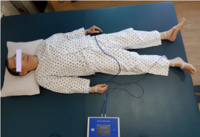
Bioelectrical bioimpedance for 20 hemiplegic stroke patients was measured at Medifarm Hospi- tal in Korea between October and November, 2015, and then for 16 hemiplegic stroke patients was measured at Gurye Nursing Hospital on September 1, 2017, using bioelectrical impedance spectroscopy (MultiScan 5000, Bodystat Ltd., Isle of Man, UK) according to the recommendations of the National Institutes of Health (NIH) Technology Assessment Statement. Before the measurement, the subjects were instructed to fast for at least 4 hours and not to consume alcohol for 24 hours. The subjects were also instructed to drink at least eight glasses of water and empty their bladders before the meas- urement was taken. Eight cutaneous electrodes (Bodystat-0525, Bodystat Ltd, Isle of Man, UK) were attached to the wrists (left, right) and the an- kles (left, right) of the hemiplegic stroke patient while they were in a supine position on a non- conductive surface in Fig. 2. The distance between the electrode used to apply current and the elec- trode used to collect voltage was maintained at least 5 cm to prevent an interactional effect be- tween them. To provide more accurate measure- ments, the anthropometric measurement was com- bined with body composition determined using BIA,
which provides accurate measurements of body composition. Prior to participation in this study, each patient received an explanation of the study purpose and method and provided written informed consent. The study was approved by the Ethics Committee of Inje University Institutional Review Board for Clinical Studies (document number:
2014250) and also approved by the IRB committee of Pusan National University Yangsan Hospital (IRB No. 03-2016-017).
3.3 Occupational Therapy Evaluation 3.3.1. Hand grip and pinch strength
Hand grip strength test is to measure the max- imum isometric strength of the hand and forearm muscles. Hand grip strength in the paretic and non-paretic upper extremity of hemiplegic stroke patients was measured using a Jamar Hand Hydraulic Dynamometer (503330J1, Jamar Ltd., USA). Hand grip strength is in the range of 0∼ 200lbs (90 kg). The subject holds the dynamometer in the hand to be tested, with the arms at right angle (90°) and the elbow by the side of the body.
The handle of the dynamometer is adjusted if Table 1. Subject’s anthropometric data (n=36) and ill-
ness duration Status
Variables Cerebral
hemorrhage Cerebral infarction Age [years] 66.8 ± 9.2 73.6 ± 8.9 Height [cm] 164.0 ± 5.6 162.3 ± 2.6 Mass [kg] 63.8 ± 8.8 55.2 ± 6.4 BMI [kg/m2] 23.4 ± 2.4 21.9 ± 2.0 Duration [years] 2.4 ± 0.5 2.0 ± 0.7 BMI: body mass index. BMI was calculated by divid- ing body mass [kg] by height squared [m2].
Fig. 2. Eight cutaneous electrodes were attached to the wrists (left, right) and the ankles (left, right) of the stroke patient who was in a supine position on a nonconductive surface. The outer electro- des (connected by red wires) are used to inject current into the human body, while the inner electrodes (connected by black wires) are used to measure voltage across the body.
required. The base should rest on first metacarpal (heel of palm), while the handle should rest on mid- dle of four fingers. When ready to measure hand grip strength, the subject should hold the dyna- mometer for about 5 seconds with the maximum isometric effect. Other body movements are not al- lowed at this time. The subject should be strongly encouraged to give a maximum effort. The hand grip strength was measured three times, and the subject was given a one-minute break between measurements. According to the occupational ther- apy assessment guide [15], there are 3 methods to measure the pinching strength of fingers. Lateral (or Key) pinch is to measure the strength between thumb pad and lateral aspect of index finger.
Palmar (or 3-Jaw Chuck) pinch is to measure the strength among thumb, index, and middle finger.
Tip-pinch is to measure the strength between thumb finger and index finger. Pinches (lateral, palmar, tip) were measured using a Jamar hydraul- ic pinch gauge (7498-05, Jamar Ltd., USA). Max- imum pinch strength of a Jamar hydraulic pinch gauge was up to 45 lbs (20 kg).
4. RESULTS AND DISCUSSION
4.1 Bioelectrical Impedance
4.1.1 Prediction Marker (or Impedance ratio) Prediction marker (PM) is defined as the ratio of the impedance (Z) measured at 200 kHz to the impedance (Z) measured at 5 kHz. When an alter- nating current (AC) having a frequency of 5 kHz is applied to the body, the current can not pass through the cell membrane but flows mainly into the extracellular fluid (ECF). Since the ECF is nar- row and the external wall of cell is composed mainly of adipose (fatty) tissues, the impedance (Z) is measured high. However, current having a fre- quency of 200 kHz has enough energy to pass through the cell membrane and can flow to both ECF and the intracellular fluid (ICF). The larger the difference between Z at 5 kHz and Z at 200
kHz, the healthier the cells in the body. The PM close to 1.00 indicates a poor cellular health or ex- cessive amount of fluid [16]. Fig. 3 shows the PM values for paretic and non-paretic upper extremity of hemiplegic stroke patients caused by cerebral hemorrhage and cerebral infarction. In both stroke patients caused by cerebral hemorrhage and stroke patients caused by cerebral infarction, mean values of PM (0.875, 0.885) in paretic regions were higher than those (0.861, 0.856) in non-paretic regions.
The standard deviation (SD) of paralysis and non- paralysis in the upper extremities of 26 stroke pa- tients caused by cerebral infarction was measured to be larger. This is due to the fact that there were many elderly subjects in 36 patients with cerebral infarction (21 females and 5 males), age dis- tribution ranged from 32 to 90 years, and cerebral infarction had progressed for a long time.
4.1.2 Phase Angle (θ)
The phase angle (θ) has long been associated with nutritional status and body cell mass and is a direct measurement of the functionality of cell membrane, It is also recognized as a global health indicator in body health assessment [17]. A higher phase angle means an increase in BCM (body cell mass) or a decrease in fluid, either recovery from infection or injury or a decrease in fluid from Fig. 3. PM values for paretic and non-paretic upper ex- tremity of hemiplegic stroke caused by cerebral hemorrhage and cerebral infarction.
dehydration. A loss of fat could also increase phase angle. On the other hand, a lower phase angle means a loss of BCM, or an increase of fluid (rehydrating, or sign of inflammation or infection [18]. Fig. 4 shows the phase angle () for paretic and non-paretic upper extremity of hemiplegic stroke patients caused by cerebral hemorrhage and cerebral infarction. In both stroke patients caused by cerebral hemorrhage and stroke patients caused by cerebral infarction, mean values (3.58°, 3.59°) of PM in paretic regions were significantly lower than those (3.90°, 4.35°) in non-paretic regions.
These resukts revealed that the loss of muscular mass in the paretic regions of stroke patient's up- per extremity had progressed considerably and that the integrity of the cell membranes in the lean mass had also deteriorated.
4.1.3 Relationship between PM and PA (θ) PM is related to the function of the cell mem- brane and PA (θ) is proportional to the lean mass (LM) in the body. Fig. 5 shows the relationship be- tween PM and PA for paretic and non-paretic up- per extremity of hemiplegic stroke patients caused by cerebral hemorrhage and cerebral infarction.
PM was found to be inversely related to PA in the paretic/non-paretic upper extremity of stroke
patients. On the other hand, patients #26, #28, #29,
#30, and #33 showed higher PM and lower PA in paretic regions (black circles, black squares) com- pared to non-paretic regions (white circles, white squares). Patient #29 is an 80-year-old male suf- fering from trauma and a traffic accident. The function of the cell membrane in the paretic region is deteriorated (PM=0.967) and the lean mass (muscles) is considerably reduced (PA=1.4°). Patient
#33 is a 32-year-old male with brain injury who is classified as stroke hemiplegia caused by cere- bral infarction. The onset of the disease was 25 years, but he was youngest (32 years) among 36 subjects. PM was 0.770 in the paretic upper ex- tremity and 0.768 in the non-paretic upper ex- tremity. PA was 6.3° in the paretic upper extremity and 6.6° in the non-paretic upper extremity.
4.1.4 Reactance (Xc) versus Resistance (R) Resistance (R) reflects the total body water con- sisting of ECF and ICF in the body, and gives the information about the function of the cell mem- sbrane. Therefore, the relationship betweenR and
for hemiplegic stroke patients with upper ex- tremity can be assessed to determine the status of physical functioning of paralysis and non-paralysis.
Fig. 6 shows the relationship between resistance Fig. 4. Phase angle [ °] for paretic and non-paretic up-
per extremity of hemiplegic stroke patients caused by cerebral hemorrhage and cerebral infarction.
Fig. 5. Relationship between PM and PA [ °] for paretic and non-paretic upper extremity of 36 hemi- plegic stroke patients caused by cerebral hem- orrhage and cerebral infarction.
(R) and reactance () for paretic and non-paretic upper extremity of 36 hemiplegic stroke patients caused by cerebral hemorrhage and cerebral infarction. The low resistances on the left reflect a lot of lean mass, while the high resistances on the right reflect a low lean mass and a high fat When the function of the cell membrane is sig- nificantly reduced, the paralysis/non-paralysis states of stroke patients are distributed below, However, when the cell membrane is healthy, the paralysis/non-paralysis states of stroke patients are distributed on the above. Therefore, the left up- per regions indicate an healthy function of the body, and the right below regions reflect a statue of stroke patients in which the bodily function is remarkably deteriorated. Overall, the status of the paralytic regions are distributed below, and the status of the paralytic parts are distributed in the
upper part. The mean value ofR and in the pa- retic upper extremity of 10 stroke patients caused by cerebral hemorrhage was (338.49, 20.19) and that in the non-paretic upper extremity of 10 stroke patients caused by cerebral hemorrhage was (337.21, 22.39). On the other hand, the mean value of R and in the paretic upper extremity of 10 stroke patients caused by cerebral infarction was (324.57, 19.53) and that in the non-paretic upper extremity of 10 stroke patients caused by cerebral infarction was (311.96, 23.03).
4.2 Occupational Therapy Evaluation 4.2.1 Hand grip strength and pinch strength
Table 2 shows hand grip strength (HGS) and pinches (lateral, palma, tip) for paretic and non-pa- retic upper extremity of 36 hemiplegic stroke pa- tients caused by cerebral hemorrhage and cerebral infarction. Hand grip strength and pinches in pa- retic region were significantly lower than those in non-paretic region for hemiplegic stroke patients.
In the clinical setting, tools used to assess re- habilitation treatment of hemiplegic stroke patients include hand grip strength and pinch strenth. The rehabilitation assessment of hemiplegic stroke pa- tients caused by cerebral hemorrhage and cerebral infarction was as follows. For stroke hemorrhage patients, a mean value of HGS was 7.2 lb in the paretic hand and 13.4 lb in the non-paretic hand.
A mean value of lateral pinch was 2.1 lb in the pa- retic fingers and 4.5 lb in the non-paretic fingers.
A mean value of palmer (three point) pinch was 1.8 lb in the paretic fingers and 3.5 lb in the non- Fig. 6. Relationship between resistance (R) and re-
actance (Xc) for paretic and non-paretic upper extremity of 36 hemiplegic stroke patients caused by cerebral hemorrhage and cerebral infarction.
Table 2. Hand grip strength (HGS) and pinch strength (lateral, palmer, tip) for paretic and non-paretic upper extremity of 36 hemiplegic stroke patients caused by cerebral hemorrhage (N=10) and cerebral infarction (N=26)
P_CH NP_CH P_CI NP_CI
HGS [lb] 7.2±6.9 13.4±8.6* 7.2±6.6 14.2±6.3*
Pinch [lb]
Lateral 2.1±2.1 4.5±1.9* 2.0±1.9 4.2±1.8*
Palmer 1.8±1.8 3.5±1.7* 1.7±1.4 3.4±1.7*
Tip 1.3±1.3 2.5±1.0* 1.1±1.1 2.3±1.1*
* Significant difference p<0.05.
paretic fingers. A mean value of tip pinch was 1.3 lb in the paretic fingers and 2.5 lb in the non-pa- retic fingers. Thus, there were significant differ- ences between the paretic upper extremity and the non-paretic upper extremity (p < 0.05). For stroke infarction patients, a mean value of HGS was 7.2 lb in the paretic hand and 14.2 lb in the non-paretic hand. A mean value of lateral pinch was 2.0 lb in the paretic fingers and 4.2 lb in the non-paretic fingers. A mean value of palmer pinch was 1.7 lb in the paretic fingers and 3.4 lb in the non-paretic fingers. A mean value of tip pinch was 1.1 lb in the paretic fingers and 2.3 lb in the non-paretic fingers. Thus, there were also significant differ- ences between the paretic upper extremity and the non-paretic upper extremity (p < 0.05).
In particular, hand grip strength (HGS) was nil in the paretic regions of eleven hemiplegic stroke patients (#10, #11, #20, #26, #28, #29, #30, #32, #34,
#35, and #36), illustrating a significant difference in neurophysiological function between paretic up- per extremity and non-paretic upper extremity.
Seven stroke patients had significantly lower phase angles (2.0° for #10, 2.7°, for #11, and 2.1° for #20, 2.1° for #26, 1.4° for #29, 2.0° for #32, 2.5° for #34) in the paretic regions, suggesting a decrease in muscle mass and deterioration in cell membrane function. Four stroke patients had relatively higher phase angles (3.5° for #28, 3.0° for #30, 4.0° for
#35, and 5.0° for #36) in the paretic regions, but their HGS score was rated as 0, indicating that brain nerve damage, motor nerve damage, or de- creased cognitive ability affected the measurement results.
4.2.2 Relationship between impedance parameters and hand grip strength
Fig. 7 shows the relationship between prediction marker (PM) and hand grip strength (HGS) for pa- retic and non-paretic upper extremity of 36 hemi- plegic stroke patients caused by cerebral hemor- rhage and cerebral infarction. In genaral, PM is
supposed to be inversely proportional to HGS.
However, as shown in the left-hand side of Fig.
7, PM values are distributed in a variety of ways in vertical line but all HGS have zero. # 29 was a hemiplegic stroke patient caused by cerebral in- farction mentioned in Fig. 5. The patient # 29 had a very high PM (0.967 in paretic region and 0.918 in non-paretic region) and a very low phase angle (1.4° in paretic region and 2.6° in non-paretic re- gion). Patient #29 shows that the reactance is very low (7.27 in paretic region and 12.33 in non-pa- retic region) in Fig. 6, indicating a significant de- crease in lean mass and a deterioration in muscular function in the paralysis and non-paralysis regions.
Patient #34 is a 56-year-old female patient with low PM values (0.848 in paretic region and 0.812 in non-paretic region) and a significantly high val- ue for PA (4.5° in paretic region and 5.9° in non-paretic region). The cause of paralysis for #34 could not be analyzed by impedance parameters (PM, PA, and PM/PA). However, in Fig. 7, HGS was 0 in paretic and non-paretic upper extremity.
This seems to be due to loss of function in the brain nervous system or the motor nerves, which makes it impossible for the hand to move at all in the pa- ralysis/paralysis area.
Fig. 7. Relationship between prediction marker and hand grip strength [ °] for paretic and non-paretic up- per extremity of 36 hemiplegic stroke patients caused by cerebral hemorrhage and cerebral infarction.
Fig. 8 shows the relationship between phase an- gle [ ° ] and hand grip strength [lb] for paretic and non-paretic upper extremity of 36 hemiplegic stroke patients caused by cerebral hemorrhage and cerebral infarction. Generally, the phase angles are exposed to be proportional to HGS. The non-pa- retic regions are mainly distributed in the upper right and the paretic regions are mainly distributed in the lower left. However, as shown in the left-hand side of Fig. 8, PM values have various values as a vertical distribution, but all HGS values were zero. Patient #29 has low phase angles (1.4°
in paretic region and 2.6° in non-paretic region).
Patient #34 had low PM (0.812 in paretic region and 0.848 in non-paretic region) and high PA (6.3° in paretic region and 6.6° in non-paretic region), but HGS was 0 in paretic and non-paretic upper extremity. As described in Fig. 7, Patient #29 and
#34 are supposed to have HGS measured as 0 for the same pathological cause.
4.3 DISCUSSION
Long-term muscle changes such as a loss in muscle mass, a reduction of fiber cross-sectional area, and an increase in intramuscular fat deposi- tion are reported to occur between 3 weeks and 6
months after stroke in both paretic and non-paretic upper extremity [19, 20]. Therefore, appropriate stroke rehabilitation is needed in a timely manner (within 3-6 months) for stroke patients. Non-in- vasive measurements are essential to evaluate pa- ralysis and non-paralysis in stroke patients ac- cording to rehabilitation therapy. Measurement of recovery after stroke is becoming increasingly im- portant with the advent of new treatment options in stroke rehabilitation research [21, 22]. The ef- fects of inpatient rehabilitation on functional re- covery of chronic stroke patients with cognitive dysfunction were investigated [5].
For example, the Fugl-Meyer scale was also de- veloped as the first quantitative assessment tool to measure stroke recovery in stroke patients [21].
In addition, among patients who had a stroke with- in 3-9 months, constraint-induced movement ther- apy resulted in statistically significant and clin- ically relevant improvements in arm motor function lasting at least one year [22]. However, these meth- ods are subjective in assessing body function in the paralysis caused by stroke, and it takes a lot of time and labor to test. On the other hand, evalu- ating paralysis and non-paralysis sites in stroke patients using BIA is a simple, non-invasive ap- proach and provides an easy way to obtain the physiological/pathological functions of muscles [23] as well as body hydration and composition.
5. CONCLUSION
Many researchers have long used biological im- pedance parameters to investigate the physical composition and physiological characteristics of tissues. However, there has been little research on the relationship between bioimpedance parameters and occupational therapy assessment tools for up- per extremity paralysis in stroke patients. In this study, we used biological impedance parameters to quantitatively evaluate paraplegic and paralytic upper limb in stroke hemiplegia patients caused by cerebral hemorrhage and cerebral infarction. The Fig. 8. Relationship between phase angle [ °] and hand
grip strength [lb] for paretic and non-paretic upper limbs of 36 hemiplegic stroke patients caused by cerebral hemorrhage and cerebral infarction.
impedance parameters (PM, PA, PM/PA, R vs. ) were compared with occupational therapy evalua- tion (hand grip and pinch strength) to determine whether the impedance parameters are related to the paretic/non-paretic status of hemiplegic stroke patients and the outcome of the rehabilitation assessment.
The results using bioimpedance and occupa- tional therapy evaluation can be summarized as follows. First, in both stroke patients caused by cerebral hemorrhage and stroke patients caused by cerebral infarction, mean values of PM (0.875, 0.885) in paretic regions were higher than those (0.861, 0.856) in non-paretic regions. Mean values (3.58°, 3.59°) of PA in paretic regions were sig- nificantly lower than those (3.90°, 4.35°) in non-pa- retic regions. PM was found to be inversely related to PA. Second, in R- graph, the status of the paralytic regions are distributed below and the sta- tus of the paralytic parts are distributed in the up- per part. The mean value ofR and in the paretic upper extremity of 10 stroke patients caused by cerebral hemorrhage was (338.49, 20.19) and that in the non-paretic upper extremity of 10 stroke patients caused by cerebral hemorrhage was (337.21
, 22.39). Third, PM is inversely proportional to hand grip strength. But, PM values are distributed in a variety of ways in vertical line and all have zero HGS. Fourth, the phase angles are propor- tional to HGS. The non-paretic regions are mainly distributed in the upper right, and the paretic re- gions are mostly distributed in the lower left. Some patients had zero HGS, regardless of the value of the impedance parameters (PM, PA, PM/PA, R vs.
). They were unable to move their hands and fingers in the paretic upper extremity because of brain nerve damage, motor nerve damage, and cognitive impairment.
The limitations of this study are as follows. The number of hemiplegic stroke patients with paretic upper extremity caused by cerebral hemorrhage was limited to 10. When the subjects are catego-
rized by gender, age, and disease states, and the impedance measurement are performed for a long time intervals in the rehabilitation therapy, im- pedance characteristics could be quantitatively distinguished as a more confident manner.
REFERENCE
[ 1 ] J. Adamson, A. Beswick, and S. Ebrahim, “Is Stroke the Most Common Cause of Disabil- ity?,”J ournal of Stroke and Cerebrovascular Diseases, Vol. 13, Issue 4, pp. 171-177, 2004.
[ 2 ] H.J. Jun, K.J. Kim, I.A. Chun, and O.K. Moon,
“The Relationship between Stroke Patients’
Socio-Economic Conditions and Their Quality of Life: The 2010 Korean Community Health Survey,” J ournal Physical Therapy Science, Vol. 27, No. 3, pp. 781-784, 2015.
[ 3 ] Hemiplegia and Hemiparesis, http://www.
stroke-rehab.com/hemiplegia.html, (accessed Jan., 25, 2017).
[ 4 ] G. Kwakkel and B. Kollen, “Predicting Impro- vement in the Upper Paretic Limb After Stroke: A Longitudinal Prospective Study,”
Restorative Neurology and Neuroscience, Vol. 25, No. 5-6, pp. 453-460, 2007.
[ 5 ] K.H. Cho and W.H. Lee, “Effects of Inpatient Rehabilitation on Functional Recovery of Stroke Patients: A Comparison of Chronic Stroke Patients with and without Cognitive Impairment,” J ournal Physical Therapy Sci- ence, Vol. 24, Issue 3, pp. 245-248, 2012.
[ 6 ] B.E. Lingwood, P.B. Colditz, and L.C. Ward,
“Biomedical Applications of Electrical Impe- dance Analysis,” Proceedings of the Fifth International Symposium on Signal Process- ing and Its Applications, Vol. 1, pp. 367-370, 1999.
[ 7 ] C.U. Yoo, Y.A. Yang, S.W. Baik, J.H. Kim, and G.R. Jeon, “Bioelectrical Impedance Analysis on the Paertic and Non-paretic Regions of Severe and Mild Hemiplegic Stroke Patients,"
Journal of Korea Multimedia Society, Vol. 20,
No. 2, pp. 115-125, 2017.
[ 8 ] L.C. Ward, “Segmental Bioelectrical Impedance Analysis: An Update,” Lippincott Williams and Wilkins, Vol. 15, Issue 5, pp. 424-429, 2012.
[ 9 ] E. Volgyi, F.A. Tylavsky, A. Lyytikainen, H.
Suominen, M. Ale, and S. Cheng, “Assessing Body Composition with DXA and Bioimpe- dance: Effects of Obesity, Physical Activity, and Age,” Obesity, Vol. 16, Issue 3, pp. 700- 705, 2008.
[10] A. Wysokiń.ski and I. Kloszewska, “Assess- ment of Body Composition Using Bioelectrical Impedance in Patients with Schizophrenia–
preliminary report,” Archives of P sychiatry P sychotherapy, Vol. 1, pp. 31-37, 2014.
[11] H.C. Lukaski and M.G. Singer, “Phase Angle as a Prognostic Indicator in Cancer,” Pro- ceeding of the AAAI Spring Symposium Technical Reports, Vol. SS-11-04, pp. 37-40, 2011.
[12] J.H. Kim, B.J. Shin, M.S. Lee, Y.J. Kim, I.S.
Jeong, and G.R. Jeon, “Detection of Intrave- nous Infiltration in Rabbit’s Ear Using Bioe- lectrical Impedance: Pilot Study,” Internatio- nal Journal of Engineering and Applied Sci- ences, Vol. 1, Issue 1, pp. 1-6, 2014.
[13] R.K. Wangsness, Electromagnetic Fields, John Wiley and Sons Incorporated, USA, pp.
455-456, 1986.
[14] P.P. Urone, College P hysics, Brooks/Cole, USA, 2001.
[15] L.C. Martinez, E.C. Ramiez, A.O. Tejeda, E.A.
Lafuente, L.P.B. Rosales, V.R. Gonzalez, et al.,
"Bioelectrical Impedance and Strength Mea- surements in Patients with Heart Failure:
Comparison with Functional Class," Nutri- tion, Vol. 23, Issue 5, pp. 412-418, 2007.
[16] J.H. Kim, S.S. Kim, S.H. Kim, S.W. Baik, and G.R. Jeon, “Comparing the Whole Body Im- pedance of the Young and the Elderly Using BIMS,” Journal of Sensors Science and Tech-
nology, Vol. 25, Issue 1, pp. 20-26, 2016.
[17] What is Phase Angle?, http://www.bodystat.
com/support/news/what-is-phase-angle, (accessed Dec., 21, 2016).
[18] A. Bosy-Westphal, S. Danielzik, R.P. RP Dörhöfer, W. Later, S. Wiese, and M.J. Muller,
"Phase Angle from Bioelectrical Impedance Analysis: Population Reference Values by Age, Sex, and Body Mass Index,"J ournal of P arenteral and Enteral Nutrition, Vol. 30, Issue 4, pp. 309-316, 2006.
[19] L. Jørgensen and B.K. Jacobsen, “Changes in Muscle Mass, Fat Mass, and Bone Mineral Content in the Legs After Stroke: A 1 Year Prospective Study,”Bone, Vol. 28, Issue 6, pp.
655-659, 2001.
[20] N. Scherbakova, S. Von Haehlingb, S.D. An- kerc, U. Dirnagla, and W. Doehn, “Stroke Induced Sarcopenia: Muscle Wasting and Disability After Stroke,”International Journal of Cardiology, Vol. 170, Issue 2, pp. 89-94, 2013.
[21] D.J. Gladstone, C.J. Danells, and S.E. Black,
"The Fugl-Meyer Assessment of Motor Recovery After Stroke: A Critical Review of Its Measurement Properties,” Neurorehabil- itation and Neural Repair, Vol. 16, Issue 3, pp. 232-240, 2002.
[22] S.L. Wolf, C.J. Winstein, J.P. Miller, E. Taub, G. Uswatte, and D. Morris, et al, “Effect of Constraint-Induced Movement Therapy on Upper Extremity Function 3 to 9 Months After Stroke: The EXCITE Randomized Clinical Trial,” The Journal of American Medical Association, Vol. 296, No. 17, pp.
2095-2104, 2006.
[23] C.U. Yoo, J.H. Kim, Y.A. Yang, J.S. Lee, and G.R. Jeon, “Bioelectrical Impedance Analysis for Severe Stroke Patients with Upper Ex- tremity Hemiplegia,”J ournal Physical Ther- apy Science, Vol. 28, No. 10, pp. 2708-2712, 2016.
Yoo Chan-Uk
He received B.S. from Inje Uni- versity, Korea, in 2006, and M.S.
degree from Yonsei University, 2008, respectively, and Ph. D degree from Inje University, Korea, 2016. He is currently pro- fessor of occupational therapy at Hanlyo University and has deep interest in Bioelectrical impedance analysis and Cognitive rehabilitation, etc.
He has been serving as a lecturer at the Korea Safety Promotion Association from 2012, and also as a lec- turer in dementia education at National Insurance Corporation from 2013.
Kim Jaehyung
He received B.S. and M. S. de- gree from Pusan National Uni- versity, Korea, in 1979 and 1981, respectively, and Ph. D degree from Kyungnam University, Ko- rea, in 1992. He was visiting scientist at Liquid Crystal Insti- tute of Kent State University, USA in 1993, and visit- ing professor at Physics Department of Portland State University, USA, in 2003. He is currently researcher at Research Institute of Nursing Science, Pusan National University and has deep interest in bio- electrical impedance, electro-dermal activity, and elec- trical stimulator, etc.
Hwang Youngjun
He received a bachelor's degree from Pusan National University, Korea, 2017. He is in the mas- ter's degree in dept. of medical science, Pusan National Univer- sity, Yangsan, Korea.
Kim Gunho
He received a bachelor's degree from Pusan National University, Korea, 2017. He is in the mas- ter's degree in dept. of medical science, Pusan National Univer- sity, Yangsan, Korea.
Shin Yong-Il
Intern and Resident, Department of rehabilitation medicine at Chonbuk National University Hospital Clinician, neurorehabi- litation, rehabilitation medicine, Chonbuk National University Hospital Associate professor &
Manager, rehabilitation medicine, Wonkwang Univer- sity School of Medicine Korean Neurosurgical Society Lee Ju-Guel academic awards (2009) World Congress in NeuroRehabilitation, the Young Investigator award (2006) The Korean Academy of Clinical Geriatrics, academic awards (2006) American Congress of Reha- bilitation Medicine(ACRM), excellent paper award (2000) Korean Academy of Rehabilitation Medicine, academic awards (1997)
Jeon Gyerok
He received B.S. and M.S. de- gree from Pusan National Uni- versity, Korea, 1978 and 1982, respectively. And doctor degree from Donga University Korea, 1993. He is currently professor at department of biomedical en- gineering, school of medicine, Busan National Univer- sity, and working at Busan national university Yangsan hospital. His major is biomedical signal proc- essing and biomedical measurement system.
![Fig. 6 shows the relationship between resistanceFig. 4. Phase angle [ °] for paretic and non-paretic](https://thumb-ap.123doks.com/thumbv2/123dokinfo/4762105.516724/6.892.464.797.145.383/fig-shows-relationship-resistancefig-phase-angle-paretic-paretic.webp)


![Fig. 8 shows the relationship between phase an- an-gle [ ° ] and hand grip strength [lb] for paretic and non-paretic upper extremity of 36 hemiplegic stroke patients caused by cerebral hemorrhage and cerebral infarction](https://thumb-ap.123doks.com/thumbv2/123dokinfo/4762105.516724/9.892.96.426.141.388/relationship-strength-extremity-hemiplegic-patients-cerebral-hemorrhage-infarction.webp)
