스마트폰 영상을 이용한 슬관절 각도 및 활보장에 대한 보행분석
A Gait Analysis Using Smart Phone Images of the Knee Joint Angle and Stride Length
장재훈
*
, 임창주, 송기호, 정성택J. H. Jang, C. J. Lim, K. H. Song, S. T. Chung
요 약
다양한 신경계 및 근골격계 질환이 있을 때 나타나는 증상으로 보행변화가 일어나며, 이에 대한 보행분 석은 병의 진행 정도를 판단하는 데 매우 중요하다. 대부분의 보행분석 방법으로는 고가의 장비 사용과 공 간의 제약을 받고 있다. 본 연구는 스마트 폰을 이용한 촬영 영상과 보행궤적 분석 프로그램을 사용하여, 보행 시 슬관절 각도의 변화와 활보장 측정을 바탕으로 보행분석을 진행하였다. 보행분석에 필요한 실험은 건강한 성인남성 7명을 대상으로 진행하였으며, 오른쪽 및 왼쪽 무릎관절 각도 및 활보장에 대한 데이터를 이용하여 보행분석이 이루어졌다. 본 연구에서 얻어진 보행분석은 기존의 보행분석 연구들과 비교하여 유사 한 결과를 획득하였다. 여기서 제안한 방법을 이용한다면 고가의 장비와 공간의 제약없이 보행 분석을 할 수 있을 것이다.
ABSTRACT
Various types of disease in the nervous and musculoskeletal system can change gait, and the gait analysis is very important in determining the progression of the disease. Most methods of analyzing gait are subject to high-priced equipment and spatial restrictions. This study used smart phone images and the walking track analysis program to make a comparative analysis with the existing gait analysis on the basis of the stride length measurements and the changes in the knee joint angle for walking.
The test necessary to analyze gait was conducted in seven healthy men, and data about the angle of right and left knee joints and stride length were used to analyze gait. The gait analysis in this study obtained the similar results to the existing ones. The use of the methods suggested in this study will enable gait analysis to be made without high-priced equipment and spatial restrictions.
Keyword : Knee Joint Angle, Stride Length, Gait Analysis
접 수 일 : 2013.11.13 심사완료일 : 2013.12.10 게재확정일 : 2013.12.20
* 장재훈 : 한국산업기술대학교 컴퓨터공학과 석사과정 [email protected] (주저자)
임창주 : 한국산업기술대학교 게임공학과 교수 [email protected] (공동저자)
송기호 : 한국산업기술대학교 컴퓨터공학과 석사과정 [email protected] (공동저자)
정성택 : 한국산업기술대학교 컴퓨터공학과 교수 [email protected] (교신저자)
※ "This research was supported by the MSIP(Ministry of Science, ICT&Future Planning), Korea, under the C-ITRC(Convergence Information Technology Research
1. INTRODUCTION
Walking is the process which can be obtained by temporal and dynamic harmony between muscular activities and mobility of joints [1-2].
Gait analysis has been made systematically since the 19th century, and the development of scientific technology, including computers and sensors, improved it steadily from the initial
Center) support program (NIPA-2013-H0401-13-1006) supervised by the NIPA(National IT Industry Promotion Agency)
clinical study to mathematical analysis and modeling through more and more complicated measurements. Gait analysis is now attracting increasing attention from such professional areas as physiotherapy, neurology, and sports science as well as from rehabilitation engineering and medical areas [3-4]. The world has been globalized rapidly since the 1960s, resulting in the constant increase in the number of lower extremity amputees due to the accompanying industrial disasters and lifestyle related diseases, such as geriatric disease and diabetes [5]. In particular, asymmetrical gait causes the lower-limb disabled to be psychologically feared, consume more energy, and get restricted in various types of daily living [6-8]. Gait analysis is being used as the most standard instrument to design the gait assessment methods and gait training systems that induce the lower-limb disabled to make near-normal gait. Measuring knee joint angle variation in analyzing gait is useful in early diagnosis of gait disturbance, and equipment for measuring the joint angle and gait includes gait-analyzing devices using medical GONIOMETER, strain gauge, the 3D-infrared camera gait analysis system, and so on [9-10].
However, these devices are expensive and spatially restricted, making it difficult to create an experimental environment [11]. This study suggested a method of analyzing the track of gait images obtained by a smart phone camera, which is easy to use.
2. MATERIALS AND METHODS
Seven men who had no neural or musculoskeletal problem likely to affect gait and who had no significant limping in walking (24.7 years old and 175.8 cm tall on the average) were selected to make a test in this study. It was used for gait analysis using. smart phone camera (SHV-E300S, Samsung Ltd, Korea) based on android platform and Integrated Analysis of Motion (IAM) program based on PC for gait analysis by Visol Ltd, Korea. This program can track gait with the unit of frame
as shown in Figure 1. An angle variation of knee joint in each walking phase is acquired by manually drawing a line segment. Subjects participating in the experiment wore clothes in the primary color in contrast to the white background to reduce pixel errors of the images.
Their walking distance was limited to 4 m, and 4.5 m, which is the optimum distance for taking gait image, was determined to be the distance between the camera and a subject.
Figure 1. Integrated Analysis of Motion (IAM) program for tracking gait
Figure 2. Measurement of knee joint angle using IAM program
To analyze gait, the IAM program was used to track walking in the unit of frame and measure the knee joint angle with the walking images taken with a smart phone camera. The IAM program was used to measure the joint
angle in each walking phase with the walking images and determine if walking was normal, as shown in Figure 2. The knee joint angle was measured around the lateral center of ankle joints, knee joints, and hip joints. The angle made by the line from the center of hip joints to that of knee joints and the line from the center of knee joints and that of ankle joints was measured to obtain the knee joint angle.
3. RESULT
3.1 Knee joint angle in walkingFigure 3. Seven gait phases for analyzing knee joint angle in walking
One of the existing gait analysis methods is to measure stride length, gait velocity, and the angle of lower limb joints [12-13]. The gait cycle was divided into a total of seven phases, as shown in Figure 3, in order to analyze the knee joint angle in walking: 1) the initial contact phase with the right heel making the initial touch, 2) the loading response phase with the entire right foot touching the ground and the left tiptoe sliding on the ground, 3) the mid stance phase with the left knee in front of the right foot, 4) the terminal stance phase with the left heel initially touching the ground and the right one off from the ground, 5) the pre swing and initial phases with the entire left foot touching the ground and the right tiptoe sliding on the ground, 6) the mid swing phase with the right knee in front of the left foot and the entire left foot touching the ground, and 7) the terminal swing phase with the right heel touching the ground, when walking began with the right foot.
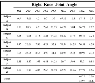
Table 1. Right knee joint angle in each gait phase
Right Knee Joint Angle
Ph1 Ph2 Ph.3 Ph.4 Ph.5 Ph.6 Ph.7 Max. Min.
Subject
A
9.5 15.01 4.2 0.7 37 67.15 10.5 67.15 0.7
Subject
B
8.59 18.5 4.9 2.67 29.75 66.77 5.04 66.77 2.67
SubjectC
7.35 10.96 5.15 3.24 34.35 60.49 5.78 60.49 3.24
SubjectD
9.47 20.04 7.96 4.29 35.8 70.54 14.28 70.54 4.29
SubjectE
6.68 22.66 8.55 4.96 31.1 60.98 2.33 60.98 2.33
SubjectF
6.88 14.87 1.65 0.88 44.28 59.7 5.93 59.7 0.88
SubjectG
7.62 15.97 6.03 2.66 30.73 67.78 11.26 67.78 2.66
Mean 64.77
±3.97 2.39
±1.17
Figure 4 shows knee joint angle variation in each phase when walking is divided into seven phases, and Tables 1 and 2 show right and left knee joint angle variation and the maximum and minimum angles. Two peak values could be found for the right knee in Table 1. The first peak was found at 16.85±3.57 degrees in Phase 2, and the second peak at 64.77±3.97 degrees in Phase 6.
Table 2. Left knee joint angle in each gait phase
Left Knee Joint Angle
Ph.1 Ph.2 Ph.3 Ph.4 Ph.5 Ph.6 Ph.7 Max. Min.
Subject
A 3.37 36.47 67.5 13.6 22.72 2.71 0.2 67.5 0.2
Subject
B 9.68 29.62 57.09 6.93 19.6 2.07 1.69 57.09 2.07
Subject
C 8.57 38.03 62.91 5.53 7.32 2.76 2.03 62.91 2.03
Subject
D 3.55 49.2 71.14 4.51 21.06 4.04 3.79 71.14 3.55
Subject
E 4.12 37.87 63.07 1.24 12.45 6.98 0.26 63.07 0.26
Subject
F 4.29 33.54 64.6 2.36 10.15 2.62 0.85 64.6 0.85
Subject
G 5.17 35.84 66.79 5.64 26.8 7.05 1.95 66.79 1.95
Mean 64.72
±4.09 1.55
±1.11
The maximum angle was 64.77±3.97 degrees and the minimum one was 2.39±1.17 degrees for the right knee in walking. Two peak values
could also be found for the left knee in Table 2:
the first peak was found at 64.73±4.09 degrees in Phase 2, and the second peak at 17.15±6.68 degrees in Phase 5. The maximum angle was 64.72±4.09 degrees and the minimum one was 1.55±1.11 degrees for the left knee in walking.
Figure 4. Knee joint angle of right and left legs in walking
3.2 Stride length and gait velocity
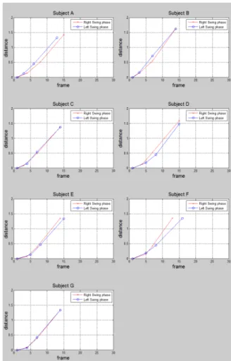
Figure 5 shows stride length of right and left legs in walking, and Table 3 shows stride length and gait velocity of right and left legs. As a result of the experiment, stride length was 1.43±0.11 m for the right leg and 1.41±0.10 m for the left one, and velocity was 1.28±0.08 m/s for the right leg and 1.23±0.11 m/s for the left one.
Figure 5. Stride length of right and left legs in walking
Table 3. Stride length and gait velocity of right and left legs
.
Stride Length(m) Gait Velocity(m/sec)
Right Leg Left Leg Right Leg Left Leg
Subject A 1.42 1.32 1.18 1.26
Subject B 1.62 1.62 1.44 1.44
Subject C 1.38 1.37 1.23 1.22
Subject D 1.59 1.48 1.33 1.23
Subject E 1.35 1.33 1.21 1.11
Subject F 1.36 1.35 1.31 1.09
Subject G 1.33 1.33 1.28 1.28
Mean 1.43±0.11 1.41±0.10 1.28±0.08 1.23±0.11
4. CONCLUSION
This study suggested a method to make gait analysis by using more convenient and less
expensive than the existing ones. The walking images laterally taken with a smart phone camera and the IAM program enabling walking track analysis were used to analyze gait. This study focused on the analysis method using several variables rather than determining any abnormality of gait by using the results of analysis in seven subjects. This study obtained the following results: First, gait analysis could be made by measuring knee joint angle variation in each gait phase, the maximum and minimum angles, and stride length and gait velocity.
Second, a smart phone camera and the IAM program in this study could make gait analysis, instead of the existing analysis methods requiring large space and costing much.
Ultimately, we will have to analyze about variation of internal rotation and exteral rotation for accuracy of gait analysis. Further gait analysis will be made with a larger number of subjects in diverse types of environment and with disabled walkers, including lower extremity amputees and those with hemiplegia, which will possibly be used as a basic research to develop a lower extremity rehabilitation training system.
REFERENCES
[1] B. O. Kim, "Methods in Clinical Gait Analysis," Journal of Korean Academy of Rehabilitation Medicine, vol. 18, no. 2, pp.
191-202, June, 1994
[2] S. H. Yune, "Analysis of Normal Gait with a 3-Dimensional Motion Analyzer," Journal of Korean Academy of Rehabilitation Medicine, vol. 16, no. 4, pp. 399-405, December, 1992 [3] W. A. Sands and M. H. Stone, “Monitoring t
he elite athlete,” USOC Olympic Coach E-Ma g., vol. 17, no. 3, Fall 2005, Available:http://co aching.usolympicteam.com/coaching/kpub.nsf/
[4] D. M. Karantonis, M. R. Narayanan, M.
Mathie, N. H. Lovell, and B. G. Celler,
“Implementation of a real-time human movement classifier using a triaxial accelerometer for ambulatory monitoring,”
IEEE Trans. Inf. Technol. Biomed., vol. 10,
no. 1, pp. 156-157, January, 2006
[5] J. H. Hong, M. S. Mun, “Prosthetic Gait and Socket Biomechanical Analyses of Transfemoral Amputee,” Journal of the Korean Society of Precision Engineering, vol.
19, no. 4, pp. 52-72, April, 2002
[6] J. H. Carr, R. B. Shepherd, "Stroke rehabilitation-guidelines for exercise and training to optimize motor skill," Oxford, Butterworth-Heinemann, 2004
[7] L. Allet, B. Leemann, E. Guyen, “Effect of different walking aids on walking capacity of patients with poststroke hemiparesis,”
Archives of Physical Medicine and Rehabilitation, vol. 90, pp. 1408-1413, August, 2009
[8] J. S. Choi, H. S. Oh, D. W. Kang,
“Comparison of Differences among Alzheimer’s Disease, Mild Cognitive Impairment and Healthy Elderly using Gait and Cognitive function,” Korean Society for Precision Engineering, vol. 10, pp. 1403-1404, 2010
[9] S. G. Kim, S. H. Shin, "Fiber-optic Gonimeter to Measure Knee Joint Angle for the Diagnosis of Gait Disturbance," Journal of the Korean Institute of Electronical Engineers, vol. 62, no. 7, pp. 1009-1013, July, 2013 [10] M. M. Patil, O. Prohaska, “Fiber-optic
sensor for joint angle measurement,” in Proc.
Annu. Int. Conf. IEEE Engineering in Medicine & Biology Society, vol. 2, pp.
803-804, November, 1988
[11] D. A. Winter, "Biomechanics and Motor Control of Human Movement," 3rd edition, NewYork, USA, Wiley&Sons, 2005.
[12] M. P. Murray, A. B. Drought and R. C.
Kory, "Walking patterns of normal men,"
Journal of Bone & Joint Surgery, vol. 46, pp.
637-650, March, 1964
[13] M. P. Murray, R. C. Kory, B. H. Clarkson,
"Walking pattern in healthy old men," Journal of Gerontology, vol. 24, pp. 169-178, April, 1969
장 재 훈
2012년 2월 한국산업기술대
학교 컴퓨터공학과
졸업(학사)
2012년 - 현재 한국산업기
술대학교 컴퓨터공
학과 석사과정
관심분야 : 헬스케어, 재활공학
임 창 주
1994년 2월 KAIST 산업공 학과 졸업(학사) 1996년 2월 KAIST 산업공 학과(HCI/VR) 졸업(석사) 2001년 2월 KAIST 산업공
학과(HCI/VR) 졸업(박사) 2004년 3월 - 현재 한국산
업기술대학교 게임
공학과 부교수 관심분야 : HCI/VR, 교육용 게임 Design
송 기 호
2012년 2월 한국산업기술대
학교 컴퓨터공학과
졸업(학사)
2012년 - 현재 한국산업기
술대학교 컴퓨터공
학과 석사과정
관심분야 : 재활공학, 생체신호처리
정 성 택
1992년 2월 KAIST 전기 및 전자공학과 졸업 (학사)
1995년 2월 KAIST 정보 및 통신공학과 졸업 (석사)
2000년 2월 KAIST 전기 및 전자공학과 졸업 (박사)
2004년 3월 - 현재 한국산
업기술대학교 컴퓨
터공학과 정교수
관심분야 : 영상처리, 헬스케어