ISSN 1225-6552, eISSN 2287-7630 https://doi.org/10.7853/kjvs.2019.42.1.9
< Original Article >
Veterinary Service
Available online at http://kjves.org
*Corresponding author: Dongseob Tark, Tel. +82-63-900-4084, Fax. +82-63-900-4012, E-mail. [email protected]
Development of monoclonal antibody capture ELISA for the detection of antibodies against transmissible gastroenteritis virus
Yeonsu Oh1, Dongseob Tark2*
1Department of Veterinary Pathology, College of Veterinary Medicine and Institute of Veterinary Science, Kangwon National University, Chuncheon 24341, Korea
2Korea Zoonosis Research Institute, Chonbuk National University, Iksan 54531, Korea (Received 9 November 2018; revised 3 February 2019; accepted 25 February 2019)
Abstract
Transmissible gastroenteritis (TGE) is a disease confined to pigs of all ages, and can be a significant cause of economic loss in breeding herds, primarily because of the very high piglet mortality. The caus- ative agent is a coronavirus, an enveloped positive strand RNA virus and closely related but non-enter- opathogenic porcine respiratory coronavirus (PRCV). Although the TGEV has declined with its innocent relative, PRCV, further genome changes could not be excluded. Therefore, the herd-level immunity against this virus is important for the prevention of disease and should be carefully monitored. The aim of this study is to develop monoclonal antibody capture enzyme-linked immunosorbent assay (MAC- ELISA) which can rapidly and accurately determine a large numbers of serum samples for surveillance purpose, and to compare the ELISA with a TGEV-specific serum neutralization test. The MAC-ELISA was sufficiently achieved, and the comparison with the virus-specific serum neutralization assays for 713 sera from pig farms showed a high correlation (r2=0.812, P<0.001). The specificity and sensitivity of MAC-ELISA for the serum neutralization test 91.9% and 91.6%, respectively, which means that the an- tibody detected by the MAC-ELISA could be said to be protective antibodies. In conclusion, the devel- oped MAC-ELISA would be very helpful in evaluating protective antibodies against TGEV.
Key words : Antibody detection, MAC-ELISA, Recombinant proteins, TGEV
INTRODUCTION
Transmissible gastroenteritis (TGE) is an acute enteric disease confined to pigs of all ages, and can be a sig- nificant cause of economic loss in breeding herds, pri- marily because of the very high piglet mortality. The causative agent is a coronavirus, an enveloped positive strand RNA virus, 60 to 220 nm in size, similar to co- ronaviruses affecting other domestic animals, including dogs and cats, with which it cross-reacts in a variety of serological tests (Dolye and Hutching, 1946; Siddel et al, 1983). The main clinical symptoms include watery diarrhea, vomiting and loss of appetite affecting all ages
of pigs. Sucking piglets are the most severely affected group. When a naïve herd is first infected, the mortality in piglets aged less than two weeks can reach 100% pri- marily due to dehydration (Saif and Wesley, 1999). This disease is on the list of diseases notifiable to the World Organisation for Animal Health (OIE).
Since TGEV was widespread in the mid 1980s, por- cine respiratory coronavirus (PRCV) which is closely re- lated but non-enteropathogenic and clinically inconse- quential (Pensaert et al, 1986), has declined together with the TGEV. However, Pritchard et al. (1999) noted clin- ically silent seroconversion to TGEV in a number of herds in the UK and it means the TGEV or PRCV ge- nomes could be possibly changed in the future. There- fore, the herd-level immunity against this virus are im-
portant for the prevention of disease and should be care- fully monitored.
The S protein of the TGEV known to be involved in cell adhesion is about 20 nm in size and highly glyco- sylated, which plays an important role to induce neutral- izing antibodies in pigs (Suñé et al, 1990; Godet et al, 1994), containing at least five antigenic determinants (Delmas et al, 1986; Correa et al, 1988; Laude et al, 1990). It could effectively be used to diagnose anti- bodies to TGEV through those antigenic determinant sites (Gebauer et al, 1991). Paired serological tests be- tween 2 and 6 months could inform when the mater- nally derived antibodies decay and the endemic TGEV or PRCV start to cause a problem in farms (Derbyshire et al, 1969; Saif and Wesley, 1999). Several serological assays found to be useful for the detection of antibodies to TGEV, including the serum neutralization test (Harada et al, 1967) and indirect fluorescent antibody assays (Benfield et al, 1978). However, the agar gel immuno- diffusion test has been complained about discrepancies with a neutralizing antibody titration due to the too high specificity (Bohac and Derbyshire, 1976; Stone et al, 1976). Fluorescent antibody assays are more sensitive but the results could be subjective. Although the serum neutralization test is widely used as the most reliable method, it requires expertise and facilities, and it is dif- ficult to test a large amount of serum.
The aim of this study is to develop the monoclonal antibody capture enzyme-linked immunosorbent assay (MAC-ELISA) which can rapidly and accurately de- termine a large numbers of serum samples for surveil- lance purpose, and to compare the ELISA with a TGEV-specific serum neutralization test.
MATERIALS AND METHODS
Cells and pig sera
The TGEV (Pyeongtaek strain; Korean isolate) was maintained in a swine testicular (ST) cell line. The
-Minimum essential medium (-MEM, GibcoBRL, NY, USA) supplemented with 5% fetal calf serum (FCS, Gemini, CA, USA), antibiotic-antimycotic (GibcoBRL,
NY, USA) was used as a cell-culture medium. The re- combinant virus, pF6AH-bac (Tark, 1998), was prolif- erated in a Spodoptera frugiperda 9 (Sf9) cell line (Invitrogen, MA, USA). The Sf9 cell line was cultured in a Grace’s medium at 28°C in a low-temperature in- cubator supplemented with 10% fetal calf serum and an- tibiotic-antimycotic. The pig serum used for the virus neutralization test and enzyme immunoassay was col- lected from a pig farm.
Concentration of recombinant TGEV spike protein To prepare the antigen for ELISA, the recombinant S protein was prepared in pF6AH-bac by deleting trans- membrane anchoring domain, rather including 6x histi- dine base toward C-terminal of the gene. The pF6AH- bac was inoculated on Sf9 cells and centrifuged for 3,000 rpm/20 min when the cytotoxic effect of 80∼
90% or more appeared after 5 days. After the super- natant was discarded, the recombinant protein was con- centrated by modification of Zinc (Zn) acetate method used by Bohac et al. (1975). Briefly, 1 M Zn acetate was added to the supernatant to a final concentration of 0.01 M to 0.08 M, and the pH was adjusted to 1 N NaOH to a pH of 7.0∼7.5. Then, it was centrifuged at 3,000 rpm for 20 min. After the supernatant was re- moved again, the precipitate was suspended by adding saturated EDTA (117 g EDTA-Na, 45 g Tris/L). Then, it was centrifuged again at the same condition and fi- nally, the supernatant was dialyzed in phosphate buf- fered saline (PBS) at 4°C for overnight. The concent- rated recombinant protein was subdivided into 0.5 mL portions and stored at −70°C for use as an antigen for ELISA.
Monoclonal antibody capture ELISA
The monoclonal antibody, 5C8 (Jang et al, 1998), specifically bound to TGEV Spike protein was diluted in 0.05 M carbonate/bicarbonate buffer to be sensitized to an ELISA plate (Maxi-sorp, Nunc, Denmark) at 37°C for 15∼18 hours. The plate, then, was washed four times with a washing solution (0.01 M Tris pH 7.5, 0.05 M NaCl, 0.05% tween 20) and blocked with a blocking
Fig. 1. Determination of the optimal monoclonal antibody (Mab) concentration. Dilution factors of each component are as follows:
1:100 for recombinant spike protein, 1:40 for porcine serum, 1:1,000 for conjugate.
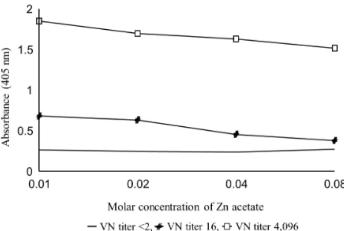
Fig. 2.Determination of the optimal molar concentration of Zn ace- tate for concentration of recombinant spike protein. Dilution factors of each component in MAC-ELISA for optimizing Zn acetate concen- tration are as follows: 1:4,000 for monoclonal antibody, 1:80 for re- combinant spike protein, 1:40 for porcine serum, 1:1,000 conjugate.
buffer (0.01 M Tris pH 7.5, 0.15 M NaCl, 1% gelatin, 20% horse serum) at 37°C for one hour. Then, the plate was washed four times and the recombinant protein was incubated at 37°C for 3 hours after being diluted in a dilution buffer (0.01 M Tris pH 7.5, 0.15 M NaCl, 1%
gelatin, 20% horse serum, 0.05% Tween 20). The plate was washed four times, and the serum samples were distributed after dilution, placed in a plate containing the recombinant protein, incubated at room temperature for 1 hour, and washed as above. Horseradish-perox- idase-conjugated goat anti-swine IgG (KPL, Gaithersburg, MD, USA) was diluted with dilution solution and added to the plate, followed by reaction at room temperature for 1 hour and then washing 4 times. Finally, 2-2’azi- no-di- (3-ethyl benzothiazoline sulfonic acid) (ABTS) (KPL, Gaithersburg, MD, USA) was added to the plate and the result was revealed at 405 nm after 10 minutes.
Virus neutralization (VN) test
The VN test was carried out by modifying the meth- od of Komaniwa et al. (1986). Neutralizing antibody tit- ers in serum were expressed as reciprocal of serum dilu- tion factor which could neutralize 100 TCID50 Pyeongtaek strain by 50%.
Statistical processing
The statistical analysis was performed by Graph Pad
Prism (version 5.0). Summary statistics were calculated for all data to assess the overall quality, including nor- mality. The values of the data from MAC-ELISA and virus neutralization test were analyzed for the correla- tion and significance. P<0.05 was considered to be sta- tistically significant. The sensitivity and specificity were compared between two methods according to the meth- od of Cho and Bohac (1985).
RESULTS
Establishment of reaction conditions for setting- up MAC-ELISA
MAC-ELISA was established with the optimal dilu- tion concentration of monoclonal antibody, 5C8, from 1:
1,000 to 1:8,000. As a result, the best reactivity between positive and negative sera was observed at 1:2,000 and 1: 4,000 (0.3∼0.6 g/mL) dilutions as shown in Fig. 1.
As shown in Fig. 2, Zn acetate at 0.01 M and 0.02 M in concentrations showed high absorbance, and it was confirmed that Zn acetate at a lower concentration per- formed effectively to concentrate the recombinant protein.
To confirm the optimal antigen concentration, the con- centrated recombinant spike protein was diluted in 1:5∼
1:640 times. As a result, the best response was obtained at a dilution of 1:20 times and the reaction was kept
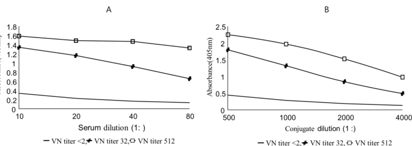
Fig. 4. Determination of the optimal serum and conjugate concentration. A; Dilution factors of each component in MAC-ELISA for optimizing serum concentration are as follows: 1:4,000 for monoclonal antibody, 1:100 for recombinant spike protein, 1:1,000 conjugate. B; Dilution factors of each component in MAC-ELISA for optimizing conjugate concentration are as follows: 1:4,000 for monoclonal antibody, 1:100 for recombinant spike protein, 1: 40 for test serum.
Fig. 3. Determination of the optimal antigen concentration. Dilution factors of each component in MAC-ELISA for optimizing recombi- nant spike protein are as follows: 1:4,000 for monoclonal antibody, 1:40 for porcine serum, 1:1,000 conjugate.
Fig. 5.Correlation between virus neutralization test and monoclonal antibody capture ELISA (MAC-ELISA). Dilution factors of each component in MAC-ELISA are as follows: 1:4,000 for monoclonal antibody, 1:100 for recombinant spike protein, 1:40 for porcine serum, 1:1,000 conjugate.
constant up to 1:160 times (Fig. 3). Therefore, 1:100-fold (total protein amount: 5 g/mL) was set to an appro- priate dilution concentration of the antigen.
The optimal dilution factors were 1:40 for the serum and 1:1,000 for the conjugate (Fig. 4). To sum it up, the optimal dilution factor of each step was set at 1:4,000 (0.3 g/mL) for monoclonal antibody, 1:100 (5 g/mL) for recombinant protein antigen, 1:40 for test serum and 1: 1,000 for conjugate.
Comparison of antibody detection efficiency bet- ween the VN and MAC-ELISA tests
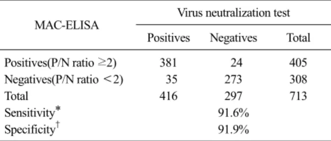
To determine an appropriate cut-off value, 713 por- cine sera were tested by paired ELISA established in this study and VN tests. The result suggested that when the P/N ratio (absorbance of the test serum/absorbance of the standard negative serum) was 2 or more, the cut- off value had the most appropriately high specificity and sensitivity when compared with the results of the VN test. The correlation between two assays were high and statistically significant (r2=0.812, P<0.0001) (Fig. 5) as shown in Table 1.
Table 1. Comparative analysis between the virus neutralization (VN) test and and MAC-ELISA to detect anti-TGEV antibodies in field porcine serum samples
MAC-ELISA Virus neutralization test Positives Negatives Total
Positives(P/N ratio ≥2) 381 24 405
Negatives(P/N ratio <2) 35 273 308
Total 416 297 713
Sensitivity* 91.6%
Specificity† 91.9%
When the VN test resulted in 2 or more, the sample was regarded as positive. All serum samples were inactivated before test and all experiments were repeated three times.
*Sensitivity (%)=(No. of MAC-ELISA positive samples / all positive samples)×100
†Specificity (%)=(No. of MAC-ELISA negative samples / all negative samples)×100
Fig. 6.Stability of the monoclonal antibody capture ELISA kit con- taining plate and other components during 7 months.
Evaluation of the stability of MAC-ELISA
To confirm the long-term stability of the developed MAC-ELISA plate, it was completely dried at 37°C, sealed with a seal tape after air was removed, and was placed at −20°C. The reproducibility of the test by the MAC-ELISA was confirmed after 7 months of storage as shown in Fig. 6.
DISCUSSION
TGEV has an antigenic determinant site capable of producing a major neutralizing antibody in the spike protein among the N, M, and S proteins. ELISA test made only with S protein will present consistent results with the commonly used serum neutralization test will reduce confusion in applying the results. Therefore, in this study, the recombinant S protein of TGEV pro- duced in the baculovirus expression system was used to develop the MAC-ELISA with high sensitivity and spe- cificity compared with the serum neutralization test.
Nelson and Kelling (1984) attempted to develop an ELISA using purified TGEV but did not obtain highly sensitive results. On the other hand, Rukhadze et al.
(1989) performed ELISA after isolating whole TGEV protein and S, M, N protein separately. As a result, when M and N proteins were used as antigens, they
showed the lowest reactivity (absorbance) to antibodies to TGEV, and even the total TGEV protein was also lower than that of S protein. It also was reported that there was a high correlation (r=0.97) between serum neutralization assays and ELISA using S protein as an antigen. Based on the above results, S protein was con- sidered to be the most useful protein for TGEV sero- logical assay, and the author produced recombinant S protein from newly isolated TGEV (Korean isolated NVRI 48 strain) in order to more easily obtain S protein.
When the recombinant baculovirus produced by cell cul- ture is used as an antigen for ELISA, the cellular com- ponent can induce a nonspecific reaction (Cho et al, 1991). Therefore, the transmembrane anchor domain portion of the recombinant S gene is removed, the half of the amino terminus containing the antibody product- ion epitope was expressed. Therefore, the recombinant S protein is secreted out of the cell so that the cell com- ponent is minimized when it is used as an antigen for ELISA. For the production of antigens for ELISA, only the cell culture supernatant was collected without any a conventional purification method such as sucrose gra- dient or ultracentrifugation steps (Nelson and Kelling, 1984; Hohdatsu et al, 1987; Rukhadze et al, 1989) and treated with 0.1M Zn acetate followed by concentration to 1/20. Lanza et al. (1993) showed a very low specific- ity (58%) when comparing the results of indirect ELISA and serum neutralizing antibody test. Therefore, in order to overcome the disadvantages of indirect ELISA, the ELISA antigen was prepared by ultracentrifugation of TGEV cultured on swine testicle cells and then attached to an ELISA plate using two kinds of monoclonal anti-
bodies specifically binding to S protein and then subject- ed to the same procedure as indirect ELISA. As a re- sult, the same effect as indirect purification of the anti- gen was obtained, and the nonspecific reaction was re- markably eliminated, and the specificity (99%) of the test method was also increased. In this study, in order to reduce nonspecific reactions, MAC-ELISA was per- formed by immobilizing the recombinant S protein on the plate by coating an ELISA plate with a monoclonal antibody, 5C8, which specifically binds to the spike pro- tein, according to the method of Lanza et al. (1993).
Antibody tests of TGEV against 713 pig sera collected from farms using established MAC-ELISA showed a high correlation with serum neutralization test (Fig. 6, Table 1). However, about 8.2% (59/713 sera) of serum did not match the results of the serum neutralization.
These discrepancies may include 1) the conjugate (HRP conjugated goat anti-swine IgG) that was used in the experiment did not react with the porcine IgM antibody produced early in the disease, 2) the presence of anti- body to the cell nucleus or cellular components in se- rum, and 3) a lot of antibodies against the antigenic site that does not have the ability to produce neutralizing antibodies (Reynolds, 1982; Cesbron et al, 1985). In or- der to put the established ELISA into practical use, the ELISA plate containing the capture monoclonal antibody and the recombinant S protein was stored at −20°C and evaluated for a period of 7 months at a constant interval.
As a result of the test, the established MACELISA showed a stable preservation and the possibility of prac- tical use was confirmed. In conclusion, the established MACELISA can be used to detect antibody levels to TGEV in pig herd, which may be useful for serological screening for TGEV.
ACKNOWLEDGEMENTS
This subject is supported by Korea Ministry of Envi- ronment (MOE) as “Public Technology Program based on Environmental Policy (No. E416-00021-0602-0)”.
REFERENCES
Benfield DA, Haelterman EO, Burnstein T. 1978. An indirect flu- orescent antibody test for antibodies to transmissible gastroenteritis virus of swine. Can J Comp Med 42: 478- 482.
Bohac J, Derbyshire JB, Thorsen J. 1975. The detection of trans- missible gastroenteritis viral antigens by immunodiffu- sion. Can J Comp Med 39(1):67-75.
Bohac J, Derbyshire JB. 1976. The detection of transmissible gas- troenteritis viral antibody by immunodiffusion. Can J Comp Med 40: 161-165.
Cesbron JY, Capron A, Ovlague G, Santoro F. 1985. Use of a monoclonal antibody in a double-sandwich ELISA for detection of IgM antibodies to Toxoplasma gondii major surface protein(P30). J Immunol Methods 83: 151-158.
Cho HJ, Bohac JG. 1985. Sensitivity and specificity of an Enzyme-linked immunosorbent assay for the detection of infectious bovine rhinotracheitis viral antibody in cattle.
Can J Comp Med 49: 189-194.
Cho HJ, Masri SA, Deregt D, Yeo SG, Thomas EJG. 1991.
Sensitivity and specificity of an enzyme-linked immuno- sorbent assay for the detection of bovine virus antibody in cattle. Can J Vet Res 55: 56-59.
Correa I, Jiménez G, Suñé C, Bullido MJ, Enjuanes L. 1988.
Antigenic structure of the E2 glycoprotein from trans- missible gastroenteritis coronavirus. Virus Res 10: 77-94.
Delmas B, Gelfi J, Laude H. 1986. Antigenic structure of trans- missible gastroenteritis virus. II. Domains in the peplom- er glycoprotein. J Gen Virol 67: 1405-1418.
Derbyshire JB, Jessett DM, Newman G. 1969. An experimental epidemiological study of porcine transmissible gastroen- teritis. J Comp Pathol 79: 445-452.
Doyle LP, Hutching LM. 1946. A transmissible gastroenteritis in pigs. J Am Vet Med Assoc 108: 257-259.
Gebauer F, Posthumus WP, Correa I, Suñé C, Smerdou C, Sánchez CM, Lenstra JA, Meloen RH, Enjuanes L.
1991. Residues involved in the antigenic sites of trans- missible gastroenteritis coronavirus S glycoprotein. Virol 183: 225-238.
Godet M, Grosclaude J, Delmas B, Laude H. 1994. Major re- ceptor-binding and neutralization determinants are lo- cated within the same domain of the transmissible gas- troenteritis coronavirus spike protein. J Virol 68: 8008- 8016.
Harada K, Kumagai T, Sasahara J. 1967. Studies on transmissible gastroenteritis in pigs. III. Isolation of cytopathogenic vi- rus and its use for serological investigation. Natl Inst Anim Health Quart 7: 127-137.
Hohdatsu T, Eiguchi Y, Ide S, Baba K, Yamagishi H, Kume T, Matumoto M. 1987. Evaluation of an enzyme-linked im- munosorbent assay for the detection of transmissible gas- troenteritis virus antibodies. Vet Microbiol 13(1): 93-97.
Jang YE, Cho SH, Kim BH, Ahn JM, Kang SY. 1998. Production
and characterization of monoclonal antibodies against porcine transmissible gastroenteritis virus. Korean J Vet Res 38(2): 336-344.
Komaniwa H, Makabe T, Fukusho A, Shimizu Y. 1986. Isolation of transmissible gastroenteritis virus from feces of diar- rheic pigs in roller culture of CPK cells in the presence of trypsin. Nihon Juigaku Zasshi 48(6): 1245-1248.
Lanza I, Rubio P, Munoz M, Carmenes P. 1993. Comparison of a monoclonal antibody capture ELISA (MACELISA) to indirect ELISA and virus neutralization test for the sero- diagnosis of transmissible gastroenteritis virus. J Vet Diagn Invest 5: 21-25.
Laude H, Rasschaert D, Delmas B, Godet M, Gelfi J, Charleyet B. 1990. Molecular biology of transmissible gastroen- teritis virus. Vet Microbiol 23: 147-154.
Nelson LD, Kelling CL. 1984. Enzyme-linked immunosorbent as- say for the detection of transmissible gastroenteritis virus antibody in swine sera. Am J Vet Res 45: 1654-1657.
Pensaert M, Callebaut P, Vergote J. 1986. Isolation of a porcine respiratory, non-enteric coronavirus related to transmis- sible gastroenteritis. Vet Q 8(3):257-261.
Pritchard GC, Paton DJ, Wibberley G, Ibata G. 1999. “Transmis- sible gastroenteritis and porcine epidemic diarrhea in Britain.” Vet Rec 144(22):616-618.
Reynolds DJ. 1982. Application of ELISA to the study of viruses.
Development and application of ELISA for veterinary diagnosis and research on enteric and other vises. pp112-
123. In: The ELISA: enzyme-linked immunosorbent as- say in veterinary research and diagnosis, ed. Wardley RC, Crowther JR. Martinus Nijhoff, The Hague, The Netherlands.
Rukhadze GG, Aliper TI, Sergeev VA. 1989. Isolation of peplom- er glycoprotein E2 of transmissible gastroenteritis virus and application in enzyme-linked immunosorbent assay.
J Clin Microbiol 27: 1754-1758.
Saif LJ, Wesley RD. 1999. Transmissible gastroenteritis and Porcine respiratory coronavirus. pp 295-325. In: Straw BE, D’Allaire S, Mengeling WL, Taylor DI(ed.). Dis- eases of swine. 8th ed. Iowa State University Press, Ames, Iowa.
Siddel S, Wege H, Ter Neulen V. 1983. The Biology of corona- viruses. J Gen Virol 64: 761-776.
Stone SS, Kemeny LJ, Jensen MT. 1976. Partial characterization of the principal soluble antigens associated with the co- ronavirus of transmissible gastroenteritis by complement fixation and immunodiffusion. Infect Immun 13: 521-526.
Suñé C, Jiménez G, Correa I, Bullido MJ, Gebauer F, Smerdou C, Enjuanes L. 1990. Mechanisms of transmissible gas- troenteritis coronavirus neutralization. Virol 177: 559-569.
Tark DS. 1998. Studies on the analysis of the spike gene of por- cine transmissible gastroenteritis virus isolated in Korea and the use of recombinant spike protein. Ph. D. Thesis, Chonnam National University, Kwangju, Korea.