Anti-proliferative Activities of Solvent Fractions of Lees Extracts in Human Colorectal HCT116 Cells
Hyung-Taek Kang
1, Seung Hoon Lee
1, Soon Young Kim
1, Mi-Sun Kim
2, Woo-Chang Shin
3, Ho-Yong Sohn
2and Jong-Sik Kim
1*
1
Department of Biological Sciences, Andong National University, Andong 760-749, Korea
2
Department of Food and Nutrition, Andong National University, Andong 760-749, Korea
3
Research Institute, Kooksoondang Brewery Co., Ltd., Seongnam 460-120, Korea
Received June 4, 2014 /Revised September 22, 2014 /Accepted September 23, 2014
In the present study, we prepared eighty-five different kinds of lees extracts and their solvent frac- tions and investigated their anti-proliferative activities against human colorectal cancer HCT116 cells.
HCT116 cells were treated with eighty-five solvent fractions of lees extracts and then cell viability was measured using MTS assay. Among the treated solvent fractions, three solvent fractions (KSD-E1-3, KSD-E2-3, and KSD-E4-3) were selected based on cell viability assay. In addition, we performed an oligo DNA microarray analysis to analyze the gene expression changes by treatment of KSD-E1-3 in HCT116 cells. Among the up-regulated genes, we selected 4 genes (NAG-1, ATF3, p21, and DDIT3) and performed RT-PCR using gene-specific primers. Among the treated solvent fractions, KSD-E1-3 dramatically induced the expressions of the four selected genes. In addition, we investigated whether the up-regulations of those genes were dependent on the transcription factor p53’s presence using p53 null HCT116 cells. The results indicate that the up-regulations of NAG-1, ATF3, and DDIT3 are not dependent on the p53 presence, whereas p21 is dependent on the p53 presence. These findings may help to understand the molecular mechanisms of the anti-proliferative activity mediated by rice wine lees in human colorectal cancer cells.
Key words : Anti-proliferation, HCT116 cells, lees, oligo DNA microarray, p53
*Corresponding author
*Tel : +82-54-820-5798, Fax : +82-54-820-7705
*E-mail : [email protected]
This is an Open-Access article distributed under the terms of the Creative Commons Attribution Non-Commercial License (http://creativecommons.org/licenses/by-nc/3.0) which permits unrestricted non-commercial use, distribution, and reproduction in any medium, provided the original work is properly cited.
Journal of Life Science 2014 Vol. 24. No. 9. 967~972 DOI : http://dx.doi.org/10.5352/JLS.2014.24.9.967
서 론
막걸리는 찹쌀 등의 전분을 원료로 하고 있고 , 발효제를 섞 어 발효시킨 탁주를 혼탁하게 제성한 한국 고유의 전통주이다 [10]. 막걸리는 다른 주류에서는 볼 수 없는 당화와 발효라는 두 가지 이화학적 , 미생물학적 변화를 혼합적으로 가지고 있 다 [14, 15]. 막걸리를 제조할 때 술을 걸러내는 과정에서 생성 되는 찌꺼기를 “주박” 혹은 “술지게미”라고 하며, 주박은 단 백질과 전분질이 주성분이며 [6], 알코올, 당류, 효모, 유기산, 무기질 등을 함유하고 있다 [20]. 주박에 대한 생리활성 연구는 면역촉진물질 활성 [4], 항산화 및 항균활성 등으로 보고되었다 [12]. 또한 누룩은 술을 빚을 때 사용하는 발효제로서 효소를 지닌 곰팡이를 곡류에 번식시킨 것이다 [11]. 누룩은 밀, 호밀, 조곡 , 현미를 기본 원료로 제조되는데 이는 지방마다 다른 생 산원료를 사용하기 때문이며 [2], 같은 원료라도 제조시 온도,
습도 등의 환경요인에 따라 차이가 나는 것으로 알려져 있다 [25]. 더욱이 누룩은 생전분을 원료로 하기 때문에 미발효성 당과 올리고당 , 식이성 섬유질이 풍부하게 함유되어 있어 웰 빙 (well being) 술의 양조가 가능하다[24].
암은 비정상적인 세포가 통제되지 못하고 과다하게 증식을 통해 확산되는 질병이다 [16]. 국내의 경우 보건복지부 중앙등 록본부의 암 등록 통계에 의하면 2011년도 기준 암 발생 현황 이 갑상선암 (17.8%), 위암(14.9%)에 이어 대장암이 세 번째 (12.8%)이며, 암으로 인한 사망율은 위암(19.6%), 대장암(15.2%), 폐암 (14.2%), 간암(11.5%) 순으로 보고되고 있다[22]. 특히 대 장암은 세계에서도 두 번째로 발병률이 높고 [3], 우리나라에서 도 상당히 발병률이 높다 .
대장암의 유발 요인은 현대화로 인한 서구형 식생활 변화에 따른 과다한 동물성 지방 섭취 및 섬유질 , 칼슘 부족 등의 환경 요인 [9]과 염증질환 및 대장 용종 등의 유전적 요인이다[1].
그러나 치료제로 사용되는 대부분의 항암제는 암세포의 apoptosis [8]는 물론, 정상 세포도 사멸을 유발한다. 그렇기 때문에 최근에는 식물로부터 추출한 천연재료를 활용한 면역 향상과 암세포의 사멸을 유도하는 항암제에 대한 연구 [17, 19]
가 진행 중이며 , 이와 더불어 가족력이 있는 환자에게는 대장 암 조기 발견을 위한 검사를 권장하고 있는 실정이다 [7].
본 연구는 8종의 주박과 3종의 누룩을 유기용매로 분획한
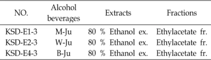
Table 1. List of three different kinds of lees extracts and their solvent fractions
NO. Alcohol
beverages Extracts Fractions KSD-E1-3
KSD-E2-3 KSD-E4-3
M-Ju W-Ju B-Ju
80 % Ethanol ex.
80 % Ethanol ex.
80 % Ethanol ex.
Ethylacetate fr.
Ethylacetate fr.
Ethylacetate fr.
총 85종에 의한 대장암 세포주의 성장 억제 활성 및 항 성장 기전을 연구하고자 하였다 . 즉, 인간 대장암 HCT116 세포주에 서 주박 추출물이 세포생존율에 미치는 영향을 확인하고 , oli- go DNA microarray 실험을 수행하여 주박 추출물 처리에 의 해 차별적으로 발현되는 유전자를 선별하였다 . 그 중 4개의 유전자를 선별하여 전사조절인자 p53의 의존성에 대해서 연 구하였다 . 이러한 연구 결과는 주박 추출물 및 분획물에 의한 대장암 세포의 성장 억제활성과 그 기전을 이해하는데 도움을 줄 것으로 기대된다 .
재료 및 방법
주박 및 누룩 추출물 제조
본 연구에서 사용된 8종의 주박과 3종의 누룩은 (주)국순당 에서 제공받아 사용하였으며 , 총 11종의 시료에서 총 85종의 분획물을 2회에 나누어 제조하였다. 제조방법은 다음의 절차 에 따라 진행하였다 . 주박의 열수 추출물은 시료 1 kg에 물 1 l를 가하여 100℃에서 30분 동안 추출하였으며, 여과한 후 (Whatman No. 2), 70℃에서 감압 농축하여 분말로 제조하였 다 . 에탄올 추출물은 시료 1.5 kg에 95% 에탄올 6 l를 가하여 상온에서 3일간 2회 추출 하였고, 여과한 후 60℃에서 감압 농축하여 분말로 제조하였다 . 또한 누룩의 에탄올 추출물은 시료 200 g에 95% 에탄올 1 l를 가하여 상온에서 7일 동안 2회 추출하였고, 여과한 후 60℃에서 감압 농축하여 분말로 제조하였다 . 각각 추출된 추출물 4 g을 물에 현탁하여 n-hex- ane, ethylacetate, butanol을 이용하여 순차적으로 분획하고 물 잔류물을 회수하여 총 85종의 추출물 및 분획물을 제조하 였다 . 추출물과 분획물은 Dimethyl sulfoxide (DMSO, sigma, USA)에 녹여 -20℃에서 저장하여 사용하였다. 총 분획물 85 종 중 cell viability assay 결과에서 선별된 분획물 KSD-E1-3, KSD-E2-3, 그리고 KSD-E4-3에 대한 자세한 정보는 Table 1 에 나타내었다 .
세포배양 및 재료
대장암 세포주 HCT116는 American Type Culture Collec- tion (ATCC, Frederick, MD, USA)에서 구입하였고, p53 null HCT116 (HCT116 p53-/-) 세포주는 Johns Hopkins 의과대학 의 Bert Vogelstein 박사로부터 분양 받았다. 두 종류의 세포주 배양은 10% FBS (Fetal Bovine Serum, Gibco, USA), 1% pen-
icillin 및 streptomycin (WelGene, Korea)이 포함된 Dulbec- co’s Modified Eagle Medium (DMEM, Gibco, USA)을 사용하 였다 . 세포의 생육정도를 관찰하면서 계대배양을 실시하여 적 정수의 세포를 유지하였다 .
세포 성장율 측정(MTS assay)
대장암 세포주 HCT116에 주박 추출물 및 분획물을 처리하 여 세포 생존율에 미치는 영향을 연구하기 위해 cell viability assay를 수행하였다. 우선 96 well plate에 1×10
3cells/well의 세포를 접종한 후 , 24시간 후에 FBS를 첨가하지 않은 serum free medium으로 배지를 교환한 뒤 DMSO에 녹인 주박 추출 물 및 분획물을 1 mg/ml 로 처리하였다. 주박 분획물의 대조 물질로는 용매인 DMSO를 처리하였다. 24시간 경과 후, cell viability assay를 수행하기 위해 MTS (3-(4,5-dimethylthia- zol-2-yl)5-(3-carboxy-methoxyphenyl)-2-(4-sulfophenyl)- 2H-tetrazolium)용액(Promega, USA)을 각 well당 20 μl씩 첨 가하여 37℃, 5% CO
2incubator에서 4시간 동안 반응시켰다.
반응이 끝난 후 NanoQuant Plate
™(Tecan Trading AG, Swit- zerland)를 사용하여 580 nm에서 흡광도를 측정하였다. 실험 결과 수치는 4개의 독립적인 well에서 수행한 값을 Microsoft EXCEL program을 이용하여 분석한 후 mean ± SD 값과 그래 프로 나타내었다 .
Total RNA 추출
Total RNA 추출은 주박 및 추출물을 처리한 대장암 세포주 로부터 RNeasy mini kit (Qiagen, USA)를 이용하여 제조사의 메뉴얼에 따라 수행하였다 . 최종적으로 정제된 total RNA는 NanoQuant Plate
TM를 이용하여 260 nm와 280 nm에서 흡광 도를 측정하여 정량한 후 , oligo DNA Microarray 실험과 RT-PCR에 사용되었다.
Oligo DNA Microarray 실험
Oligo DNA microarray 실험과 데이터 분석은 지노믹트리 사 (Genomictree, Inc, Korea)에 위탁하여 수행하였다. 사용한 microarray는 Agilent사의 Agilent Human GE 4 X 44K (V2) arrays (Agilent Techonlogies, Palo Alto, USA)를 사용하였다
Reverse transcription-PCR
수확한 세포주로부터 추출한 total RNA 2 μg을 주형으로 PrimeScript
TMRT-PCR Kit (TaKaRa, Japan)를 이용하여 제조 사의 메뉴얼에 따라 cDNA를 합성하였다. 합성된 cDNA를 주 형으로 하여 유전자 특이적인 oligonucleotide primer (Table 2)를 이용하여 PCR (Polymerase Chain Reaction) 과정을 수행 하였다 . PCR 반응은 94℃에서 5분 denaturation시키고, 94℃
에서 30초, 58℃에서 30초, 72℃에서 30초의 cycle을 총 30번
반복한 뒤 , 마지막으로 72℃에서 10분간 반응시켰다. 최종적
인 PCR product는 1.5% agarose gel에서 전기영동 하고 ethi-
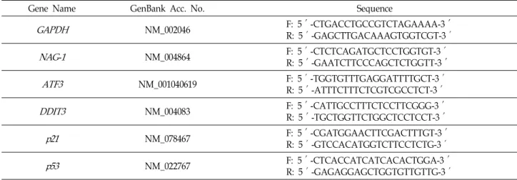
Table 2. Sequences of oligo-nucleotide primers used for RT-PCR
Gene Name GenBank Acc. No. Sequence
GAPDH NM_002046 F: 5′-CTGACCTGCCGTCTAGAAAA-3′
R: 5′-GAGCTTGACAAAGTGGTCGT-3′
NAG-1 NM_004864 F: 5′-CTCTCAGATGCTCCTGGTGT-3′
R: 5′-GAATCTTCCCAGCTCTGGTT-3′
ATF3 NM_001040619 F: 5′-TGGTGTTTGAGGATTTTGCT-3′
R: 5′-ATTTCTTTCTCGTCGCCTCT-3′
DDIT3 NM_004083 F: 5′-CATTGCCTTTCTCCTTCGGG-3′
R: 5′-TGCTGGTTCTGGCTCCTCCT-3′
p21 NM_078467 F: 5′-CGATGGAACTTCGACTTTGT-3′
R: 5′-GTCCACATGGTCTTCCTCTG-3′
p53 NM_022767 F: 5′-CTCACCATCATCACACTGGA-3′
R: 5′-GAGAGGAGCTGGTGTTGTTG-3′
A
B
Fig. 1. Effects of eighty-five kinds of lees extracts and their solvent fractions on HCT116 cell viabilities. HCT116 cells were plated at 1×10
3cells/well in a 96-well plate and treated with eighty-five kinds of solvent fractions of lees extracts (1 mg/ml (A) and 1 mg/ml (B)) for 24 hr. After treatment, cell viability was measured using MTS proliferation assay kit. Bars are mean values ± SE from 4 independent experiments.
dium bromide (EtBr, Bioneer, Korea)로 염색하여 Gel Image Analysis System (CoreBio, Korea)을 이용하여 사진 촬영하였다.
통계 분석
모든 실험은 최소한 3회 이상 실험을 실시하였다. p<0.05 값에서 유의성을 조사하기 위하여 Student’s t-test로 평균값을 분석하였다 .
결과 및 고찰
85종의 주박 추출물 및 분획물이 세포생존율에 미치는 영향
주박 추출물 및 분획물이 대장암 세포주 HCT116의 생존율
에 미치는 영향을 확인하기 위하여 40종의 1차 추출물 및 분획
물을 1 mg/ml의 농도로 처리한 다음 cell viability assay를
수행하였다 . 주박 추출물 및 분획물의 대조물질로는 DMSO를
이용하였다 . 그 결과 대부분의 추출물 및 분획물에 의해 cell
Fig. 2. Effects of three kinds of solvent fractions on human HCT116 cell viabilities. HCT116 cells were plated at 1×10
3cells/well in a 96-well plate and treated with three kinds of solvent fractions (KSD-E1-3, KSD-E2-3 and KSD-E4-3) of different concen- trations (0.25, 0.5, and 1 mg/ml) for 24 hr. After treatment, cell viability was measured using MTS proliferation assay. Bars are mean values ± SE from 4 independent experiments.
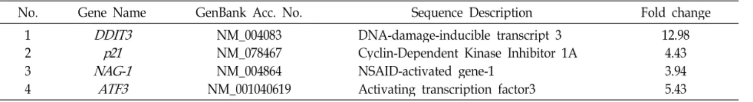
Table 3. Selected up-regulated genes by KSD-E1-3 treatment in HCT116 cells
No. Gene Name GenBank Acc. No. Sequence Description Fold change
1 2 3 4
DDIT3 p21 NAG-1
ATF3
NM_004083 NM_078467 NM_004864 NM_001040619
DNA-damage-inducible transcript 3 Cyclin-Dependent Kinase Inhibitor 1A NSAID-activated gene-1
Activating transcription factor3
12.98 4.43 3.94 5.43 viability 생성이 감소되는 것을 확인할 수 있었다. 특히 KSD-
E3-2, KSD-E3-3, 그리고 KSD-E1-3등의 시료에 의해 생존율이 크게 감소되는 것을 확인할 수 있었다 (Fig. 1A).
2차 주박 추출물 및 분획물 45종에 의한 세포생존율을 측정 하기 위해 1 mg/ml의 농도로 처리한 뒤 동일한 조건으로 cell viability assay를 수행하였다. 그 결과 KSD-E4-3, KSD-E4-4 그리고 KSD-W7-5 등의 분획물에서 세포생존율의 큰 감소를 확인 할 수 있었다 (Fig. 1B). 본 연구에서는 총 85종의 주박 추출물 및 분획물에 의한 cell viability assay에 결과와 마우스 대식 세포인 RAW264.7 세포주에 대한 세포 사멸결과(data not shown)를 종합하여 최종적으로 3종의 분획물(KSD-E1-3, KSD-E2-3, KSD-E4-3)을 선정하여 추후 실험을 진행하였다 (Table 1).
선별된 3종의 시료의 농도별 처리에 따른 세포생존율 측정 선별된 3종의 주박 추출물 및 분획물(KSD-E1-3, KSD-E2-3, KSD-E4-3)의 농도에 따른 처리가 HCT116 세포 생존율에 미 치는 영향을 연구하였다 . 연구방법은 HCT116 세포주에 선별 된 주박 추출물 및 분획물 3종을 0.25, 0.5, 1 mg/ml의 농도로 24시간 처리한 후, cell viability를 측정하였다. 그 결과 3종의 시료에 의해 농도 의존적으로 cell viability가 감소함을 확인할 수 있었으나 , 이 중 KSD-E1-3에 의한 세포 생존율 감소가 가장 크게 일어남을 확인하였다 (Fig. 2). KSD-E1-3에 의한 유전체수 준에서의 발현을 확인하기 위하여 oligo DNA microarray 실
험을 진행하였다 .
Oligo DNA microarray를 이용한 유전체 수준에서의 유 전자 발현 분석
선별된 3종의 시료 중에서 가장 높은 항성장 활성을 보여준 KSD-E1-3 분획물을 1 mg/ml 농도로 HCT116 세포주에 처리 한 후 , oligo DNA microarray analysis를 수행하였다. DNA microarray 실험 결과, 2배 이상 발현이 증가되는 유전자중 항암 유전자 4개(Non-steroid Anti-inflammatory drug-acti- vated gene-1 [NAG-1], Activating Transcription Factor 3 [ATF3], DNA-damage-inducible Transcript 3 [DDIT3], Cyclin- dependent kinase inhibitor 1A [CDKN1A, p21])를 선별하였 다 (Table 2). 세포 생존율 실험 결과에 따라 선별된 3종의 분획 물을 1 mg/ml의 농도로 처리한 후, 4가지의 유전자 발현을 확인하였다 . 그 결과, p21 유전자의 경우 KSD-E1-3에 의해서 만 발현이 증가된 반면 , DDIT3, ATF3 그리고 NAG-1유전자는 처리한 모든 분획물에 의해 발현이 유도되었다 (Fig. 3). 또한, 처리한 분획물 중 KSD-E1-3에 의해 모든 항암 유전자의 발현 이 가장 높게 증가됨을 확인하였다 .
NAG-1 유전자는 TGF-β (Transforming Growth Factor-β)
superfamily의 한 종류로써, pro-apoptotic 및 anti-tumori-
genic의 기능을 가지는 것으로 알려져 있으며, 최근 파이토케
미칼과 같은 천연물에 의해 다양한 기작을 통하여 NAG-1 유
전자의 발현이 조절된다는 총설이 보고된 바 있다 [23]. ATF3
Fig. 3. Up-regulation of NAG-1, ATF3, DDIT3 and p21 gene by three kinds of solvent fractions. HCT116 cells were treat- ed with three kinds of solvent fractions of lees extracts (KSD-E1-3, KSD-E2-3 and KSD-E4-3) for 24 hr. And then, total RNAs were extracted from treated cells and used for RT-PCR with NAG-1, ATF3, DDIT3 and p21 gene spe- cific primers.
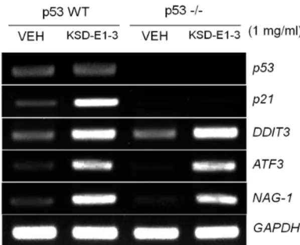
Fig. 4. Effect of p53 on gene expression by KSD-E1-3. HCT116 cells or p53 null HCT116 were treated with 1 mg/ml KSD-E1-3 for 24 hr and then RT-PCR was performed us- ing p53, p21, ATF3, DDIT3 and NAG-1 gene specific primers.
유전자는 전사조절인자로서 ATF/cAMP responsive element binding protein (CREB) family에 속하며, 천연물에 의해 발현 이 증가되며 세포사멸과 관련이 있는 것으로 보고 되었다 [5, 21]. DDIT3 유전자는 endoplasmic reticulum stress에 의해 활 성화되며 세포사멸과 관련이 있는 것으로 알려져 있다 [18].
p21WAF1/CIP1유전자는 대표적인 cyclin-dependent kinase inhibitor 중 하나로서, 세포주기 진행을 억제하는 역할을 수행 한다 .
KSD-E1-3에 의해 발현이 유도된 유전자의 p53 의존성 검증
암 억제 유전자인 p53은 DNA 손상으로 인해 발생할 수
있는 치명적인 영향으로부터 세포를 보호하는 역할 이외에 세포주기의 정지 , 노화, apoptosis 등 다양한 역할을 수행하는 전사조절인자이다 . 또한, 천연물의 처리에 의해 발현이 증가 되며 , 암세포 사멸과 직접적 관련성이 있는 것으로 알려져 있 다 [13]. 본 연구에서는 oligo DNA microarray 실험에 의해 선 별된 4개의 유전자 발현이 전사조절인자 p53에 의존적인지 확인하고자 p53 유전자가 null인 HCT116 세포주를 이용하였 다 . p53 wild type HCT116 세포주는 p53의 발현이 되는 것을 확인 하였고 , KSD-E1-3의 처리 유무에 관계없이 p53 null 세포 주에서는 p53이 발현되지 않았다. 선별된 4개의 유전자 중 NAG-1, ATF3, DDIT3 유전자는 p53 발현 유무에 관계없이 KSD-E1-3에 의해 발현이 증가됨을 확인하였다. 반면, p21 유 전자의 경우 p53에 의해서만 KSD-E1-3에 의해 발현이 증가되 는 것을 확인하였다 (Fig. 4).
연구결과를 종합하면 주박 추출물 및 분획물이 여러 항암 유전자 발현을 다양한 경로를 통하여 증가시킴으로써 암세포 에 대한 항성장 활성을 나타냄을 증명하였다 . 그러나 선별된 KSD-E1-3 분획물의 작용기전을 이해하고, 항성장 활성의 핵 심물질을 찾기 위해서는 추가 분획물을 제조하고 , 추가적인 기전연구가 수행되어야 할 것으로 생각된다 .
감사의 글
본 연구는 2012년도 농림수산식품부 고부가가치식품기술 개발사업 (과제번호 112073-3)에 의해 수행되었으며 이에 감사 드립니다 .
References