Treatment with ultra-dilutions of Arnica montana increases COX-2 expression and PGE2 secretion in mouse chondrocytes
Yun Kyu Kim1, Myeong Gu Yeo2*
1Researcher, Department of Medicine, University of Ulsan College of Medicine,
2Professor, Department of Integrative Medical Sciences, Nambu University
생쥐 연골세포에 Arnica montana 처리에 따른 COX-2 발현과 PGE2 분비 비교
김윤규1, 여명구2*
1울산대학교 의과대학 전임연구원, 2남부대학교 통합의료학과 교수
Abstract Objective: We studied the effects of 4x, 30x, 30c, and 200c homeopathic dilutions of A. montana on inflammation in primary cultured mouse chondrocytes.
Methods: Examined expression of Coll-2 and COX-2, and secretion of PGE2.
Results: Treatment with 4x, 30x, and 30c A. montana decreased mRNA expression of Coll-2 and 30x A. montana increased mRNA expression of COX-2, while treatment with 30x and 30c A. montana increased protein expression of COX-2. Treatment with the 30c A. montana increased release of PGE2.
Conclusion: Treatment with A. montana induces dedifferentiation and inflammatory responses, including increased COX-2 expression and PGE2 secretion.
Key Words :Convergence, Arnica montana, Homeopathic medicine, COX-2, PGE2.
요 약 연구목적 : 4x, 30x, 30c, and 200c 농도의 동종약물 Arnica montana (A. montana)를 1차 배양된 생쥐 연골세포에 적용하여 염증관련 인자의 변화를 관찰하고자 하였다.
연구방법 : 본 연구는 collagen type II (Coll-2), cyclooxygenase-2 (COX-2) 발현 그리고 prostaglandin 2 (PGE2) 분비에 대해 조사하였다.
결과: 4x, 30x 그리고 30c의 A. montana를 처리하였을 때, Coll-2의 mRNA 발현이 감소하였으며, 30x A. montana의 경우 COX-2 mRNA의 발현이 증가하였다. 또한 COX-2 단백질 발현은 30x와 30c의 A. montana 처리 시 증가함을 보였다.
PGE2 분비 또한 30c에서 증가함을 관찰하였다.
결론 : A. montana 처리에 따라 생쥐의 연골세포의 분화 억제를 확인하였으며, 염증관련 인자인 COX-2 및 PGE2의 발현이 증가함을 확인하였다.
주제어 : 융합, Arnica montana, 동종의약물, COX-2, PGE2
*This study was supported by research funds from Nambu University (2016) and by the Basic Science Research Program through the National Research Foundation of Korea (NRF) funded by the Ministry of Education (NRF-2018R1D1A1B07050192).
*Corresponding Author : Myeong Gu Yeo ([email protected]) Received January 15, 2019
Accepted February 20, 2019
Revised February 8, 2019 Published February 28, 2019
1. Introduction
Products derived from medicinal herbs are used to treat and prevent diseases. Various natural plant compounds are biologically active and thus of pharmacological importance[1]. The homeopathic medicine A. montana elicits anti- inflammatory effects and has been used to reduce pain relief in various inflammatory conditions such as sprains, bruises, gingivitis, and rheumatic complaints[2,3]. Homeopathic medicines are dilutions of active ingredients or are administered at reduced doses, and are prescribed according to the similia principle[4]. One of the main criticisms of homeopathic medicines is the lack of stringent quality control parameters, and symptomatic analogy must be identified. Many trials have investigated the effects of homeopathic medicines involving its mechanisms[5,6]. A. montana 6cH modulates acute inflammation in animal models[8].
Inflammation is a complex biological response of tissues to harmful stimuli, and its symptoms include pain, heat, redness, swelling, and loss of function.
Although various homeopathic remedies elicit anti-inflammatory effects and there by alleviate pain and reduce swelling in animal models, the underlying molecular mechanisms are poorly understood.
Many studies have demonstrated the anti-inflammatory and immunomodulatory effects of homeopathic remedies using experimental animal models[7]. Homeopathic dilutions of Rhus toxicodendron elicit anti-inflammatory effects, which involve histamine and prostaglandin, and thereby improve arthritis. We previously reported that Rhus toxicodendron modulates anti- inflammatory mechanisms in primary cultured mouse chondrocyte[5].
Here, we investigated the effects of A. montana on inflammation in mouse chondrocytes. We performed reverse-transcriptase polymerase chain reaction (RT-PCR) to examine mRNA expression of Coll-2, which is a marker protein of differentiated chondrocytes. In addition, we examined mRNA and
protein expression of COX-2, which is a major mediator of inflammation in arthritis. COX-2 is primarily involved in inflammation and is responsible for synthesis of prostanoids (prostaglandins and thromboxanes) involved in pathological processes [8-10]. COX-2 promotes release of the pro-inflammatory mediator PGE2, while COX-2 inhibitors suppress production of PGE2[11]. The present study demonstrates that treatment with A.
montana increased mRNA and protein expression of COX-2 and secretion of PGE2 in primary cultured mouse chondrocytes, suggesting that this remedy modulates the inflammatory process.
2. Materials and methods
2.1 Preparation of reagents
Liquid dilutions of A. montana at 4x, 30x, 30c, and 200c were purchased from Boiron (Newtown Square, PA, USA) and used to 1:10 dilution according to the manufacturer’s instructions. The liquid form of A.
montana was supplied in 20% ethanol (EtOH). The cell culture medium was supplemented with EtOH alone at a final concentration of 0.5% (v/v) as a control. Unless otherwise mentioned, all chemicals were purchased from Sigma-Aldrich (St. Louis, MO, USA).
2.2 Primary culture of mouse chondrocytes The mice were maintained under specific pathogen-free conditions, and experimental treatment was approved through Institute of Asan Life Science (Seoul, Korea, Animal Care and Use Committee).
Articular chondrocytes were obtained from 8-day-old mice[12]. Cartilage was isolated from the femoral head, femoral condyle, and tibial plateau, and then digested with 0.2% (w/v) collagenase type II. Cells were grown in Dulbecco’s modified Eagle’s media (Invitrogen, Carlsbad, CA, USA) containing 10% (v/v) fetal bovine serum, 100 units/ml penicillin, and 100 µg/ml streptomycin.
2.3 Cell proliferation and toxicity assays Cell proliferation and toxicity were assessed using a Cell Titer 96 Non-radioactive Cell Proliferation Assay, which uses MTT (3-(4,5-di- methylthiazol-2-yl)-2,5- diphenyltetrazolium bromide), and a Lactate Dehydrogenase (LDH) Assay Kit (Promega, Madison, WI, USA), respectively, according to the manufacturer’s instructions. Briefly, cells were plated into 96-well plates at a density of 1.0×10⁴cells/wells and cultured for upto 24hr. Thereafter, cells were treated with 2%
EtOH or the 4x, 30x, 30c, or 200c homeopathic dilution of A. montana for 48 hr at 37°C in a humidified atmosphere containing 5% CO2. Absorbance at 570 and 490 nm was measured in the MTT and LDH assays, respectively, using an ELISA reader (BioTek Instruments, Winooski, VT, USA). Data are the average results of three wells in one independent experiment, which was repeated fourtimes.
2.4 RT-PCR
Chondrocytes were grown in medium containing 0.5% EtOH or the 4x, 30x, 30c, or 200c homeopathic dilution of A. montana for 48 hr. Total RNA was extracted using a RNeasy Kit (Qiagen, Austin, TX, USA) according to the manufacturer’s instructions and reverse- transcribed into cDNA. Reverse transcription was performed using 1 µg total RNA and TOPscript RT DryMIX (Enzynomics, Seoul, Korea). PCR was performed using a GeneAmp PCR System 9700 (Applied Biosystems, Carlsbad, CA, USA) and AmpliTaq DNA polymerase (PE Applied Biosystems, Foster City, CA, USA) according to the manufacturer’s instructions. The following primers (Macrogen, Seoul, Korea) were used for RT-PCR: glyceraldehyde phosphate dehydrogenase (GAPDH, 587 bp), sense 5’-TCACGCCACCCAGAAGAC-3’ and antisense 5’-TCACTGCCACCCAGAAGAC-3’; and COX-2, sense 5’-GGTCTGGTGCCTGGTCTGATGAT-3’ and antisense 5’-GTCCTTTCAAGGAGAATGGTGC-3’.
The PCR conditions were denaturation (95°C for 3 min), amplification and quantification (22 cycles of 95°C
for 20 sec, 62°C for 10 sec, and 72°C for 30 sec to analyze GAPDH, and 28 cycles of 95°C for 20 sec, 63°C for 10 sec, and 72°C for 30 sec to analyze COX-2), followed by a final elongation (72°C for 5 min). The amplified PCR products were visualized by electrophoresis on 1.5% agarose gels. RT-PCR was performed in triplicate. Amplification of the target gene was normalized against that of GAPDH in the same reaction.
2.5 Immunoblot analysis
Chondrocytes were treated with 0.5% EtOH or the 4x, 30x, 30c, or 200c homeopathic dilution of A.
montana for 48 hr and lysed in lysis buffer (1% Triton X-100, 50 mM HEPES (pH 7.5), 150 mM NaCl, 1 mM EDTA, 10 mM NaF, 1 mM pyrophosphate, and 2 mM Na3VO4) supplemented with a protease inhibitor cocktail (Sigma-Aldrich) on ice for 10min. The protein concentration was determined using the Bradford method (Bio-Rad Laboratories, Hercules, CA, USA).
Proteins were separated by SDS-PAGE. Immunoblot analysis was performed using antibodies against COX-2 (R&D Systems, Minneapolis, MN, USA) and actin (Millipore, Billerica, MA, USA). Bound antibodies were detected by enhanced chemiluminescence (Pierce Chemical, Rockford, IL, USA) using a LAS 4000 mini biomolecular imager (GE Healthcare, Uppsala, Sweden).
2.6 PGE2 assay
Chondrocytes were stimulated with 0.5% EtOH or the 4x, 30x, 30c, or 200c homeopathic dilution of A.
montana for 48 hr. The level of PGE2 in the culture medium was assessed using a PGE2 Assay Kit (R&D Systems) according to the manufacturer’s instructions.
Data represent the average results of triplicates in one independent experiment, which was repeated four times.
2.7 Statistical analysis
Experiments were performed at least four times.
Values are presented as the mean ± standard error of the mean (SEM). Data were statistically analyzed using the independent samples t-test and an analysis of variance. p<0.05 was considered statistically significant.
3. Results
3.1 Effect of A. montana on the proliferation and viability of chondrocytes
Fig. 1. Treatment with homeopathic dilutions of A.
montana does not affect proliferation of chondrocytes or elicit cytotoxic effects.
Cells were assessed by the MTT(A) and LDH(B) assays. Graphs show the mean ±SEM.
*p<0.05. Con: untreated chondrocytes.
The effect of A. montana on proliferation of primary cultured mouse chondrocytes and its cytotoxic effects were evaluated using the MTT and LDH assays, respectively. Cells were analyzed following treatment with 0.5% EtOH or the 4x, 30x, 30c, or 200c homeopathic dilution of A. montana for 48 hr.
Treatment with homeopathic dilutions of A. montana did not affect the survival or proliferation of chondrocytes Fig. 1A and did not elicit cytotoxic effects, although 4x dilution showed cytotoxicity Fig.
1B.
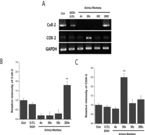
3.2 Effect of A. montana on mRNA expression of Coll-2 and COX-2 in chondrocytes
Coll-2 and COX-2 function in joint inflammation in arthritis[13]. Cells were treated with 0.5% EtOH or the 4x, 30x, 30c, or 200c homeopathic dilution of A.
montana for 48 hr, and then mRNA expression of COX-2 and Coll-2 was examined by RT-PCR.
Treatment with 4x, 30x, and 30c homeopathic dilutions of A. montana decreased mRNA expression of Coll-2 Fig. 2, suggesting that it induced dedifferentiation of chondrocytes. Moreover, treatment with the 30x homeopathic dilution of A. montana significantly increased mRNA expression of COX-2 Fig. 2A and Fig. 2C.
Fig. 2. Treatment with the 30x homeopathic dilution of A. montana increases mRNA expression of COX-2 in chondrocytes. (A) cDNA was electrophoresed on an agarose gel. The band densities of Coll-2(B) and COX-2(C) were determined by densitometry. Target gene expression was normalized against that of GAPDH. Graphs show the mean ±SEM.
** p<0.01. Con: untreated chondrocytes.
3.3 Effects of A. montana on protein expression of COX-2 in chondrocytes
We next investigated the effects of A. montana on protein expression of COX-2 in chondrocytes. Cells were treated with 0.5% EtOH or the 4x, 30x, 30c, or 200c homeopathic dilution of A. montana for 48 hr, and then protein expression of COX-2 was examined by
immunoblotting. Treatment with 4x, 30x and 30c homeopathic dilutions of A. montana significantly increased protein expression of COX-2 in chondrocytes Fig. 3A and Fig. 3B. This result indicates that homeopathic dilutions of A. montana increase protein expression of COX-2 and thereby affect the inflammatory response.
Fig. 3. Treatment with 4x, 30x, and 30c homeopathic dilutions of A. montana increases protein expression of COX-2 in chondrocytes. (A) Protein expression of COX-2 was analyzed by immunoblotting with an anti-COX-2 polyclonal goat antibody. The membrane was re-probed with an anti-actin antibody.
(B) The band densities of COX-2 were determined by densitometry. The graph shows the mean ±SEM. * p<0.05, ** p<0.01.
Con: untreated chondrocytes.
3.4 Effects of A. montana on PGE2 release by chondrocytes
Induction of COX-2 expression is closely associated with release of PGE2, and treatment with a COX-2 inhibitor reduces inflammation in animal models[9]. The level of PGE2 in the culture medium was examined following treatment of chondrocytes with homeopathic dilution of A. montana for 48 hr. Treatment with 30x and 30c homeopathic dilutions of A. montana increased the level of PGE2 in the culture medium Fig. 4, which was consistent with the effects on mRNA and protein expression of COX-2.
Fig. 4. Treatment with the 30c homeopathic dilution of A. montana increases release of PGE2 by chondrocytes. The level of PGE2 in the culture medium was measured. The graph shows the mean ±SEM. *p<0.05. Con:
untreated chondrocytes.
4. Discussion
Many clinical trials and studies have demonstrated the effectiveness of homeopathic remedies using experimental animal models [14,15]. A. montana, which is one of the most common homeopathic remedies, has been used to alleviate joint pain. In its original, undiluted form, A. montana typically causes muscular, articular, and ligament pain and induces dermatitis[7].
However, the mechanisms underlying the effects of A.
montana are unknown.
This study investigated the effects of A. montana on primary cultured mouse chondrocytes. To determine the effects of homeopathic dilutions of A. montana on inflammation, we measured mRNA expression of COX-2 by RT-PCR. Treatment with the 30x homeopathic dilution of A. montana increased mRNA expression of COX-2, while treatment with 4x, 30x, and 30c homeopathic dilutions of A. montana decreased mRNA expression of Coll-2. COX-2 is dramatically upregulated during inflammation in patients with rheumatoid arthritis. A COX-2 inhibitor reduces inflammation[15, 16], and treatment with highly potent homeopathic remedies was reported to elicit a similar effect[15]. Although homeopathic dilutions of A.
montana also affected chondrocytes in the current study, the underlying mechanism is unclear.
Treatment with 30x and 30c homeopathic dilutions of A. montana increased PGE2 release. Previous studies suggested that a broad spectrum of inflammation mediators regulate COX-2 expression[8] and that PGE2, the major COX-2 product, is upregulated in various cell lines[17,18]. The results of the present study are consistent with previous findings demonstrating a correlation between COX-2 expression and PGE2 production.
Collectively, our data show that treatment with homeopathic dilutions of A. montana increases mRNA and protein expression of COX-2 in primary cultured mouse chondrocytes and thereby increases PGE2 secretion. These findings support the effectiveness of homeopathic remedies, particularly A. montana, in vitro and provide a basis for future research. Further studies are required to investigate the effectiveness of A.
montana in vivo and the underlying mechanism.
REFERENCES
[1] N. Verma, S. K. Tripathi, D. Sahu, H. R. Das & R. H.
Das. (2010). Evaluation of inhibitory activities of plant extracts on production of LPS-stimulated pro-inflammatory mediators in J774 murine macrophages. Molecular and Cellular biochemistry 336, 127-135.
[2] C. Lass, M. Vocanson, S. Wagner, C. M Schempp, J. F.
Nicolas, I. Merfort &, S. F. Martin. (2008).
Anti-inflammatory and immune-regulatory mechanisms prevent contact hypersensitivity to Arnica montana L.
Experimental Dermatology 17, 849-857.
[3] P. P. Alfredo, C. A. Anaruma, A. C. Pião, S. M. João
& R. A. Casarotto (2009). Effects of phonophoresis with Arnica montana onto acute inflammatory process in rat skeletal muscles: an experimental study. Ultrasonics 49, 466-471.
[4] F. Wiegant & R. Van Wijk. (2010). The similia principle:
Results obtained in a cellular model system.
Homeopathy 99, 3-14. 2010.
[5] Y. H. Huh, M. J. Kim & M. G. Yeo. (2013). Homeopathic
Rhus toxicodendron treatment increased the expression of cyclooxygenase-2 in primary cultured mouse chondrocytes. Homeopathy 102, 248-253..
[6] A. P. Kawakami, C. Sato, T. N. Cardoso & L. V.
Bonamin. (2011). Inflammatory Process Modulation by Homeopathic Arnica montana 6CH: The Role of Individual Variation. Evidence-Based Complementary and Alternative Medicine 11, 12.
[7] C. R. Patil, A. D. Rambhade, R. B. Jadhav, K. R. Patil, V. K. Dubey, B. M. Sonara & S. S. Toshniwal. (2011).
Modulation of arthritis in rats by Toxicodendron pubescens and its homeopathic dilutions. Homeopathy 100, 131-137.
[8] B. Hinz & K. Brune. (2002). Cyclooxygenase-2—10 Years Later. Journal of Pharmacology and Experimental Therapeutics 300, 367-375.
[9] C. S. Williams, M. Mann & R. N. DuBois. (1999). The role of cyclooxygenases in inflammation, cancer, and development. Oncogene 18, 7908-7916.
[10] A. W. M. Cheng, T. V. Stabler, M Bolognesi, & V. B.
Kraus. (2011). Selenomethionine inhibits IL-1β inducible nitric oxide synthase (iNOS) and cyclooxygenase 2 (COX2) expression in primary human chondrocytes.
Osteoarthritis and cartilage /OARS, Osteoarthritis Research Society 19, 118-125.
[11] A. C. Abrahao, R. M. Castilho, C. H. Squarize, A. A.
Molinolo, D. D. Santos-Pinto & J. S. Gutkind. (2010). A role for COX2-derived PGE2 and PGE2-receptor subtypes in head and neck squamous carcinoma cell proliferation. Oral oncology 46, 880-887.
[12] Y. H. Huh, J.-H. Ryu & J. S. Chun. (2007). Regulation of Type II Collagen Expression by Histone Deacetylase in Articular Chondrocytes. Journal of Biological Chemistry 282, 17123-17131.
[13] F. Denizot & R. Lang. (1986). Rapid colorimetric assay for cell growth and survival: Modifications to the tetrazolium dye procedure giving improved sensitivity and reliability. J Immunol Methods 89, 271-277.
[14] D. E .Trentham, A. S. Townes & A. H. Kang. (1997).
Autoimmunity to type II collagen an experimental model of arthritis. The Journal of Experimental Medicine 146, 857-868.
[15] D. A.Willoughby, A. R. Moore & P. R. Colville-Nash.
(2000). COX-1, COX-2, and COX-3 and the future treatment of chronic inflammatory disease. The Lancet 355, 646-648.
[16] H. K. Oh, E. Y. Do & H. R. Park. (2015). Convergence Studies of NO Homeostasis in Cellular Signalling.
Journal of Digital Convergence, 13(12), 461-467.
[17] A. Y. Jang, Y. C. Sueng & J.G. Ji. (2016). The comparative study on physiological activity of White ginseng, Red ginseng and Black ginseng extract. Journal of Digital Convergence, 14(5), 459-471.
[18] B. Hinz, K. Brune & A. Pahl.. (2000). Prostaglandin E2 upregulates cyclooxygenase-2 expression in lipopolysaccharide-stimulated RAW 264.7 macrophages.
Biochemical and Biophysical Research Communications 272, 744-748.
김 윤 규(Kim, Yun Kyu) [정회원]
․2009년 2월 : 우석대학교 생명공 학과(이학사)
․2011년 8월 : 전북대학교 해부학 과(이학석사)
․2012년 8월 ~ 현재 : 서울아산병 원 연구원
․관심분야 : 재생의학, 면역학
․E-Mail : [email protected]
여 명 구(Yeo, Myeong Gu) [정회원]
․1989년 2월 : 조선대학교 유전공 학과(이학사)
․1991년 2월 : 조선대학교 유전자 과학과(이학석사)
․1995년 2월 : 조선대학교 유전자 과학과(이학박사)
․2011년 3월 ~ 현재 : 남부대학교 통합의료학과 교수
․관심분야 : 세포신호전달계, 세포이동
․E-Mail : [email protected]