Protective Effects of Korean Red Ginseng against Alcohol-induced Hepatosteatosis
Sun Ju Kim
1, Sung Hwan Ki
2and Sangkyu Lee
1*
1
College of Pharmacy, Research Institute of Pharmaceutical Sciences, Kyungpook National University, Daegu 702-701, Korea
2
College of Pharmacy, Chosun University, Gwangju 501-759, Korea
Received December 22, 2014 /Revised March 9, 2015 /Accepted March 27, 2015
Alcohol-induced fatty liver (steatosis) results from excessive generation of reducing equivalents by ethanol metabolism. Generally, chronic ethanol treatment causes hepatosteatosis by regulating sterol regulatory element-binding protein 1c (SREBP-1c), which increases the synthesis of hepatic lipids. The effect of ethanol on SREBP-1c is mediated through mammalian sirtuin-1 (SIRT-1), a NAD
+-dependent protein deacetylase that regulates hepatic lipid metabolism. Ginseng is a widely used herbal medicine that is used in Asia for its anti-diabetes and anti-obesity effects. The pharmacological and therapeutic effects of ginseng are primarily produced by bioactive constituents known as ginsenosides. Here, we examined the regulatory effects of Korean red ginseng (KRG) extracts on SREBP-1c and SIRT-1 on lip- id homeostasis in AML-12 mouse hepatocytes. AML-12 cells were treated with ethanol and/or KRG extracts (0 - 1,000 μg/ml). Lipid droplets were assayed using Oil red O staining, and western blotting was used to measure SIRT-1 and SREBP-1 expression. Treatment with KRG extracts restored SIRT-1 expression and reduced SREBP-1c expression in ethanol-treated cells. We also showed that KRG ex- tract and ginsenosides Rb
2and Rd significantly decreased SREBP-1 acetylation in ethanol-treated cells.
These results show that treatment with KRG extract and its active ginsenoside constituents Rb
2and Rd protected against alcohol-related hepatosteatosis via regulation of SIRT-1 and downstream acetyla- tion of SREBP-1c, which altered hepatic lipid metabolism.
Key words : Acetylation, Ginsenoside Rd, Korean red ginseng extract, SIRT-1, SREBP-1c
*Corresponding author
*Tel : +82-53 950 8571, Fax : +82-53 950 8557
*E-mail : [email protected]
This is an Open-Access article distributed under the terms of the Creative Commons Attribution Non-Commercial License (http://creativecommons.org/licenses/by-nc/3.0) which permits unrestricted non-commercial use, distribution, and reproduction in any medium, provided the original work is properly cited.
ISSN (Online) 2287-3406 Journal of Life Science 2015 Vol. 25. No. 3. 317~322 DOI : http://dx.doi.org/10.5352/JLS.2015.25.3.317
Introduction
The liver is the main organ that metabolizes absorbed ethanol, and is therefore prone to diverse forms of liver dam- age, including fatty liver, steatohepatitis, hepatic fibrosis, and cirrhosis. Fatty liver (steatosis) is the most common form of hepatic disease that is caused by chronic alcohol con- sumption, as well as the earliest form to appear. Alcohol-in- duced fatty liver is believed to result from excessive gen- eration of reducing equivalents from ethanol metabolism, which enhance fat accumulation. The production of reducing equivalents by hepatic lipid metabolism is controlled by lev- els of enzymes involved in fatty acid oxidation and syn- thesis, which are primarily regulated by 2 transcription fac- tors: peroxisome proliferator-activated receptor-α (PPAR-α)
and sterol regulatory element-binding protein 1c (SREBP-1c) [2]. The role of ethanol in the progression of fatty liver has been well documented and is mediated by alteration of NADH/NAD
+redox potential by ethanol metabolism, inter- ference with PPAR-α, and induction of SREBP-1c. In addi- tion, chronic ethanol administration impaired signaling via the hepatic sirtuin 1 (SIRT-1)-AMP-activated kinase (AMPK) axis, which is also involved in the control of lipid metabo- lism [1, 19, 22].
SREBP-1c is a transcription factor that regulates cholester- ol and lipid synthesis and is involved in the development of alcoholic fatty liver in animals [15, 24]. SREBP-1c protein stability and activity are regulated by reversible acetylation, and many previous studies have suggested that SIRT-1, an NAD
+-dependent class III protein deacetylase, regulates the acetylation of SREBP-1c [3, 15, 24]. The acetylation of SREBP-1c at Lys-289 and Lys-309, which increases the stabil- ity of SREBP-1c and prevents ubiquitination, is mediated by cAMP response element binding protein (CBP)/p300 [5].
SREBP- 1c is a key lipogenic activator, and prolonged
SREBP-1c activation stimulates lipid synthesis by inducing
genes encoding lipogenic enzymes such as fatty acid syn-
thase, stearoyl-coenzyme A desaturase, mitochondrial glyc- erol-3-phosphate acyltransferase, and acetyl-CoA carbox- ylase [20].
Ginseng is a traditional Asian herbal medicine that con- tains bioactive constituents with therapeutic effects. Korean red ginseng (KRG) is a processed form of ginseng that has been reported to have more potent pharmacological effects than the unprocessed form. The main bioactive constituents of ginseng are ginsenosides, which are active saponins that are believed to be responsible for the pharmacological activ- ity of Panax ginseng, including its hepatoprotective effect [9].
In several previous studies, ginsenosides reduced hepatic fat accumulation via regulation of AMPK [10]. Ginsenosides Rb
1, Re, and compound K decreased hepatic lipid accumu- lation via AMPK activation in human hepatoma cells and obese rats [12, 16, 18].
Although the effects of KRG on alcohol-induced liver damage have been reported, and KRG is known to prevent fat accumulation in the liver, the mechanisms underlying the action of ginseng extracts and ginsenosides against alco- hol-induced fatty liver are not clear [17]. The role of signal- ing via AMPK/SIRT-1 and SIRT-1/SREBP-1c should be clarified. Here, we examined the protective effect of ginseno- sides against ethanol-induced hepatosteatosis in mouse hep- atoma cells and the role of SREBP-1 acetylation in this effect.
Materials and Methods
Materials
KRG extract from the roots of 6-year-old specimens of Panax ginseng C.A. Meyer was purchased from the Korea Ginseng Corporation (Daejeon, Korea). Ginsenosides Rb
1, Rb
2, Rd, Rf, and Rg
1were obtained from Sigma-Aldrich (St.
Louis, MO, USA). The SIRT-1 and SREBP-1 antibodies were obtained from Sigma-Aldrich (St. Louis, MO, USA). All other chemicals used were of analytical grade and were used as received.
Cell culture and ethanol treatment
AML-12 cells were purchased from the ATCC (American Type Culture Collection, Manassas, VA, USA). Cells were plated at 3×10
5per well in 60-mm dishes, and cells were used at 70-80% confluency. Cells were maintained in Dulbecco's modified Eagle’s medium (Nutrient Mixture F-12; DMEM/F12) containing 10% fetal bovine serum (FBS) (Hyclone, Logan, UT, USA), 50 units/ml penicillin, 50 g/ml
streptomycin, 5 μ g/ml insulin, 5 μ g/ml transferrin, 5 ng/ml selenium, and 40 ng/ml dexamethasone at 37°C in a humidi- fied 5% CO
2atmosphere. The KRG extract and ginsenosides were dissolved separately in phosphate-buffered saline (PBS) and added to the appropriate cells, which were in- cubated at 37°C for the indicated time period. After in- cubation, cells were washed twice with ice-cold PBS before sample preparation.
Immunoblotting and immunoprecipitation
For western blot analysis, cells were lysed in RIPA buffer (50 mM Tris-HCl at pH 8.0 containing 150 mM NaCl, 1.0%
NP-40, 0.5% sodium deoxycholate, and 0.1% SDS) containing 10 μl/ml protease inhibitor cocktail (Merck Millipore, Darmstadt, Germany) and cellular debris was cleared by centrifugation. The protein concentration in the lysate was determined using the BCA
TMprotein assay kit (Thermo Scientific, Rockford, IL) according to the manufacturer’s instructions. Total protein (40 μg) was separated by SDS- PAGE on an 8% gel and transferred to PVDF membranes (Roche, Indianapolis, IL). After blocking in 5% BSA over- night, proteins were detected by incubation with primary antibodies overnight at 4°C, followed by incubation with HRP-conjugated secondary antibodies for 2 hr, and detection by an ImageQuant LAS-4000 mini (GE Healthcare, Little Chalfont, UK). Primary antibodies were used to detect SIRT1 (120 kDa), SREBP-1c (60-70 kDa, cleaved form), PPAR-α (52.5 kDa), and β-actin (42 kDa). For immunoprecipitation, the protein extracts were incubated overnight with the appro- priate antibody at 4°C, followed by incubation overnight at 4°C with a 50% slurry of protein G magnetic beads. After washing the immunoprecipitates 3 times with immun- oprecipitation buffer, SDS-PAGE and western blotting were performed for the immunoprecipitated proteins as described above.
Oil red O staining
To analyze fat accumulation in the liver, cells were grown on a 6-well plate. After treatment, the cells were fixed in 4% formaldehyde in PBS for 1 hr and rinsed with 60% iso- propanol, after which they were stained with Oil Red O solution.
SIRT1 activity assay
SIRT1 activity was measured using a SIRT1 fluorometric
assay kit (Abcam, Cambridge, MA, USA) as recommended
by the manufacturer. The cell lysate was mixed with SIRT1 assay buffer, fluoro-substrate peptide, and NAD
+at room temperature for 60 min. The reaction was stopped by the addition of 5 μl of developer reagent, and the fluorescence was subsequently monitored for 60 min at 350 nm (excita- tion) and 460 nm (emission), which was used to calculate the rate of the reaction.
Statistics
The mean value ± standard error (S.E.) was determined for each treatment group in a given experiment. Differences between the means of the individual groups were assessed by one-way analysis of variance (ANOVA) with Duncan’s multiple range test (SPSS 12.0; IBM Corp., Armonk, NY, USA). The threshold for significance was set at P <0.05.
Results and Discussions
Ethanol induced steatosis in hepatocytes has been asso- ciated with alcoholic fatty liver disease by in vitro and in vivo experiments. In this study, we used the AML-12 cell line, which are murine hepatocytes that express alcohol de- hydrogenase and aldehyde dehydrogenase 2 proteins [8].
Therefore, ethanol can be metabolized in AML-12 cells, which are thus a model that can be used to investigate the protective effect of KRG against alcoholic fatty liver disease.
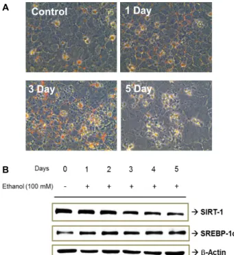
Treatment of AML-12 cells with 100 mM ethanol for 5 days caused significant lipid accumulation in the cells, as revealed by Oil red O staining (Fig. 1A), and also down-regulated SIRT1 and up-regulated SREBP-1c in a time-dependent man- ner (Fig. 1B). This down-regulation of SIRT1 and up-regu- lation of SREBP-1c by ethanol exposure was similar to the results of previous studies [14, 24].
Ethanol-induced lipid accumulation was significantly de- creased by co-treatment with KRG extract at 100, 500, and 1,000 μg/ml (Fig. 2A). SIRT1 expression was significantly decreased by ethanol-treatment (100 mM), but ethanol-in- duced down-hrregulation of SIRT1 was dose-dependently restored by co-treatment with KRG extract at concentrations of 10-1,000 μg/ml (Fig. 2B). Moreover, acetyl-SREBP-1c was significantly and dose-dependently decreased by KRG ex- tract, although this treatment did not change total SREBP-1c expression. KRG restored AMPK activity in AML-12 cells after it was reduced by ethanol treatment (Fig. 3). When KRG at a concentration of 500 μg/ml was co-administered with 100 mM ethanol for 24 hr, AMPK expression was incr
A
B
Fig. 1. Alcohol-induced lipid accumulation in AML-12 cells.
AML-12 cells were treated with 100 mM ethanol for 5 days. Oil red O staining was performed after ethanol treatment (A), and the effects of ethanol on SIRT-1 and SREBP-1c expression were assayed via Western blotting (B).
Fig. 2. The effect of KRG on acetyl-SREBP-1c in AML-12 cells.
Oil red O staining (A) and Western blots for SIRT-1,
SREBP-1c, and acetyl-SREBP-1c (B) after treatment with
100 mM ethanol for 3 days in combination with KRG
extract at 0, 10, 100, or 1,000 μg/ml.
Æ AMPK Æ p-AMPK
Ethanol (100 mM) - + + + +
- + + +
KRG (500 μg/ml) +
Time (hr) - 3 6 12 24
Fig. 3. The stimulatory effect of KRG on AMP-activated kinase (AMPK). Western blots for phospho-AMPK and AMPK after treatment with 100 mM ethanol and 500 μg/ml KRG extract for 24 hr.
Table 1. Effects of ginsenosides on SIRT-1 activity in hepato- cytes
Group Relative SIRT-1 activity (%)
Control Ethanol
Ethanol + ginsenoside Rb
2Ethanol + ginsenoside Rd Ethanol + ginsenoside Rg
1100±2.8
a96.2±0.2
a,b123.3±2.2
c116.0±1.9
d90.5±1.5
a,bSIRT-1 activity was calculated in AML-12 cells after treatment with ginsenosides Rb
2, Rd, or Rg
1(10 μg/ml) and 100 mM etha- nol for 3 days.
The mean value ± standard error (S.E.) was determined for each treatment group (n = 3). The threshold for significance was p
<0.05.
A
B
Fig. 4. The effects of individual ginsenosides on SIRT-1-medi- ated SREBP-1c deacetylation in AML-12 cells via Western blotting (A) and Oil red O staining (B). Western blots were used to assay SIRT-1, SREBP-1c, and acetyl- SREBP-1c expression in AML-12 cells after co-admin- istration of 100 mM ethanol with ginsenoside Rb
2, Rd, or Rg
1(1 and 10 μg/ml) for 3 days.
eased and p-AMPK expression was restored in a time-de- pendent manner.
AMPK is a molecular switch that can activate catabolic pathways and deactivate anabolic pathways [10]. The benefi- cial effects of ginseng and its active components on fat accu- mulation have been shown to be produced by activation of AMPK. For example, activation of the AMPK pathway medi- ated decreased hepatic fat accumulation produced by ginse- noside Rb
1in HFD-fed rats [18], and also reduced hepatic triglycerides and cholesterol levels in HepG2 cells produced by ginsenoside Rc [13]. In accordance with the activating effect of KRG extract on AMPK, KRG extract restored AMPK activation in ethanol-exposed hepatocytes. Moreover, AMPK, as a key regulator of cellular energy, controls the expression of genes involved in energy metabolism by acting in coordi- nation with SIRT1 [1]. In AML-12 cells treated with 100 mM ethanol, restored SIRT1 expression might be mediated by activation of AMPK by KRG extract.
Among the bioactive saponins, polyacetylenes, sesqui- terpenes, and polysaccharides found in ginseng, ginseno- sides are known as the primary mediators of its pharmaco- logical effects [6, 10]. Ginsenosides have been evaluated for their effects on molecular functions, but their effects on the SIRT1/SREBP-1c pathway have not been reported. Therefore, we investigated the effects of ginsenosides Rb
2, Rd, and Rg
1at concentrations of 1 and 10 μg/ml in AML-12 cells treated with 100 mM ethanol for 3 days. As shown in Fig. 4A, Rb
2and Rd restored SIRT1 expression and reduced expression of SREBP-1c and acetyl-SREBP-1c in ethanol-treated AML-12 cells. The ethanol-induced expression of acetyl-SREBP-1c was significantly decreased by co-treatment with ginseno- sides Rb
2and Rd, but not by treatment with Rg
1. Additional- ly, SIRT1 activity was significantly increased by ginsenosides Rb
2and Rd (Table 1). The recovery of SIRT1 activity is an important mechanism in recovery from alcoholic liver stea-
tosis, and deacetylation of SREBP-1c by SIRT1 reduces the stability of SREBP-1c. The hepatoprotective effects of ginse- noside Rb
2and Rd were confirmed by Oil red O staining in AML-12 cells, which showed that these compounds po- tently prevented lipid accumulation (Fig. 4B).
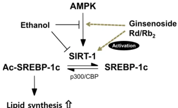
The pathology of alcoholic liver steatosis is initiated by
ethanol-induced abnormal regulation of lipid metabolic
processes involved in fatty acid oxidation and synthesis in
the liver [4, 21, 23]. Several regulatory molecules are in-
volved in this hepatotoxic effect of ethanol, including PPAR-
SIRT-1
Lipid synthesis Ac-SREBP-1c
Ginsenoside Rd/Rb
2SREBP-1c AMPK
p300/CBP
Ethanol
Activation