342
AOT is a rare, non-invasive, benign (hamartomatous) epi- thelial lesion of odontogenic origin and accounts for 2% to 7% of all odontogenic tumors. AOT usually affects young patients, mostly during their second to third decades of life.
Women are affected more frequently than men (male:female ratio=1:1.9), and the lesions tend to occur in the anterior max- illary region5. There are three types of AOT: a follicular type (73% of all cases), which has a central lesion associated with an impacted tooth; an extrafollicular type (24% of all cases), which has no relation with an impacted tooth; and a periph- eral type (3% of all cases)6. Of the follicular types of AOT, 36% occur in the mandible. In one study of AOT affecting the mandible, only 28% of cases involved the mandibular incisor area7. Here, we present a case of AOT involving the mandible with an unerupted lateral incisor.
II. Case Report
An 11-year-old female patient visited the Department of Oral and Maxillofacial Surgery of Dankook University Dental Hospital (Cheonan, Korea) with the chief complaint of a two-year history of swelling in the mandibular anterior region. There was mild swelling in the chin area, which was non-tender and had a firm consistency with ambiguous mar- gins.
Intraorally, the patient had distinct swelling in the anterior
I. Introduction
Adenomatoid odontogenic tumor (AOT) was first de- scribed by Steensland in 1905. In 1907, Dreibladt called this lesion a pseudo-adenoameloblastoma1. Adenoameloblastoma, ameloblastic adenomatoid tumor, adamantinoma, epithe- lioma adamantinum, and teratomatous odontoma have also been used to describe the lesions that we currently know as AOT. It was not until 1948 that Stafne considered the lesion as a distinct entity2, and Philipsen and Birn3 proposed the name “adenomatoid odontogenic tumor”. In 1971, the World Health Organization (WHO) adopted Philipsen and Birn’s term, and the WHO currently defines AOT as composed of odontogenic epithelium in various histoarchitectural patterns, embedded in mature connective tissue stroma, and character- ized by slow but progressive growth4.
CASE REPORT
Chul-Hwan Kim
Department of Oral and Maxillofacial Surgery, Dankook University Dental Hospital, College of Dentistry, Dankook University, 119 Dandae-ro, Dongnam- gu, Cheonan 31116, Korea
TEL: +82-41-550-0271 FAX: +82-41-551-8988 E-mail: [email protected]
ORCID: http://orcid.org/0000-0002-5199-2420
This is an open-access article distributed under the terms of the Creative Commons Attribution Non-Commercial License (http://creativecommons.org/licenses/by-nc/4.0/), which permits unrestricted non-commercial use, distribution, and reproduction in any medium, provided the original work is properly cited.
CC
Adenomatoid odontogenic tumor associated with an unerupted mandibular lateral incisor: a case report
Won-Gyo Seo, Chul-Hwan Kim, Hae-Seo Park, Jong-Won Jang, Woo-Yeol Chung Department of Oral and Maxillofacial Surgery, Dankook University Dental Hospital,
College of Dentistry, Dankook University, Cheonan, Korea
Abstract(J Korean Assoc Oral Maxillofac Surg 2015;41:342-345)
Adenomatoid odontogenic tumor (AOT) is a rare, benign odontogenic tumor that predominantly appears in the second decade of life in female patients.
Most AOTs occur in the anterior part of the maxilla and are usually associated with impacted anterior teeth. There are three types of AOT, follicular, extrafollicular, and peripheral, which are classified based on the location of the lesion and its association with the impacted tooth. We report a rare case of AOT associated with an impacted right mandibular lateral incisor in an 11-year-old female patient.
Key words: Adenomatoid odontogenic tumor, Follicular, Impacted lateral incisor, Mandible
[paper submitted 2015. 7. 24 / revised 1st 2015. 10. 14, 2nd 2015. 11. 5 / accepted 2015. 11. 7]
Copyright Ⓒ 2015 The Korean Association of Oral and Maxillofacial Surgeons. All rights reserved.
http://dx.doi.org/10.5125/jkaoms.2015.41.6.342 pISSN 2234-7550·eISSN 2234-5930
AOT associated with an unerupted mandibular lateral incisor
343 associated with expansion of the cortical bone. Significant radiological features in patients aged 30 years and older were root resorption and lesions that crossed the midline8.
As mentioned above, there are three pathologic types of AOT, intraosseous follicular, intraosseous extrafollicular, and peripheral, all of which have the same histological identity.
The follicular type is a central intraosseous lesion associated with an impacted tooth, whereas intraosseous extrafollicular AOT is similar to the follicular type but has no relation with an unerupted tooth. It usually develops around or is superim- posed onto adjacent teeth. The peripheral type usually looks like a gingival fibroma or epulis9.
Radiographically, AOT is usually unilocular, although a few multilocular cases have been reported. In addition to AOT, the differential diagnosis should include a dentigerous cyst. Radiographically, the pericoronal radiolucency of a den- tigerous cyst occurs most frequently in the jaws, and does not extend over the cement-enamel-junction of the tooth. How- ever, an AOT often envelops the crown as well as the root past the cemento-enamel-junction, which distinguishes AOTs from dentigerous cysts. AOTs have numerous, variable- shaped radiopaque foci, which also distinguish them from dentigerous cysts; 78% of AOTs have these foci. Tumor ex- pansion causes displacement of the adjacent teeth, and tooth displacement is more common than root resorption. Irregular root resorption is rare10.
This case describes an 11-year-old female patient with pain associated with one of her lateral mandibular incisors. Corti- cal expansion and dislocation of the adjacent tooth were also present, and there was no sign of root resorption. In cone beam computed tomography, the lesion extended slightly over the cemento-enamel-junction of the impacted tooth and region of the mandible from the left to the right mandibular
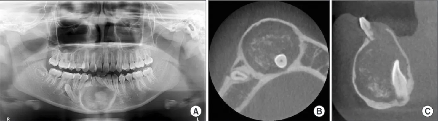
canines. The overlying mucosa was normal, and there was no paresthesia in the mandibular region. Because of the swell- ing, the central and lateral mandibular incisors were deviated to the left and the patient had retained her right deciduous lat- eral mandibular incisor and canine. A panoramic radiograph and computed tomography image were taken, which revealed a well-defined unilocular radiolucency of the anterior man- dibular region, which contained an impacted right lateral mandibular incisor with radiopaque foci and an impacted right mandibular canine.(Fig. 1) The differential diagnosis based on the clinical and radiographic findings included a dentigerous cyst, unicystic ameloblastoma, and AOT.
Intraorally, a mucoperiosteal flap was elevated to expose the lesion. The lesion was carefully separated from the sur- rounding bone, and the lesion and impacted lateral incisor were enucleated. Intraoperatively, the specimen exhibited a solid property and sand-like texture inside. The impacted right mandibular canine was also extracted, but the deciduous teeth were not removed in order to maintain space between the left central mandibular incisor and the right first premolar.
The specimen was then sent to the department of pathology for histopathological observation.
Written informed consent was obtained from the patient for publication of this case report and any accompanying images.
III. Discussion
In a review of 272 AOTs by Becker et al.8, the patients’
ages at the time of diagnosis ranged from 3 to 82 years (mean, 18.4 years). The maxilla-to-mandible ratio was 1.7:1. In 77%
of the lesions, small opacities were present, and most were
A B C
Fig. 1. A. Panoramic view shows lesion of mandible anterior region and impacted mandibular right canine. B. Axial view of computed tomography (CT) scan reveals lesion containing mandibular right lateral incisor and radiopaque foci. C. Sagittal view of CT scan reveals le- sion containing mandibular right lateral incisor and radiopaque foci.
Won-Gyo Seo et al: Adenomatoid odontogenic tumor associated with an unerupted mandibular lateral incisor: a case report. J Korean Assoc Oral Maxillofac Surg 2015
J Korean Assoc Oral Maxillofac Surg 2015;41:342-345
344
conducted Pan-CK staining.(Fig. 2. C) The stimulus that trig- gers proliferation of AOT is still unknown, although recent studies indicate a histological similarity of AOT to the dental lamina and reduced enamel epithelium. Its resemblance to dental laminae or enamel epithelium remnants implies that AOT is odontogenic in origin. It is usually a hamartomatous lesion rather than a neoplasm17.
In conclusion, all types of AOT show encapsulation and benign behavior. The treatment of choice is conservative surgical enucleation, with a very low recurrence rate9. In the present case, the lesion and impacted lateral incisor were sur- gically removed and the patient was without recurrence over the six months of follow-up.
Conflict of Interest
No potential conflict of interest relevant to this article was reported.
ORCID
Won-Gyo Seo, http://orcid.org/0000-0002-6097-1322 Chul-Hwan Kim, http://orcid.org/0000-0002-5199-2420 Hae-Seo Park, http://orcid.org/0000-0002-6207-0931 Jong-Won Jang, http://orcid.org/0000-0003-1515-032X Woo-Yeol Chung, http://orcid.org/0000-0001-8031-1711
References
1. Lingen MW. Lucas' pathology of tumors of the oral tissues. Arch Pathol Lab Med 2000;124:475.
2. Lee JK, Lee KB, Hwang BN. Adenomatoid odontogenic tumor: a case report. J Oral Maxillofac Surg 2000;58:1161-4.
3. Philipsen HP, Birn H. The adenomatoid odontogenic tumour.
Ameloblastic adenomatoid tumour or adeno-ameloblastoma. Acta Pathol Microbiol Scand 1969;75:375-98.
contained radiopaque foci. Judging from the association of the impacted tooth, we concluded that this was an intraosse- ous follicular AOT.
According to the WHO, the histological properties of AOT are as follows11: “A tumor of odontogenic epithelium with duct-like structures and varying degrees of inductive changes in the connective tissue. The tumor may be partly cystic and in some cases, the solid lesion may present as a mass in the wall of a large cyst. It is generally believed that the lesion is not a neoplasm.”
Histologically, AOT originates from the odontogenic epi- thelium and exhibits a solely solid growth pattern or a mixed proportion of solid and cribriform patterns. The histology all AOT types is identical and shows remarkable consistency.
The most visible pattern is various solid nodules of columnar or cuboidal epithelial cells that form a rosette-like configura- tion in the center at low magnification. Most AOTs contain structures with a tubular or duct-like appearance that consist of convoluted structures of epithelium with areas of ductal patterns mixed with globular masses of calcified material12. Eosinophilic, amorphous, and uncalcified material (“tumor droplets” or “tumor deposits”) can be found. Philipsen and Reichart13 and Bravo et al.14 showed that the amyloid-like eo- sinophilic deposits represent electron-dense plaques or some form of enamel matrix.
In this case, we found a solid nodule of cuboidal cells and duct-like structures at low magnification (Fig. 2. A) and a tu- mor droplet at high magnification.(Fig. 2. B)
The AOT phenotype is characterized by cytokeratin stain- ing immunohistochemically. According to Larsson et al.15, AOT shows positive staining for CK5, CK17, and CK19, and shows negative staining for CK4, CK10, CK13, and CK18.
Crivelini et al.16 found CK14 in AOT and suggested that its origin was from reduced dental epithelium. In this case, we
A B
100 m 100 m 100 m
C
Fig. 2. A. Duct-like structure (arrow) and solid nodule of epithelial cells (arrowheads) (H&E staining, ×100). B. Tumor droplet (arrowheads) (H&E staining, ×400). C. Pan-CK staining, ×200.
Won-Gyo Seo et al: Adenomatoid odontogenic tumor associated with an unerupted mandibular lateral incisor: a case report. J Korean Assoc Oral Maxillofac Surg 2015
AOT associated with an unerupted mandibular lateral incisor
345
11. Handschel JG, Depprich RA, Zimmermann AC, Braunstein S, Kübler NR. Adenomatoid odontogenic tumor of the mandible:
review of the literature and report of a rare case. Head Face Med 2005;1:3.
12. Vera Sempere FJ, Artes Martínez MJ, Vera Sirera B, Bonet Marco J.
Follicular adenomatoid odontogenic tumor: immunohistochemical study. Med Oral Patol Oral Cir Bucal 2006;11:E305-8.
13. Philipsen HP, Reichart PA. The adenomatoid odontogenic tumour:
ultrastructure of tumour cells and non-calcified amorphous masses.
J Oral Pathol Med 1996;25:491-6.
14. Bravo M, White D, Miles L, Cotton R. Adenomatoid odontogenic tumor mimicking a dentigerous cyst. Int J Pediatr Otorhinolaryngol 2005;69:1685-8.
15. Larsson A, Swartz K, Heikinheimo K. A case of multiple AOT-like jawbone lesions in a young patient: a new odontogenic entity? J Oral Pathol Med 2003;32:55-62.
16. Crivelini MM, de Araújo VC, de Sousa SO, de Araújo NS. Cyto- keratins in epithelia of odontogenic neoplasms. Oral Dis 2003;9:1- 17. Philipsen HP, Samman N, Ormiston IW, Wu PC, Reichart PA. Vari-6.
ants of the adenomatoid odontogenic tumor with a note on tumor origin. J Oral Pathol Med 1992;21:348-52.
4. Baskaran P, Misra S, Kumar MS, Mithra R. Adenomatoid odonto- genic tumor: a report of two cases with histopathology correlation.
J Clin Imaging Sci 2011;1:64.
5. Philipsen HP, Reichart PA, Siar CH, Ng KH, Lau SH, Zhang X, et al. An updated clinical and epidemiological profile of the adenoma- toid odontogenic tumour: a collaborative retrospective study. J Oral Pathol Med 2007;36:383-93.
6. Krishnamurthy K, Balaji RS, Devadiga S, Prasad RG. Adenoma- toid odontogenic tumor in the maxillary antrum: a rare case entity.
J Pharm Bioallied Sci 2014;6 Suppl 1:S196-9.
7. Leon JE, Mata GM, Fregnani ER, Carlos-Bregni R, de Almeida OP, Mosqueda-Taylor A, et al. Clinicopathological and immunohis- tochemical study of 39 cases of adenomatoid odontogenic tumour:
a multicentric study. Oral Oncol 2005;41:835-42.
8. Becker T, Buchner A, Kaffe I. Critical evaluation of the radiologi- cal and clinical features of adenomatoid odontogenic tumour. Den- tomaxillofac Radiol 2012;41:533-40.
9. Dayi E, Gürbüz G, Bilge OM, Ciftcioğlu MA. Adenomatoid odon- togenic tumour (adenoameloblastoma): case report and review of the literature. Aust Dent J 1997;42:315-8.
10. Neville BW. Update on current trends in oral and maxillofacial pa- thology. Head Neck Pathol 2007;1:75-80.