Introduction
Calanthe plants are distributed across cool and subtropi- cal regions of Africa (Madagascar), Asia (Korea, Japan, China, Taiwan, India, and Nepal), the Indian Ocean, Australia (New Guinea), and the southwest Pacific, and approximately 29 species are known to occur in Asia (Gale and Drinkell, 2007;
Govaerts et al., 2007). In Korea, Calanthe is narrowly dis- tributed in Ulleungdo (Gyeongsangbuk-do), Anmyeondo (Chungcheongnam-do), Sinan, Jindo, Wando (Jeollanam-do),
and the Jeju islands (Lee, 2011). The genus Calanthe includes approximately 171 to 200 species (Godo et al., 2010; Kurzweil, 2007; Suetsugu and Fukushima, 2014), but only five species (Calanthe discolor, C. bicolor, C. sieboldii, C. coreana, and C.
reflexa) are known to occur in Korea (Lee and Kwack, 1983; So et al., 2012). Calanthe, similar to other orchid genera, is sub- ject to infection by several plant viruses. So far, four viruses (Bean yellow mosaic virus, BYMV; Cucumber mosaic virus, CMV;
Cymbidium mosaic virus, CymMV; and Odontoglossum ringspot virus, ORSV) have been reported in Calanthe spp. in Korea.
Of these, CymMV (genus: Potexvirus, family: Flexiviridae) is the most prevalent and has caused significant economic losses (Wong et al., 1994; Zettler et al., 1990). CymMV has a
©The Korean Society of Plant Pathology
cc
This is an open access article distributed under the terms of the Creative Commons Attribution Non-Commercial License (http://creativecommons.org/
licenses/by-nc/4.0/), which permits unrestricted non-commercial use, distribution, and reproduction in any medium, provided the original work is properly cited.
Research Article Open Access
Survey of the Incidence of Viral Infections in Calanthe spp. and Characterization of a GW Isolate of Cymbidium mosaic virus in Korea
*Corresponding author Tel: +82-53-950-5763 Fax: +82-53-950-6758 E-mail: [email protected]
Chung Youl Park 1 , Da Some Baek 1 , Jonghee Oh 1 , Jong-Yoon Choi 1 , Dae Hyeon Bae 1 , Jeong-Seon Kim 3 , Gil-Hun Jang 4 , and Su-Heon Lee 1,2 *
1
School of Applied Biosciences, Kyungpook National University, Daegu 41566, Korea
2
Institute of Plant Medicine, Kyungpook National University, Daegu 41566, Korea
3
Agricultural Microbiology Division, National Academy of Agricultural Sciences, Rural Development Administration, Wanju 55365, Korea
4
Korean Institute of Calanthe, Gwangju 61999, Korea
Received February 1, 2016 Revised April 28, 2016 Accepted June 1, 2016
Cymbidium mosaic virus (CymMV) is a major virus infecting orchid plants and causing economic loss.
In this study, the incidence of viral infection in Calanthe spp. at the Korean Institute of Calanthe was investigated using reverse transcription polymerase chain reaction. The CymMV infection rate was 42%, and the two viruses Odontoglossum ringspot virus and Cucumber mosaic virus had frequencies of 8% and 2%, respectively. Additionally, we characterized an isolate of CymMV, CymMV-GW, using biological tests and examined the nucleotide sequence properties of its complete genome. CymMV- GW induced chlorotic ringspots and chlorotic spot symptoms in inoculated leaves of Chenopodium amaranticolor and Nicotiana benthamiana, respectively. In this study, we have for the first complete genome sequence of CymMV-GW in Korea. The CymMV-GW genome was 6,225 nucleotides in length, excluding the poly-(A) tail, and showed whole-genome nucleotide and amino acid sequence identities of 97.7% and 100%, respectively, with the NJ-1 isolate of CymMV. Here, we report the complete genome sequence of the CymMV-GW isolate and viral infection rates for Calanthe spp. in Korea.
Keywords: Bioassay, Calanthe, CymMV
Research in Plant Disease pISSN 1598-2262, eISSN 2233-9191 www.online-rpd.org
65
monopartite, positive single-strand RNA genome that con- sists of five open reading frames (ORFs) containing genes en- coding putative RNA-dependent RNA polymerase (RdRp), tri- ple gene block (TGB) proteins, and coat protein (CP) (Wong et al., 1997). Some plant viruses containing the TGB movement protein are used to confirm cell-to-cell movement via protein interactions (Chou et al., 2013; Lim et al., 2008). CymMV has been used to study floral gene functions and lifespan using virus-induced gene silencing as a tool (Hsieh et al., 2013). For virus detection, various diagnostic methods have been devel- oped, such as enzyme-linked immunosorbent assays (Vejarat- pimol et al., 1998), polymerase chain reaction (PCR) (Lim et al., 1993), reverse transcription (RT)-PCR (Seoh et al., 1998), real- time PCR (Eun et al., 2000), and RT loop-mediated isothermal amplification assays (Lee et al., 2011).
Nevertheless, the virus has been studied with respect to its genetic diversity, homogeneity, relationships, distribution, phenotype, morphological characteristics, and taxonomy in Korea (Cho et al., 2007; Chung et al., 2014; Hyun et al., 1999; Kim et al., 2013; Lee et al., 2008; Srikanth et al., 2012).
Although Jang (1998) studied viral disease in many kinds of orchids, including Calanthe, these studies were limited to an electron microscope analysis, biological tests, and diagnoses.
Thus, we report the complete genome sequence and charac-
terization of CymMV isolated from a Calanthe plant in Korea and the incidence of viral infections caused by different vi- ruses at the Korean Institute of Calanthe.
Materials and Methods
Sample collection. From 2012 to 2013, Calanthe spp.
samples were provided by the Korean Institute of Calanthe (Gwangju, Korea). All 35 samples showed typical symptoms of viral infection, such as mild mosaic, mosaic, and malformation (Fig. 1), and were named GW isolates. The leaves with these symptoms were cut using a razor and the samples stored in a deep freezer (–80
oC).
Virus diagnosis. In order to identify the infecting virus, to- tal RNA was extracted from 35 samples of Calanthe spp. using Tri-Reagent (MRC Reagent, Cincinnati, OH, USA) following the manufacturer’s instructions. The cDNA was synthesized using Oligo (dT6) primers with SuperScript™ II Reverse Transcriptase (Invitrogen, Carlsbad, CA, USA) according to the manufactur- er’s protocol. All samples were tested for eight viruses (BYMV, CMV, CymMV, ORSV, Clover yellow mosaic virus [ClYMV], Clover yellow vein virus [ClYVV], Turnip mosaic virus [TuMV], and Or
chid fleck virus [OFV]) using specific primer sets by PCR with
A B C
D E F
Fig. 1. Naturally infected Calanthe plants showing typical symptoms of viral infection. (A) Chlorosis, (B, C) mosaic, (D) yellowing, (E) necro-
sis, and (F) malformation.
Elongase
®Enzyme Mix (Invitrogen) (Table 1). The amplifica- tion conditions were as follows: an initial denaturation at 94
oC at 2 minutes, 26 cycles of 94
oC for 30 seconds, 55
oC for 30 sec- onds, and 68
oC for 1 minute, and a final extension at 68
oC for 5 minutes. Amplified PCR products were confirmed by 1.0%
agarose gel electrophoresis with ethidium bromide.
Biological test. To obtain a biological and molecular char- acterization of CymMV, a singly infected GW isolate was used (No. 1). The host range for CymMV-GW was determined by mechanical transmission with indicator plants for 34 species representing 7 families (Aizoaceae, Amaranthaceae, Brassica
ceae, Chenopodiaceae, Cucurbitaceae, Leguminosae, and So
lanaceae). Sap extracted from symptomatic leaf samples was used as the inoculum with 0.1 M potassium phosphate (pH 7.0), and tests were repeated three times per indicator plant.
All indicator plants were maintained at 25
oC–30
oC in a green- house. To confirm local or systemic infections, at 8 weeks post-inoculation, inoculated and non-inoculated leaf samples were harvested and examined by RT-PCR as described above.
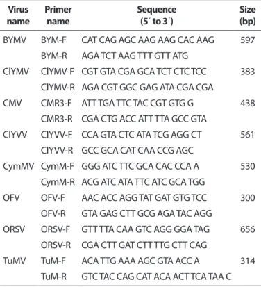
Complete genome sequencing and genome analy- sis. To determine the complete genome sequence, six primer sets were designed using the nucleotide sequences of CymMV isolates (GenBank accession Nos. HQ681906, EF125180, EF125179, AF016914, and JQ860108) from Nation- al Center for Biotechnology Information (NCBI) (Table 2). PCR products with expected sizes were obtained from CymMV- GW, and these products were cloned into a TA cloning vec- tor (RBC Bioscience, Taipei, Taiwan) and sequenced (Solgent, Daejeon, Korea). To confirm the 5´- and 3´-terminal regions, extracted total RNA was subjected to rapid amplification of Table 1. Primers used to diagnosis Calanthe plants
Virus
name Primer
name Sequence
(5´ to 3´) Size
(bp) BYMV BYM-F CAT CAG AGC AAG AAG CAC AAG 597
BYM-R AGA TCT AAG TTT GTT ATG
ClYMV ClYMV-F CGT GTA CGA GCA TCT CTC TCC 383 ClYMV-R AGA CGT GGC GAG ATA CGA CGA
CMV CMR3-F ATT TGA TTC TAC CGT GTG G 438 CMR3-R CGA CTG ACC ATT TTA GCC GTA
ClYVV ClYVV-F CCA GTA CTC ATA TCG AGG CT 561 ClYVV-R GCC GCA CAT CAA CCG AGC
CymMV CymM-F GGG ATC TTC GCA CAC CCA A 530 CymM-R ACG ATC ATA TTC ATC GCA TGG
OFV OFV-F AAC ACC AGG TAT GAT GTG TCC 300 OFV-R GTA GAG CTT GCG AGA TAC AGG
ORSV ORSV-F GTT TTA CAA GTC AGG GGA TAG 656 ORSV-R CGA CTT GAT CTT TTG CTT CAG
TuMV TuM-F ACA TTG AAA AGC GTA ACC A 314 TuM-R GTC TAC CAG CAT ACA ACT TCA TAA C BYMV, Bean yellow mosaic virus; ClYMV, Clover yellow mosaic virus;
CMV, Cucumber mosaic virus; ClYVV, Clover yellow vein virus; Cym- MV, Cymbidium mosaic virus; OFV, Orchid fleck virus; ORSV, Odonto
glossum ringspot virus; TuMV, Turnip mosaic virus.
Table 2. Primer sets used to amplify the complete genome of Cymbidium mosaic virus (CymMV) isolated from Calanthe spp.
Primer sets Primer name Sequence (5´ to 3´) Position Size (bp)
1 CymM F1 GGC ACT CGT AAA CTG CCC TT 174–193 924
CymM R1 ACC AAG TGG GTC ACC TCC AC 1078–1097
2 CymM F2 GTG AGG CAA TTG ATC CCG AC 1033–1052 1,327
CymM R2 AGG CAC TCT GCA TAG GAT CC 2340–2359
3 CymM F3 GGG TAC ATC GAG ACC ATG GT 2119–2138 1,614
CymM R3 AAG GAA CCA GAT AGG CAC AT 3714–3733
4 CymM F4 GAC CAG TCA CAA GAT GCT GC 3598–3617 1,268
CymM R4 AAA GTG CCG GGA ACC TGC TG 4845–4864
5 CymM F5 TTG CTT GAC CCT AGC AAC CG 4506–4535 1,143
CymM R5 GGC CAA GGT TGT TAA CCC AC 5628–5647
6 CymM F6 ACT CCA GCT GCC ACT TAC TC 5497–5506 589
CymM R6 CCT CGG CAA TGT TGG TGA TG 6066–6085
cDNA ends. First-strand cDNA was synthesized using prim- ers (CymMV-R0, 5´- GAT TGA CTG TCA CGC CGA GA -3´) and (Oligo-dT15-Adaptor primer, 5´- GAC CAC GCG TAT CGA TGT CGA T
16-3´). The cDNA was purified using the Exgene
TMPlant SV Kit (GeneALL, Seoul, Korea). At the 5´ ends, a poly-(G) tail was added using purified cDNA with terminal deoxynucleoti- dyl transferase (Takara, Shiga, Japan). Each cDNA was ampli- fied by PCR with primers (Oligo-dCD, 5´- GAC CAC GCG TAT CGA TGT CGA C17D -3´ and CymMV-R0) and (CymMV-F0, 5´- AGC CTG CTG AAT GGC AGC GC -3´ and Adaptor primer). PCR
products were purified, cloned, and sequenced.
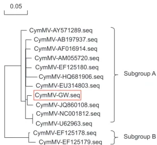
Phylogenetic analyses were conducted to examine the re- lationships among isolates with in the species CymMV, and to identify the most closely related isolates using DNAMAN software ver. 5.2.10 (Lynnon Biosoft, Quebec, QC, Canada).
Sequence data for 12 CymMV isolates were obtained from NCBI. Multiple alignments were generated using complete genome sequences, and a phylogenetic tree was constructed using the maximum likelihood technique. Bootstrapping was performed using 1,000 replicates.
Results
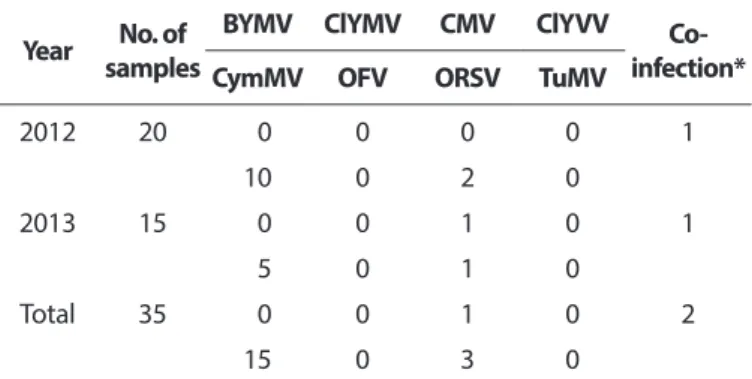
To identify the infecting virus, all 35 collected samples were tested using RT-PCR. Among them, 15 were positive for CymMV, 3 were infected with ORSV, and 1 was infected with CMV. Two samples, GW isolate numbers 19 and 21, were co- infected with CymMV and ORSV. Five viruses (BYMV, ClYMV, ClYVV, TuMV, and OFV) were not detected in this test (Table 3).
To confirm the host range of CymMV GW isolate, the bioas- say carried out using various test plants. After three weeks, chlorotic ringspots were detected on inoculated leaves of Chenopodium amaranticolor. Nicotiana benthamiana showed chlorotic spots on inoculated leaves at two weeks post- inoculation. N. glutinosa produced mosaic symptoms on non- Table 3. Viral infection rate in Calanthe plants collected at the Ko-
rean Institute of Calanthe Year No. of
samples
BYMV ClYMV CMV ClYVV Co-
infection*
CymMV OFV ORSV TuMV
2012 20 0 0 0 0 1
10 0 2 0
2013 15 0 0 1 0 1
5 0 1 0
Total 35 0 0 1 0 2
15 0 3 0
BYMV, Bean yellow mosaic virus; ClYMV, Clover yellow mosaic virus;
CMV, Cucumber mosaic virus; ClYVV, Clover yellow vein virus; Cym- MV, Cymbidium mosaic virus; OFV, Orchid fleck virus; ORSV, Odonto
glossum ringspot virus; TuMV, Turnip mosaic virus.
*Infection of both viruses=CymMV+ORSV.
A B
C D
Fig. 2. Mechanical inoculation of Cymbi
dium mosaic virus from Calanthe plants to several indicator plants. (A) Chenopodium amaranticolor produced chlorotic ring spot symptoms on inoculated leaves. (B) Nico
tiana benthamiana showed chlorotic spots
on inoculated leaves. (C) Systemic mosaic
symptoms in N. glutinosa. (D) Left: healthy
plant, right: malformation symptoms on
upper leaves in N. tabacum var. Turkish.
inoculated leaves. N. tabacum var. Turkish and N. tabacum cv.
Samsun showed malformation symptoms on upper leaves (Fig. 2). The remainder of the test plants is without symptoms.
Two species (C. amaranticolor and N. benthamiana) yielded RT-PCR products consistent with the expected size of CymMV, while the virus was not detected for 32 species whose indica- tor plants showed symptoms of viral infection.
The genome of the CymMV-GW isolate consisted of 6,225 nucleotides (nt), excluding the poly-(A) tail. The virus con- tained five ORFs, ORF1 (RdRp, nucleotide positions 73–4,326), ORF2 (TGB1, 4,332–5,033), ORF3 (TGB2, 5,008–5,346), ORF4 (TGB3, 5,201–5,476), and ORF5 (CP, 5,479–6,150). The 5´ and 3´
untranslated regions were 72 and 75 nt long, respectively (Fig.
3). CymMV-GW was most closely related to the CymMV NJ-1 isolate, which was isolated from Phalaenopsis spp. in China (GenBank accession No. JQ860108). According to a previous study by Vaughan et al. (2008), the GW isolate clustered into a group referred to as subgroup A (Fig. 4). The complete nucle- otide sequence of CymMV-GW showed the highest identity with the NJ-1 isolate (nt identity, 97.7%; amino acid identity, 100%). Other CymMV isolates shared more than 95% identity at the nucleotide and amino acid levels, except for two USA isolates. The CymMV 18-1 and 18-30 isolates (GenBank acces- sion Nos. EF125178 and EF125179), which were isolated from Dendrobium spp. in the United States, showed nucleotide sequence identities of 88.7% and 87.5%, respectively, with CymMV-GW (data not shown).
Discussion
Calanthe spp. are expected to be new economically impor- tant horticultural crops. A great deal of research has been carried out to develop export-quality crops, but viral disease
is a major cause of low quality. Here, we surveyed cultivated Calanthe plants in Korea and obtained 35 samples to test for the presence of the viruses BYMV, ClYVV, ClYMV, CMV, CymMV, OFV, ORSV, and TuMV. CymMV had the highest infec- tion rate (42%) among the eight viruses (BYMV: 0%, ClYVV:
0%, ClYMV: 0%, CMV: 2%, OFV: 0%, ORSV: 8%, and TuMV: 0%).
These results imply that CymMV is a major virus infecting Calanthe plants, similar to other orchid plants (Hu et al., 1993).
In the host range test, among the 34 species examined, symptoms of viral infection were observed in 6 species (C.
amaranticolor, N. benthamiana, N. glutinosa, N. rustica, N.
tabacum cv. Samsun, and N. tabacum var. Turkish). RT-PCR results for C. amaranticolor and N. benthamiana were positive for CymMV, but four indicator plants tested negative for the virus. Additionally, harvested indicator samples were tested using other virus-specific primers (Table 1) and the results were negative. The present results suggest that identification
1 kb 2 kb 3 kb 4 kb 5 kb 6 kb
73
ORF1
RdRp
4,326 5,008 5,346 5,479 6,150
ORF2 ORF3
ORF4 ORF5
4,332 5,033 5,201 5,476 TGB1 TGB2 TGB3 Coat protein