․Correspondence to: Joon-seok Byun 136, Sincheondong-ro, Suseong -gu, Daegu, Korea
Dept. of Internal Medicine, College of Oriental Medicine, Daegu Haany University TEL: 053-770-2192 FAX: 053-770-2055 E-mail: [email protected]
․본 논문은 2014년 대구한의대학교 대학원 학의학 석사학위 논문입니다.
Gastric Protective Effects of Banhasasim-tang on Indomethacin-treated Rats
Su-wan Park, Joon-seok Byun
Dept. of Internal Medicine, College of Oriental Medicine, Daegu Haany University
Gastric Protective Effects of Banhasasim-tang on Indomethacin-treated Rats
Su-wan Park, Joon-seok Byun
Dept. of Internal Medicine, College of Oriental Medicine, Daegu Haany University
ABSTRACT
Purpose :
Banhasasim-tang (BHSST) has been applied for treating the symptom of gastric stuffiness, which is similar to dyspepsia.
The object of this study was to observe the healing effect of BHSST on the indomethacin (IND)-induced gastric ulcer in rats.
Methods :
Three different dosages of BHSST (400, 200 and 100 mg/kg) were orally administered 30 min before IND treatment;
6 hrs after IND treatment, the changes on the gross lesion scores, fundic histopathology, myeloperoxidase (MPO) activity, lipid peroxidation and antioxidant defense system (glutathione contents, catalase (CAT) and superoxide dismutase (SOD) activities) were observed, and compared with the activity of the synthetic anti-ulcer drug, a representative proton pump inhibitor omeprazole (OME) 10 mg/kg.
Results :
All three different dosages of BHSST treatment in the IND-induced gastric ulcer rats, significant and dose dependent decreased gastric damages - hemorrhagic gross lesions, gastric mucosa MPO levels and histopathological gastric ulcerative lesions - were detected as compared with the IND treated control rats. BHSST also strengthened the antioxidant defense systems – decreased the level of lipid peroxidation and CAT activity but increased the level of GSH and SOD activity, and BHSST 200 mg/kg showed similar anti-ulcerative effect as compared with OME 10 mg/kg.
Conclusions :
The results obtained in this study suggest that BHSST has favorable effects against IND-induced gastric damages, through significant and dose-dependent decreasing gastric damages and the strengthening of the body’s antioxidant defense systems with direct anti-inflammatory effects.
Key words :
Banhasasim-tang (BHSST), gastric ulcer, indomethachin (IND), omeprazole (OME)
Ⅰ. Introduction
A GASTRIC ULCER is a multi-etiologic chronic
disease
1. Because of favorable prevention effects on occurrences of malignant tumors, stroke, eclampsia, Alzheimer’s dementia and cardiovascular diseases related to hyperlipidemia, the demands of nonsteroidal anti-inflammatory drugs (NSAIDs) were remarkably increased in recent years
2-4. However, about 25%
of urgent gastric ulcer are known as related to
NSAIDs administration
5, and various factors like
stress, empty and Helicobacter pylori infections
exacerbated NSAIDs related gastric ulcers
6. Various synthetic anti-ulcer drugs are presently available and some of these like misoprostol are specifically used to cure the NSAID induced gastric ulcer. However, each of these drugs confers simpler to severe side effects
7, such as antifungal agent metabolism inhibitory effects of proton pump inhibitor (PPI), headaches and antiandrogenic effects of H2 receptor blockers, dizziness of sucralfate, stillbirth and melena of misoprostol in pregnant, warranting a search for non-toxic and inexpensive antiulcer medication
8. Disorders or decreases of gastric mucosa antioxidant defense systems have been involved in the pathogenesis and progression of various gastric ulcers, and also associated with NSAIDs
9.
Banhasasim-tang (BHSST; Hanzung Pharm.
Co., Daejeon, Korea) is one of the herbal formulas described in “Treatise on Cold Damage and Miscellaneous Diseases (Shan-han-za-bing-lin)”
10, the Chinese authoritative monographs. This formula is composed of eight herbs like Pinelliae Rhizoma, Scutellariae Radix, Ginseng Radix Alba, Glycyrrhizae Radix, Zingiberis Rhizoma Siccus, Coptidis Rhizoma, Zingiberis Rhizoma and Zizyphi Fructus
10,11. In traditional Korean medicine, this formula has been applied for treating the symptom “gastric stuffiness”
10, which is similar to dyspepsia.
Recently, several studies have elucidated the gastric function and related mechanisms of BHSST
10-14. Moreover, BHSST can be obtained as an over-the -counter herbal formula in Korea or prescribed for dyspeptic symptoms by the Traditional Korean Medicine doctors, and the safety and effectiveness of BHSST granules in patient suffering from functional dyspepsia are also reported through randomized, double-blind, placebo-controlled clinical trial
10. And up to date, several animal experimental studies were
preceded
15,16, they are insufficient to explain machnism of BHSST. Whereas we have experimented with various aspects of histopathological, immunological, endocrine. In this study we compared the anti-ulcer effects of synthetic anti-ulcer drug and BHSST granule directly. As a result, we had found a appropriate dose of the BHSST, which is having similar effect with the synthetic anti-ulcer drug.
To verify the effect on the IND-induced gastric ulcer in rats, the researcher carried out oral administration with capacity of BHSST 400, 200, 100 mg/kg. Then, the experiment result were utilized to carry out comparative analysis and evaluation of the group that got omeprazole (OME) 10 mg/kg administered.
Ⅱ. Materials & Methods
1. Test materials: BHSST and OME
Light brown granules of BHSST, produced according to Korean Good Manufacturing Practice (GMP) and permitted and regulated by the Korean Food & Drug Administration (KFDA; Seoul, Korea) were used in this experiment, and OME was used as reference drug as listed follows. Individual compositions of 8 kinds of herbs in BHSST were listed in Table 1. OME and BHSST were stored in a refrigerator at 4 ℃ until use.
1) Test material
(1) Name : Banhasasim-tang [Herbal formulas for treating dyspepsia; Table 1]
(2) Source : Hanzung Pharm. Co., Daejeon, Korea [http://www.hzpharm.co.kr]
(3) Confirmed test article dosages : 400, 200 and 100 mg/kg (Single oral treatment)
(4) Solubility in vehicle : well soluble at least, 40 mg/ml concentration in sterilized distilled water
2) Reference drug
(1) Name : Omeprazole [PPI reference drug]
(2) Systematic (IUPAC) name : (RS)-5-methoxy -2-((4-methoxy-3,5-dimethylpyridin-2-yl) methylsulfinyl) -1H-benzo[d]imidazole
(3) Source : Sigma-Aldrich, St. Louise, MO, USA (4) Chemical formula : C
17H
19N
3O
3S
(5) Molecular weights : 345.4 g/mol
(6) Confirmed test article dosages : 10 mg/kg (Single, oral administration)
(7) Appearance : Off-white powders
(8) Solubility in vehicle : well soluble up to 1 mg/ml in sterilized distilled water, at least
Herbs Scientific names Amounts (g)
Pinelliae Rhizoma Pinellia ternata (Thunb.) Breitenb. 1.34
Scutellariae Radix Scutellaria baicalensis Georgi 1.80
Ginseng Radix Alba Panax ginseng C.A.Meyer. 0.96
Glycyrrhizae Radix Glycyrrhiza uralensis Fisch 1.08
Zingiberis Rhizoma Siccus Zingiber officinale Roscoe 0.59
Coptidis Rhizoma Coptis japonica (Thunb.) Makino 0.24
Zingiberis Rhizoma Zingiber officinale Roscoe 0.26
Zizyphi Fructus Zizyphus jujuba Miller var. inermis Rehder 1.67
Total 8 types 7.94
Table 1. Composition of BHSST Used in This Study.
2. Animals and husbandry
A total of 48 virgin, Sprague-Dawley, specific pathogen-free female rats (6 wk old upon receipt;
Harlan, Udine, Italy; Body weight ranged in 170~190 g upon receipt) were used after acclimatization for 67 days. Animals were allocated four per polycarbonate cage in a temperature (20-25 ℃) and humidity (50-55%) controlled room. Light : dark cycle was 12 hrs : 12 hrs and feed (Samyang, Seoul, Korea) and water were supplied free to access. 40 rats were used as IND-induced gastric ulcer rats and 8 rats were used as sterilized distilled water treated intact control, instead of IND, in this study. All animals were overnight fasted (about 24 hrs) before IND or test material administration, and they were treated according to the national regulations of the usage and welfare of laboratory animals, and approved by the Institutional Animal Care and Use Committee in Daegu Haany University
[Approval No DHU2013-018]. Six groups, total 48 rats were selected base on the body weights (mean 271.42±20.68 g, ranged in 235-313 g at 67 days after acclimatization,) and used in this experiment as follows.
1) Experimental groups (Six groups, 8 rats per group were used)
(1) Intact control : Vehicle (distilled water 5 mg/kg) administered rats
(2) IND control : Vehicle and IND treated control rats
(3) OME : OME 10 mg/kg and IND treated rats (4) BHSST 400 : BHSST 400 mg/kg and IND treated rats
(5) BHSST 200 : BHSST 200 mg/kg and IND treated rats
(6) BHSST 100 : BHSST 100 mg/kg and IND
treated rats
3. BHSST and OME treatment
After subdivided into aforementioned six groups as eight rats/group, BHSST were once orally administered at 30 min before IND treatment in a volume of 5 ml/kg, dissolved in sterilized distilled water at dose levels of 400, 200 or 100 mg/kg by gastric gavages using a stainless Zonde attached to 3 ml-syringe, respectively. OME was also single orally administered at a dose level of 10 mg/kg, dissolved in sterilized distilled water in a volume of 10 mg/kg. In intact and IND control rats, only sterilized distilled water was administered, once orally, instead of BHSST or OME. The dosage of OME was selected based on the previous efficacy test
17, and the lowest dosage of BHSST was selected as 100 mg/kg based on the previous brief efficacy test on the cisplatin-induced gastric dysmotility
11. In addition, 400 and 200 mg/kg were also selected as higher and middle dosages of BHSST in this experiment using common ratio 2.
4. IND-induced gastric ulcer
30 min after administration of vehicle, three different dosages of BHSST or OME on 24 hrs fasted rats, IND was single orally administered in a volume of 5 ml/kg dissolved in sterilized distilled water at a dose level of 25 mg/kg according to previous report
18. In intact control rats, only sterilized distilled water was once treated by gastric gavages instead of IND.
1) Inducer agent
(1) Name : Indomethacin [NSAIDs, inducer agent]
(2) Systematic (IUPAC) name : 2-{1-[(4-chlorophenyl) carbonyl]-5-methoxy-2-methyl-1H-indol-3-yl}acetic acid
(3) Source : Sigma-Aldrich, St. Louise, MO, USA (4) Chemical formula : C
19H
16ClNO
4(5) Molecular weights : 357.787 g/mol
(6) Confirmed test article dosages : 25 mg/kg
(Single, oral administration)
(7) Appearance : Beige colored powders (8) Solubility in vehicle : well soluble up to 5 mg/ml in sterilized distilled water, at least
5. Quantification of gross lesions
The animals were sacrificed at 6 hrs after IND or vehicle, sterilized distilled water treatment by cervical dislocation. Excised stomach was opened out along with greater curvature and fixed in 10%
neutral buffered formalin for 24 hrs and acquired digital images. Ulcer areas on the stomachs’ surface were examined macroscopically and measured by computer based automated image analysis process (iSolution FL ver 9.1, IMT i-solution Inc., Quebec, Canada) according to the method of Süleyman et al.
1with some modifications. Any macroscopically visible lesions were measured to calculate the gastric damage score. For this purpose, the total areas of the ulcerous stomach regions were calculated as mm
2.
6. Myeloperoxidase (MPO) activity
The tissue samples (about 0.2 g) were homogenized in 10 volumes of ice-cold potassium phosphate buffer (50 mM K
2HPO
4, pH6.0; Sigma-Aldrich, St.
Louise, MO, USA) containing hexadecyltrimethyl- ammonium bromide (HETAB; 0.5% w/v; Sigma- Aldrich, St. Louise, MO, USA)
19. The homogenate was centrifuged at 12000 rpm for 10 min at 4 ℃, and the supernatant was discarded. The pellet was then re-homogenized with an equivalent volume of 50 mM K
2HPO
4containing 0.5% (w/v) HETAB and 10 mM EDTA (Sigma-Aldrich, St.Louise, MO, USA).
MPO activity was assessed by measuring the H
2O
2-dependent oxidation of o-dianizidine 2 HCl.
One unit (U) of enzyme activity was defined as the
amount of the MPO present/g tissue weight that
caused a change in absorbance of 1.0 ml at 460 nm and 37 ℃ using UV-vis spectrophotometer (UV-3600, Shimadzu Scientific Instruments, Columbia, MD, USA)
20.
7. Determination of lipid peroxidation (MDA) formation
The concentrations of gastric mucosal lipid peroxidation were determined by estimating MDA using the thiobarbituric acid test
21. The corpus mucosa was scraped, weighed, and homogenized in 10 ml of 100 g/ℓ KCl (Sigma-Aldrich, St. Louise, MO, USA). The homogenate (0.5 ml) was added to a solution containing 0.2 ml of 80 g/ℓ sodium lauryl sulfate (Sigma-Aldrich, St. Louise, MO, USA), 1.5 ml of 200 g/ℓ acetic acid (Merck, West Point, PA,, USA), 1.5 ml of 8 g/ℓ 2-thiobarbiturate (Sigma -Aldrich, St. Louise, MO, USA), and 0.3 ml of distilled water. This mixture was heated at 98 ℃ for 1 hr and after it had cooled, 5 ml of n-butanol:pyridine (15:l) (Sigma-Aldrich, St. Louise, MO, USA) was added.
The mixture was vortexed for 1 min and centrifuged for 30 min at 4000 rpm. The supernatant’s absorbance was measured at 532 nm. The standard curve was obtained by using 1,1,3,3-tetramethoxypropane (Sigma -Aldrich, St. Louise, MO, USA). The results were expressed as nM MDA per gram of wet tissue (nM/g tissue).
8. Total glutathione (GSH) determination
The gastric mucosa’s GSH content was measured according to the method of Sedlak and Lindsay
22. The stomach’s mucosal surface was collected by scraping, weighed, and homogenized in 2 ml of 50 mM Tris-HCl buffer containing 20 mM EDTA and 0.2 mM sucrose (Merck, West Point, PA, USA), at pH 7.5. The homogenate was immediately precipitated
with 0.1 ml of 25% trichloroacetic acid (Merck, West Point, PA, USA), and the precipitate was removed after centrifugation at 4200 rpm for 40 min at 4 ℃.
The supernatant was used to determine GSH using 5,5-dithiobis (2-nitrobenzoic acid; Sigma-Aldrich, St. Louise, MO, USA). Absorbance was measured at 412 nm using a spectrophotometer. The results of the test for GSH content in the gastric mucosa were expressed as nM/mg tissue.
9. Tissue catalase (CAT) activity
CAT was determined according to the method of Evans and Diplock
23. Homogenate of rat gastric mucosa was diluted with buffer, as described before, in order to obtain an adequate dilution of the enzyme. Then, 2 ml of the enzyme dilution were added to the cuvette and mixed with 1 ml of 30 mM H
2O
2, measuring the absorbance at 240 nm for 100 sec.
Initial absorbance of the reaction mixture must be around 0.5. The enzyme activity is expressed as the first order constant that describes the decomposition of H
2O
2at room temperature, mM/min/mg tissue.
10. Tissue superoxide dismutase (SOD) activity
Gastric SOD activity was determined by the
modified version from the method of Minami and
Yoshikawa
24. Briefly, 15 μl of gastric homogenate
were mixed with 450 μl of cold deionized water,
125 μl of chloroform, and 250 μl of ethanol. The
mixture was then, centrifuged at 8000 rpm for 2
minutes at 4 ℃. 500 μl of the extracts were added
to the reaction mixture containing 500 μl of 72.4
mM triscacodylate buffer with 3.5 mM diethylene
pentaacetic acid (pH 8.2; Sigma-Aldrich, St. Louise,
MO, USA), 100 μl of 16% Triton X-100, and 250 μl
of 0.9 mM nitroblue tetrazolium (Sigma-Aldrich,
St. Louise, MO, USA). The reaction mixture was
incubated for 5 min at 37 ℃ before adding 10μl of 9 mM of pyrogallol dissolved in 10 mM HCl.
Then, it was incubated for exactly 5 min at 37 ℃.
The reaction was stopped with the addition of 300 μl of 2 M formic buffer (pH 3.5) containing 16%
Triton X-100 (Sigma-Aldrich, St. Louise, MO, USA).
The absorbance was measured at 540 nm in a spectrophotometer. One unit of SOD enzymatic activity is equal to the amount of enzyme that diminishes the initial absorbance of nitroblue tetrazolium by 50% (mM/min/mg tissue).
11. Histopathology
Approximated regions of individual stomach (between cardiac and pylorus, the fundus) were sampled. All trimmed fundus were fixed in 10%
neutral buffered formalin for 24 hrs, at least. After paraffin embedding, 3-4 μm sections were prepared.
Representative sections were stained with hematoxylin and eosin (H&E) for light microscopically examination.
To more detail changes, the total thicknesses of fundic mucosa, from luminal mucosal surface to muscularis mucosa on the periulcerative regions of the crossly trimmed histological specimens, were measured using computer based automated image analysis process as described by Ku et al.
25. In addition, lesion invasive percentages in fundus (%) were also calculated as follow Equation [1] according to the method of Ku et al.
25, and semiquantative scoring as divided into four degrees; 0 = normal intact mucosa, 1 = slight surface erosive damages, 2 = moderate muocsa damages and 3 = severe total mucosa damages, based on general and histomorphometrical analysis, aforementioned in this experiment.
EQUATION [1]. Invasive Percentages of Lesions (%)=(Length of lesions on the crossly trimmed
fundic walls/total thickness of crossly trimmed fundic walls)×100
12. Statistical Analyses
Variance homogeneity was examined using the Levene test
26. If the Levene test indicated no significant deviations from variance homogeneity, the obtain data were analyzed by one way ANOVA test followed by least-significant differences (LSD) multi-comparison test to determine which pairs of group comparison were significantly different. In case of significant deviations from variance homogeneity were observed at Levene test, a non-parametric comparison test, Kruskal-Wallis H test was conducted.
When a significant difference is observed in the Kruskal-Wallis H test, the Mann-Whitney U (MW) test was conducted to determine the specific pairs of group comparison, which are significantly different.
Statistical analyses were conducted using SPSS for Windows (Release 14.0K, IBM SPSS Inc., Armonk, NY, USA)
27. In addition, the percent changes between intact and IND control were calculated to observe the severities of gastric mucosa damages including ulcerative lesions induced in this study, and the percent changes as compared with IND control and BHSST or OME treated rats were also calculated to help the understanding of the efficacy of test substances as follow Equation [2] and [3], respectively.
EQUATION [2]. Percentage Changes as Compared with Intact Control (%)=[((Data of IND control -Data of intact control rats)/Data of intact control rats)×100]
EQUATION [3]. Percentage Changes as Compared
with IND Control (%)=[((Data of test substance
treated rats-Data of IND control rats)/Data of
IND control rats)×100]
Ⅲ. Results
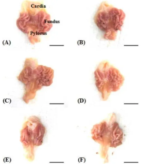
1. Changes on the gastric mucosa gross lesions Focal hemorrhagic ulcerative lesions were dispersed throughout whole gastric mucosa in all IND treated rats, but slight negligible restricted ulcerative lesions were grossly observed in intact control rats. However, Noticeable inhibitions of the gross gastric damages were observed in OME and all three different dosages of BHSST, dose-dependently. Accordingly, significant (p<0.01) increases of gastric mucosa gross lesion areas were detected in IND control as compared with intact control rats, but significantly (p<0.01) and dose-dependently decreased by treatment of BHSST and also by OME 10 mg/kg as compared with IND control rats (Fig. 1, 2).
The gastric mucosa gross lesion areas in IND control were changed as 3158.95% as compared with intact control, and changed as -45.29, -69.70, -43.51 and -21.83% in OME 10 mg/kg, BHSST 400, 200 and 100 mg/kg treated rats as compared with IND control rats, respectively.
Fig. 1. Representative gross images, taken from intact
or indomethacin-treated rats.
A=Intact control rats, B=Indomethacin control rats, C=Omeprazole 10 mg/kg treated rats, D=BHSST 400 mg/kg treated rats, E=BHSST 200 mg/kg treated rats, F=BHSST 100 mg/kg treated rats
Scale bars=11 mm
Fig. 2. Changes on the gastric mucosa gross lesion areas in IND-treated rats.
Values are expressed as mean ± SD of eight rats.
ap<0.01 as compared with intact control by LSD test
bp<0.01 as compared with IND control by LSD test
2. Changes on the MPO activity
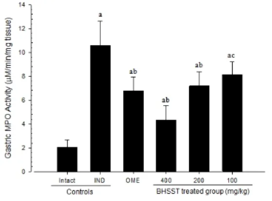
Significant (p<0.01) increases of gastric MPO activities were detected in IND control rats, but these increases were significantly (p<0.01) inhibited after single oral administration of OME and dose- dependently BHSST 400, 200 and 100 mg/kg (Fig. 3).
The MPO activities in IND control were changed
as 423.47%, and changed as -36.00, -58.99, -32.12
and -23.16% in OME 10 mg/kg, BHSST 400, 200
and 100 mg/kg treated rats.
Fig. 3. Changes on the gastric MPO activities in IND-treated rats.
Values are expressed as mean ± SD of eight rats.
ap<0.01 as compared with intact control by MW test
bp<0.01 and cp<0.01 as compared with IND control by MW test
3. Effects on the lipid peroxidation
Significant (p<0.01) increases of gastric lipid peroxidation, the increases of MDA contents were detected in IND control rats, but these increases were significantly (p<0.01) inhibited after single oral administration of OME and dose-dependently BHSST 400, 200 and 100 mg/kg (Table 2).
The MDA contents in IND control were changed as 707.99%, and changed as -31.64, -63.37, -32.79 and -15.06% in OME 10 mg/kg, BHSST 400, 200 and 100 mg/kg treated rats.
4. Effects on the gastric GSH contents
Significant (p<0.01) decreases of gastric GSH contents were observed in IND control rats, but
these decreases were significant (p<0.01 or p<0.05) and dose-dependently normalized by BHSST 400, 200 and 100 mg/kg. In addition, OME 10 mg/kg was also significant (p<0.01) (Table 2).
The GSH contents in IND control were changed as -70.33%, and changed as 55.72, 147.50, 53.01 and 34.63% in OME 10 mg/kg, BHSST 400, 200 and 100 mg/kg treated rats.
5. Changes on the CAT activities
Significant (p<0.01) increases of gastric CAT activities were demonstrated in IND control rats, but these increases were significantly (p<0.01) inhibited after single oral administration of OME and dose- dependently BHSST 400, 200 and 100 mg/kg (Table 2).
The CAT activities in IND control were changed as 107.78%, and changed as -32.01, -42.12, -32.86 and -21.20% in OME 10 mg/kg, BHSST 400, 200 and 100 mg/kg treated rats.
6. Effects on the SOD activities
Significant (p<0.01) decreases of gastric SOD activities were detected in IND control rats, but these decreases were significantly (p<0.01) and dose- dependently increased by treatment of BHSST and OME (Table 2).
The SOD activities in IND control were changed
as -44.64%, and changed as 42.50, 60.00, 44.84 and
29.84% in OME 10 mg/kg, BHSST 400, 200 and
100 mg/kg treated rats.
Groups
Antioxidant defense systems Lipid peroxidation
(nM of MDA/g tissue) Glutathione
(nM/mg tissue) CAT
(nM/min/mg tissue) SOD (nM/min/mg tissue)
Controls Intact 2.38±0.59 4.98±1.07 85.13±15.79 144.50±11.03
IND 19.21±2.18
a1.48±0.42
d176.88±21.53
a80.00±13.94
aOME 10 mg/kg 13.13±2.40
ac2.30±0.42
df120.25±16.21
ac114.00±13.47
acBHSST
400 mg/kg 7.04±2.00
ac3.65±0.71
ef102.38±13.69
bc128.00±12.31
bc200 mg/kg 12.91±1.86
ac2.26±0.39
de118.75±16.40
ac115.88±14.40
ac100 mg/kg 16.32±2.04
ac1.99±0.20
dg139.38±13.94
ac103.88±14.23
ac Values are expressed as mean ± SD of eight rats.ap<0.01 and bp<0.05 as compared with intact control by LSD test
cp<0.01 as compared with IND control by LSD test
dp<0.01 and ep<0.05 as compared with intact control by MW test
fp<0.01 and gp<0.05 as compared with IND control by MW test
Table 2. Changes on the Antioxidant Defense Systems.
7. Changes on the gastric mucosa histopathology Severe focal extensive superficial epithelial damage, desquamation of focal epithelium, neutrophil infiltrations and necrosis of gastric glands, the ulcerative lesions were detected on the fundus after treatment of IND.
However, they were markedly inhibited by pre-treatment of OME and BHSST (Fig. 4). At histomorphometrical and semiquantative analysis, significant (p<0.01) increases of invaded percentages of lesions and semiquantative histological scores, decreases of peri-ulcerative mucosa thicknesses were observed in IND control, but they were significantly (p<0.01 or p<0.05) normalized by BHSST and OME (Table 3).
The lesions in IND control were changed as 3402.16%, and changed as -63.83, -79.85, -67.63 and -29.87% in OME 10 mg/kg, BHSST 400, 200 and 100 mg/kg treated rats.
The peri-ulcerative mucosa thicknesses were changed as -63.73%, and changed as 63.73, 100.73, 74.17 and 52.99% in OME 10 mg/kg, BHSST 400, 200 and 100 mg/kg treated rats.
The semiquantative histological scores were changed as 633.33%, and changed as -40.91, -63.64, -40.91 and -27.27% in OME 10 mg/kg, BHSST 400, 200 and 100 mg/kg treated rats.
Fig. 4. Representative histological images of fundus, taken from intact or IND-treated rats.
A=Intact control rats, B=IND control rats, C=OME 10 mg/kg treated rats, D=BHSST 400 mg/kg treated rats, E=BHSST 200 mg/kg treated rats, F=BHSST 100 mg/kg treated rats Lu=lumen, Mu=mucosa, Mm=muscularis mucosa, Sm=submucosa, ML=muscle layer
Scale bars = 160 μm
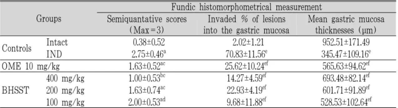
Groups
Fundic histomorphometrical measurement Semiquantative scores
(Max=3)
Invaded % of lesions into the gastric mucosa
Mean gastric mucosa thicknesses (μm)
Controls Intact 0.38±0.52 2.02±1.21 952.51±171.49
IND 2.75±0.46
a70.83±11.56
e345.47±109.16
eOME 10 mg/kg 1.63±0.52
ac25.62±10.24
ef565.63±94.62
efBHSST
400 mg/kg 1.00±0.53
bc14.27±4.59
ef693.48±82.14
ef200 mg/kg 1.63±0.74
ac22.93±4.19
ef601.71±91.89
ef100 mg/kg 2.00±0.53
ad9.68±11.88
ef528.53±102.64
efValues are expressed as mean ± SD of eight rats.
ap<0.01 and bp<0.05 as compared with intact control by LSD test
cp<0.01 and dp<0.05 as compared with IND control by LSD test
ep<0.01 as compared with intact control by MW test
fp<0.01 as compared with IND control by MW test