Palmul-tang, a Traditional Herbal Formula, Protects against Ethanol-induced Acute Gastric Injury in Rats
In-Sik Shin, Mee-Young Lee, Chang-Seob Seo, Hye-Sun Lim, Jung-Hoon Kim, Woo-Young Jeon, Hyeun-Kyoo Shin
Herbal Medicine EBM Research Center, Korea Institute of Oriental Medicine
Original Article
⋅Received:9 October 2011 ⋅Revised:10 November 2011 ⋅Accepted:10 November 2011
⋅Correspondence to:Hyeun-Kyoo Shin
Herbal Medicine EBM Research Center, Korea Institute of Oriental Medicine 483 Expo-ro, Yusung-gu, Daejeon 305-811, Republic of Korea.
Tel:+82-42-868-9464, Fax:+82-42-864-2120, Email:[email protected]
Objectives: Palmul-tang (hachimotsu-to in Japanese and bawu-tang in Chinese) is a mixture of eight herbs. It is traditionally used for the treatment of anemia, anorexia, general weakness, and female infertility in China, Japan, and Korea. In this study, we investigated the protective effects of Palmul-tang water extract (PTE) against ethanol-induced acute gastric injury in rats.
Material and Methods: Acute gastric lesions were induced by intragastric administration of 5mL/kg body weight of absolute ethanol to each rat. Control group rats were given PBS orally and the ethanol group (EtOH group) received absolute ethanol (5mL/kg) by oral gavage. The positive control group and the PTE group were given oral doses of omeprazole (50mg/kg) or PTE (400mg/kg), respectively, 2 h prior to the administration of absolute ethanol. The stomach of each animal was excised and examined for gastric mucosal lesions. To confirm the protective effects of PTE, we evaluated the degree of lipid peroxidation, the level of reduced glutathione (GSH), and the activities of the antioxidant enzymes catalase, glutathione-S-transferase, glutathione peroxidase, and glutathione reductase in the stomach.
Results: PTE reduced ethanol-induced hemorrhage and hyperemia in the gastric mucosa. PTE reduced the increase in lipid peroxidation associated with ethanol-induced acute gastric lesions and increased mucosal GSH content and the activities of antioxidant enzymes.
Conclusion: These results indicate that PTE protects gastric mucosa against ethanol-induced acute gastric injury by increasing antioxidant status. We suggest that PTE could be developed as an effective drug for the treatment of acute gastric injury.
Key Words : Palmul-tang, herbal formula, ethanol, acute gastric injury, antioxidative effect
Introduction
Oxidative stress is generally defined as excess formation or insufficient removal of reactive oxygen species (ROS) and plays a critical role in the development of various diseases
1,2). Oxidative stress induces degradation of cell membrane lipids, resulting in cell damage. This process is known as lipid per- oxidation and produces malondialdehyde (MDA) as
an end product. Organisms have antioxidant defense systems to reduce oxidative stress and lipid peroxidation.
The human antioxidant defense system includes reduced
glutathione (GSH), catalase, glutathione-S-transferase
(GST), glutathione peroxidase (GPx), glutathione
reductase (GR), and superoxide dismutase (SOD)
3).
Oxidative stress plays an important role in gastric
injury because the gastric mucosa is continuously
exposed to factors such as ethanol, stress, and non-
Scientific name Amount (g) Company of purchase Source
Ginseng Radix Alba 4.5 Omniherb Korea
Cnidii Rhizoma 4.5 Omniherb Korea
Rehmanniae Radix Preparata 4.5 Omniherb Korea
Hoelen 4.5 HMAX China
Glycyrrhizae Radix 4.5 HMAX China
Angelicae Gigantis Radix 4.5 Omniherb Korea
Paeoniae Radix 4.5 Omniherb Korea
Atractylodis Rhizoma 4.5 Omniherb Korea
Total amount 36.0
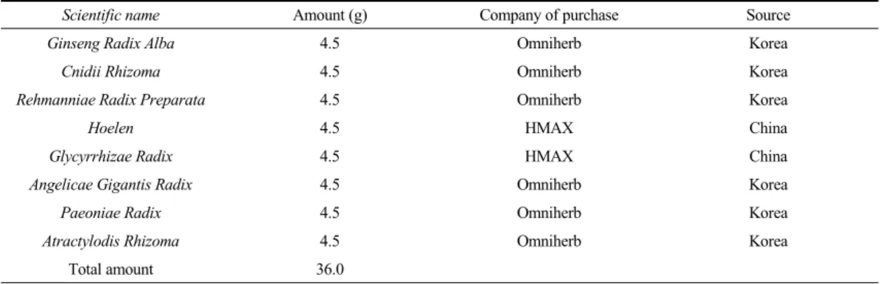
Table 1. Composition of PTE
steroidal anti-inflammatory drugs
4-6). These agents Induce oxidative stress, which increases lipid peroxi- dation and decreases the activity of antioxidant enzymes, resulting in acute gastric damage, including hemorrhage, congestion, edema, erosion, and ulcers
7,8). Treatment of acute gastric injury is focused on improving antioxidant status and reducing lipid peroxidation.
Indeed, many antioxidants exhibit protective effects against ethanol-induced acute gastric injury
9,10).
Palmul-tang (Bawu-tang in Chinese, Hachimotsu-to
in Japanese), an oriental traditional herbal formula, is composed of eight herbs (Table 1). The effects of
Palmul-tang extract (PTE) have been confirmed byseveral researchers. Previous studies have shown that PTE enhances female reproductive function by preven- ting apoptosis and DNA damage, promoting cell proliferation
11), conferring an antiallergic effect
12), and protecting against hypoxic damage in H9c2 cardiomyoblast cells
13). Its constituent crude herbs and compounds also showed pharmacological effects. Atractylodes
rhizome14), Angelicae gigantis radix
15), Ginseng radix
16),
Glycyrrizae radix17), Hoelen
18), and Paeoniae radix
19)showed anti-inflammatory and antioxidative effects.
Atractylodes rhizome20)
, Glycyrrhizae radix
21), and
Ginseng radix22)attenuated experimentally induced gastric injury. In addition, glycyrrhizin
23), ferulic acid
24), liquiritin
25), and paeoniflorin
26)have antioxidative effects. Previous studies showed that glycyrrhizin
27)and paeoniflorin
28)protect against irritant-induced gastric mucosal injury. In oriental medicine, PTE is used for the treatment of symptoms such as dizziness, pale or sallow complexion, discomfort of digestion which are caused by the deficiency of Qi and blood.
Because Qi and blood are produce in the spleen, Qi and blood are sufficiently maintained in body when
Qi of the spleen is intense. The spleen is also closelyrelated with digestion and dietary absorption. If stomach diseases such as gastritis or gastric ulcer occur, the production of Qi and blood in the spleen is insufficient. Therefore, PTE supplements the Qi and blood in the body via tonifying the Qi of the spleen, resulting in attenuation of gastric damage.
Considering this information, we hypothesized that PTE would attenuate absolute ethanol-induced gastric injury via enhancement of antioxidative status. Therefore, we evaluated the protective effects of PTE on ethanol- induced acute gastric injury in rats.
Materials and Methods
1. Preparation of PTE
The PTE was prepared in our laboratory (Table 1)
from a mixture of chopped crude herbs. Before
performing the study, the identity of each crude herb
was confirmed by a pharmacist and by professors at
the College of Oriental Medicine. PTE was extracted
in distilled water at 100℃ for 2 h. The solution was evaporated to dryness and freeze-dried (yield: 25.9%).
Lyophilized PTE (200mg) was dissolved and mixed in distilled water (20mL). The solution was filtered through a SmartPor GHP syringe filter (0.2μm pore size, Woongki Science, Seoul, Korea).
2. Reagents and materials
Ferulic acid and 5-hydroxymethylfurfural (HMF) were purchased from Sigma-Aldrich (St Louis, MO, USA). Albiflorin, paeoniflorin, and glycyrrhizin were produced by Wako (Osaka, Japan). Liquiritin and nodakenin were purchased from NPC BioTechnology Inc. (Daejeon, Korea). The purity of all reference standards was greater than 98.0%. HPLC-grade methanol, acetonitrile, and water were obtained from J.T. Baker (Phillipsburg, NJ, USA). Glacial acetic acid was of analytical reagent grade (Junsei, Tokyo, Japan). The materials for the PTE were purchased from Omniherb (Yeongcheon, Korea) and HMAX (Chungbuk, Korea).
A voucher specimen (2008-KE05-1~KE05-8) has been deposited at the Herbal Medicine EBM Research Center, Korea Institute of Oriental Medicine.
3. Preparation of standard solutions and calibration curves
Standard stock solutions of 5-HMF, albiflorin, paeoniflorin, liquiritin, ferulic acid, nodakenin, and glycyrrhizin (all 1,000μg/ml) were prepared in methanol and stored below 4℃. Working standard solutions were prepared by serial dilution of stock solutions with methanol. Calibration curves were obtained by assessment of peak areas from standard solutions with the following concentration ranges: 5-HMF, 6.25- 50.00μg/mL; albiflorin, ferulic acid, and glycyrrhizin, 3.13-25.00μg/mL; paeoniflorin, 9.38-150.00μg/mL; li- quiritin, 12.50-100.00μg/mL; and nodakenin, 4.69- 75.00μg/mL.
4. HPLC analysis
We performed simultaneous analysis using a Shimadzu LC-20A HPLC system (Shimadzu Co., Kyoto, Japan), consisting of a solvent delivery unit, an inline degasser, a column oven, an autosampler, and a photodiode array (PDA) detector. The data processor used LC solution software (Version 1.24).
The analytical column was a Gemini C18 (250mm×
4.6mm; particle size, 5μm; Phenomenex, Torrance, CA, USA) and was maintained at 40℃. The mobile phases consisted of 1.0% (v/v) aqueous acetic acid (A) and 1.0% (v/v) acetic acid in acetonitrile (B).
The gradient flow was as follows: (A)/(B)=95/5 (0min)
→(A)/(B)=40/60 (40min)→(A)/(B)=0/100 (45min; hold for 5min)→(A)/(B)=95/5 (55min). The analysis was carried out at a flow-rate of 1.0mL/min with PDA detection at 230nm (for albiflorin and paeoniflorin), 254nm (for glycyrrhizin), 280nm (for 5-HMF and liquiritin), 320 nm (for ferulic acid), and 330 nm (for nodakenin). The injection volume was 10μL.
5. Acute gastric injury
Specific pathogen-free male Sprague-Dawley rats, weighing 200-250g (aged 6 weeks) were purchased from the Orient Co. (Seoul, Korea) and used after a week of quarantine and acclimatization. The animals were kept in a room at 23±3°C with a relative humidity of 50% under a controlled 12h/12h light/
dark cycle. The rats were given a standard rodent chow and sterilized tap water ad libitum. All experimental procedures were performed in compliance with the NIH Guide for the Care and Use of Laboratory Animals and the National Animal Welfare Law of Korea
Acute gastric lesions were induced by intragastric
administration of absolute ethanol in according to a
previously described method
29). Twenty-eight rats
were allocated to one of four groups and fasted for
18 h before the experiment. Rats in the control group
were given PBS orally (5ml/kg body weight), and
the absolute-ethanol group (EtOH group) received absolute ethanol (5mL/kg body weight) by oral gavage. Rats in the positive control group were given omeprazole (50 mg/kg body weight) by oral administration 2 h prior to the administration of absolute ethanol. Omeprazole was used as a positive control drug because it possesses anti-inflammatory and antioxidant activities and has been widely used for the treatment of gastritis
3). The dose of PTE was based on the preliminary study. In the preliminary study, PTE was administered to SD rats at dose levels of 200 and 400mg/kg. The reduction of gastric mucosal lesions was detected in 400 mg/kg treated animals more than 200mg/kg treated animals in macroscopic examination. The fourth group received PTE (400mg/kg body weight) 2 h prior to absolute ethanol administration.
6. Macroscopic examination of gastric mucosa Animals were sacrificed using an overdose of 50mg/kg pentobarbital administered 1 h after they received the absolute ethanol treatment. The stomach was removed from each animal, and opened along the greater curvature. The tissue was gently rinsed in PBS. The stomach was stretched out a piece of cork with the mucosal surface facing upward, and was examined in a standard position for gastric mucosal lesions.
Photographs of hemorrhagic erosions in the stomach were taken with a Photometric Quantix digital camera.
After the gastric lesions were photographed, the stomach tissue was stored at -70℃ for biochemical analysis.
7. Biochemical analysis
The stomach tissue was cut into small pieces and homogenized (1/10w/v) with tissue lysis/extraction reagent and a protease inhibitor (Sigma, MI, USA).
The homogenates were centrifuged at 12,000 rpm for 10min at 4℃ to precipitate cell debris, and the supernatant was used to measure levels of malondial-
dehyde (MDA), GSH, catalase, GST, GPx, and GR.
Total protein was determined using a protein assay reagent (Bio-Rad Laboratories).
Lipid peroxidation was estimated by determination of MDA using a thiobarbituric acid-reactive substances (TBARS) assay kit (BioAssay Systems, CA, USA).
In brief, a 100μL aliquot of homogenate was mixed with 100μL of 10% trichloroacetic acid and incubated for 15min on ice. The mixture was centrifuged at 12,000rpm for 5min at 4℃ and then 200μL of the supernatant was mixed with 200μL of thiobarbituric acid and incubated at 100℃ for 60min. After the mixture had cooled, absorbance at 535nm was measured.
The results are expressed as nmol of MDA/mg protein.
The levels of reduced glutathione (GSH) were measured using a GSH assay kit (Cayman, Ann Arbor, MI), which involves an optimized enzymatic recycling method and glutathione reductase. The sulfhydryl group of GSH reacts with 5, 5-ditho-bis-2-nitrobenzoic acid (DTNB) and produces a yellow-colored 5-thio-2- nitrobenzoic acid (TNB). A mixed disulfide, GSTNB (between GSH and TNB), is produced concomitantly and is reduced by glutathione reductase to recycle the GSH, producing more TNB. The rate of TNB production is directly proportional to this recycling reaction which is in turn directly proportional to the concentration of GSH in the sample. The absorbance of TNB at 410nm was used to estimate the amount of GSH in the sample. The GSH level was expressed as μmol/mg protein.
Glutathione-S-transferase (GST) activity was de-
termined by measuring the conjugation of 1-chloro-2,
4-dinitrobenzene (CDNB) with reduced glutathione
using GST assay kit (Cayman). Conjugation is accom-
panied by an increase in absorbance at 340nm and
rate of increase is directly proportional to the GST
activity in the sample. Glutathione peroxidase (GPx)
activity was measured indirectly via a coupled reaction
with glutathione reductase using a GPx assay kit
(Cayman). Oxidized glutathione, produced upon reduction
of hydroperoxide by GPx, is recycled to its reduced
Fig. 1.
state by GR and nicotinamide adenine dinucleotide phosphate (NADPH). The oxidation of NADPH to NADP
+is accompanied by a decrease in absorbance at 340nm. Glutathione reductase (GR) activity was determined by measuring the rate of NADPH oxidation using a GR assay kit (Cayman). Oxidation of NADPH to NADP
+is accompanied by a decrease in absorbance at 340 nm. Catalase activity was measured according to the peroxidatic function of catalase using a catalase assay kit (Cayman). This method is based on the reaction of the enzyme with methanol in the presence of an optimal concentration of hydrogen peroxide. The formaldehyde produced is measured colorimetrically at 540nm with 4-amino-3-hydrazino- 5-mercapto-1, 24-triazole (Purpald) as the chromagen.
The activities of catalase, GST, GPx, and GR, were expressed as U/mg protein.
8. Statistical analysis
Data are expressed as means ± standard deviations (SDs). Statistical significance was determined using analysis of variance (ANOVA). If a test showed a significant difference between groups, the data were analyzed by a multiple comparison procedure using Dunnett’s test
30). Statistical analysis was performed
using SYSTAT version 10. The levels of significance were set as p<0.05 and p<0.01.
Results
1. HPLC analysis of PTE
A three-dimensional chromatogram was obtained using an HPLC-PDA detector. Under optimized chro- matography conditions, seven components were eluted within 45min using mobile phases consisting of solvent A (1.0% v/v aqueous acetic acid) and solvent B (1.0% v/v acetic acid in acetonitrile). The three- dimensional HPLC chromatogram for PTE is shown in Fig. 1. The retention times of the components were 8.166min (5-HMF), 16.21min (albiflorin), 17.16min (paeoniflorin), 19.23min (liquiritin), 19.87min (ferulic acid), 20.36min (nodakenin), and 39.96min (glycyrrhizin).
The linearity of the peak area (y) versus concen-
tration (x, μg/mL) curve for each component was
used to calculate the contents of the main components
in the PTE. The correlation coefficients (r2) of
calibration curves for the seven constituents was
greater than 0.9998. The concentrations of the seven
components in PTE ranged from 0.63 to 6.22 mg/g
(Table 2).
(A) Normal control
(C) Omeprazole (D) PTE-200 (B) EtOH
Fig. 2.
Batch (#)
Content (mg/g)
5‐HMF Albiflorin Paeoniflorin Liquiritin Ferulic acid Nodakenin Glycyrrhizin Mean SD RSD (%) Mean SD RSD (%) Mean SD RSD (%) Mean SD RSD (%) Mean SD RSD (%) Mean SD RSD (%) Mean SD RSD(%) 1 1.82 0.002 0.135 1.23 0.007 0.580 6.16 0.079 1.287 3.80 0.064 1.681 0.63 0.006 0.928 3.03 0.013 0.426 2.91 0.002 0.052 2 1.82 0.003 0.155 1.29 0.010 0.746 6.10 0.051 0.835 3.77 0.020 0.533 0.64 0.004 0.622 3.07 0.024 0.778 2.92 0.003 0.105 3 1.82 0.002 0.133 1.30 0.006 0.488 6.22 0.071 1.146 3.79 0.012 0.322 0.64 0.005 0.783 3.00 0.007 0.219 2.91 0.002 0.072 Table 2. The contents of seven marker compounds in PTE (n=3).
2. PTE reduced ethanol-induced acute gastric injury
Gross examination of gastric mucosa showed that the EtOH group had gastric mucosal injuries (hemor- rhage and hyperemia) (Fig. 2), whereas no abnormalities or lesions were found in the normal control group.
In contrast, PTE pretreatment attenuated gastric mucosal injury compared with the EtOH group to a degree similar to that in the omeprazole-treated group.
3. PTE reduced the increase in lipid peroxidation and GSH content in ethanol-induced acute gastric lesions
The concentration of MDA, an end product of lipid peroxidation, was greater in the EtOH group (13.32±1.32nmol/mg protein, p<0.05) than in control rats (10.09±0.66 nmol/mg protein) (Fig. 3A). The MDA level in PTE-treated rats differed significantly (9.84±1.25nmol/mg protein, p<0.05) from those in
the EtOH group. Also, in the omeprazole-treated group (8.73±1.08nmol/mg protein, p<0.01) was observed a significant reduction compared with the EtOH group.
Conversely, the content of GSH in the stomachs of the EtOH group (32.30±1.09μmol/mg protein, p<0.01) was less than that in the control group (45.99±2.05 μ mol/mg protein), and that in the PTE-treated groups was significantly greater (44.19±2.05μmol/mg protein, p<0.01) than that in the EtOH group (Fig. 3B).
4. PTE attenuated the ethanol-induced decrease in the activities of antioxidant enzymes
As shown in Fig. 4A, catalase activity in the
EtOH group (238.13±28.07U/mg protein, p<0.01) was
less than that in the control (326.23 ± 29.08 U/mg
protein). However, PTE treatment resulted in a
significant increase (301.41±24.88U/mg protein, p<0.01)
in catalase activity compared with that of the EtOH
group. Also, the omeprazole-treated group (283.55±
20 16 12 80 4 0 (A)
NC EtOH Ome PTE NC EtOH Ome PTE
80
60
40
20
0 (B)
Glutathione(umon/mg protein)
MIDA level (nmol/mg protein)
Fig. 3.
NC EtOH Ome PTE NC EtOH Ome PTE
16
12
8
4
0 (B)
GST activity (U/mg protein)
500 400 300 200 100 0 (A)
Catalase activity (U/mg protein)
NC EtOH Ome PTE NC EtOH Ome PTE
120
90
60
30
0 (D)
GRactivity (U/mg protein)
120
90
60
30
0 (C)
GPx activity (U/mg protein)
Fig. 4.