Curcumin Induces Recovery from Indomethacin-Induced Gastric Mucosal Lesions in Rats
Jeong-Hwan Kim
1, Byung-Woo Kim
1,3, Hyun-Ju Kwon
1,3, Yeon-Hee Kim
2,4and Soo-Wan Nam
1,2,4*1
Blue-Bio Industry RIC,
2Department of Biotechnology and Bioengineering, College of Engineering,
3Department of Life Science and Biotechnology, College of Natural Sciences,
4Department of Smart Bio-Health, Dong-Eui University, Busan 614-714, Korea
Received October 15, 2013 /Revised November 16, 2013 /Accepted November 21, 2013In the present study, the curative effect of curcumin on indomethacin-induced gastric mucosal lesions in rats was investigated. Indomethacin is a nonsteroidal anti-inflammatory drug (NSAID), with serious side effects, including erosion, ulcerative lesions, and petechial bleeding in the mucosa of the stomach.
Gastric mucosal lesions were caused by oral administration of 25 mg/kg of indomethacin. Various doses (10, 50, and 100 mg/kg) of curcumin were treated for 3 days by oral gavage. Indomethacin clearly increased the gastric ulcer area in the stomach, and curcumin significantly decreased the gas- tric ulcer area in a dose-dependent manner. Curcumin also markedly inhibited lipid peroxidation in the gastric mucosa and activated radical scavenging enzymes, such as superoxide dismutase (SOD), catalase, and glutathione peroxidase, in a dose-dependent manner. These results suggest that curcu- min can induce recovery from indomethacin-induced gastric mucosal lesions through inhibition of lip- id peroxidation and activation of radical scavenging enzymes, such as SOD, catalase, and glutathione peroxidase. Curcumin appears to be a powerful free radical quencher, and it may offer an attractive strategy for healing gastric mucosal lesions in humans.
Key words : Curcumin, gastric mucosal lesions, lipid peroxidation, oral gavage, radical scavenging enzymes
*Corresponding author
*Tel : +82-51-890-2276, Fax : +82-51-890-2632
*E-mail : [email protected]
This is an Open-Access article distributed under the terms of the Creative Commons Attribution Non-Commercial License (http://creativecommons.org/licenses/by-nc/3.0) which permits unrestricted non-commercial use, distribution, and reproduction in any medium, provided the original work is properly cited.
Journal of Life Science 2014 Vol. 24. No. 1. 20~25 DOI : http://dx.doi.org/10.5352/JLS.2014.24.1.20
Introduction
Indomethacin is a non-steroidal anti-inflammatory drug (NSAID) with anti-inflammatory, antipyretic, and pain-re- lieving properties, which is known to produce erosions, ul- cerative lesions, and petechial bleeding in the mucosa of stomach as serious side effect [8, 15]. The oral gavage of indomethacin in rats causes ulcerative lesions in the gastric mucosa [6]. The development of the gastric mucosal lesions induced by indomethacin is mainly mediated through gen- eration of oxygen free radicals and lipid peroxidation [4, 19, 22-25]
Curcumin is a natural phenolic component derived from the plant Curcuma longa, which is used in some cultures for the treatment of diseases associated with oxidative stress and inflammation [14]. Recently, great attention has been
paid to the medical applications of curcumin in the treat- ment of human diseases [10, 20, 21]. Curcumin has been rec- ognized as a powerful anticancer drug due to its efficient induction of proliferation arrest and cell death including apoptosis and necrosis in a variety of tumor cells [2, 5, 7, 13]. However, the curative properties of curcumin against gastric mucosal lesions are not well understood. In this study, the curative effect of curcumin against indomethacin- induced gastric mucosal lesions in rats was evaluated by measuring the amount of lipid peroxidation and by compar- ing the activities of enzymatic scavengers such as SOD, cata- lase, and glutathione peroxidase.
Materials and Methods Chemicals
Curcumin and indomethacin were purchased from Sigma
Chemicals (St Louis, MO., USA). Curcumin was dissolved
in saline immediately before use and administered intra-
gastrically to rats in a volume of 5 ml/kg. Indomethacin was
dissolved in 5% sodium bicarbonate (Sigma, St Louis, MO.,
USA) and administered to rats in a dose of 25 mg/kg by
oral gavage, with an appropriate feeding needle as a volume
of 5 ml/kg.
Animals
Male Sprage-Dawley rats (200~250 g, 7 weeks old) were purchased from Daehan Biolink Co., Ltd. Rats were placed singled in cages with wire-net floors in a controlled room (temperature 22~24
oC, humidity 70~75%, lighting regimen of 12 hr light and 12 hr dark), and they were fed a normal laboratory diet. Rats were fasted for 24 hr before ex- perimental use, but allowed free access to tap water throughout. The animal experiment was performed in ac- cordance with guidelines established by the Animal Care and Use Committee of Dong-Eui University and approved by the committee.
Experimental design
Gastric mucosal lesions were induced by a single oral dose of 25 mg/kg indomethacin. The optimal concentration of indomethacin for induction of gastric mucosal lesions was determined on the basis of the previous our study [11]. Rats were divided into six groups (n=6 rats per group). The nor- mal group received only 5% sodium bicarbonate orally in a volume of 5 ml/kg. The control group received only 25 mg/kg indomethacin. Each of the remaining four groups was treated with a vehicle (curcumin, 0 mg/kg) or three doses (10, 50, and 100 mg/kg) of curcumin for 3 days after induction of gastric ulcers by pretreatment with 25 mg/kg indomethacin. All the rats were killed under deep ether an- esthesia 4 hr after the last oral administration. The rat stom- achs were promptly excised, weighed, and chilled in ice-cold 0.9% NaCl. After washing with 0.9% NaCl, the mucosa was homogenized in 50 mM potassium phosphate buffer at pH 7.5. Mitochondria and cytosol fractions were prepared ac- cording to the method of Hogeboom [9]. The quantitative analysis of protein was measured by Bradford protein assay [3].
Malondialdehyde levels
Lipid peroxidation was determined by measuring malo- nylaldehyde (MDA) production by using a thiobarbituric acid reaction [17, 18]. Briefly, the stomach homogenate was supplemented with 8.1% sodium dodecyl sulfate, 20% acetic acid (pH 3.5), and 0.8% thiobarbituric acid, and boiled at 95
oC for 1 hr. After cooling with tap water, the reactants were supplemented with n-butanol and pyridine (15:1 v/v), shaken vigorously for 1 min, and centrifuged for 10 min at 3,500 g. Absorbance was measured at 532 nm. Lipid peroxide level was calculated from the standard curve using the MDA
tetrabutylammonium salt. MDA (Sigma, St Louis, MO., USA) concentrations were expressed as nmoles/g of tissue.
Activities of enzymatic scavengers
The activity of SOD in the gastric mucosa was measured according to the method of McCord and Fridovich [16]. The standard assay was performed in 3 ml of 50 mM potassium phosphate buffer at pH 7.8 containing 0.1 mM EDTA in a cuvette thermostated at 25
oC. The reaction mixture con- tained 0.1 mM ferricytochrome c, 0.1 mM xanthine, and suf- ficient xanthine oxidase to produce a reduction rate of ferri- cytochrome c at 550 nm of 0.025 absorbance unit per min.
Tissue homogenate was mixed with the reaction mixture (50 mM potassium phosphate buffer, pH 7.8 containing 0.1 mM EDTA, 0.1 mM ferricytochrome c, and 0.1 mM xanthine).
Kinetic spectrophotometric analysis was started adding xan- thine oxidase at 550 nm. Under these conditions, the amount of SOD required to inhibit the reduction rate of cytochrome c by 50% was defined as 1 unit of activity. The results were expressed as units/mg of protein.
The activity of catalase in the gastric mucosa was meas- ured according to the method of Aebi [1]. The standard as- say was performed in 3 ml of 50 mM potassium phosphate buffer at pH 7.0 (1.9 ml) containing 10 mM H
2O
2(1 ml) and tissue homogenate (100 μl). Under these conditions, the amount of catalase required to decompose 1.0 μM of H
2O
2per min at pH 7.0 at 25
oC was defined as 1 unit of activity.
Absorbance was measured at 240 nm for 2 min, and the re- sults were expressed as units/mg of protein.
The activity of glutathione peroxidase in the gastric muco- sa of rats was determined by a modified method of Lawrence and Burk [12]. The reaction mixture consisted of glutathione peroxidase assay buffer (50 mM potassium phos- phate buffer pH 8.0, 0.5 mM EDTA) and NADPH assay re- agent (5 mM NADPH, 42 mM reduced glutathione, and 10 units/ml glutathione reductase). A supernatant of homoge- nate in 50 mM potassium phosphate buffer at pH 7.5 was prepared by centrifuging it at 1,000 g for 10 min at 4
oC.
Subsequently, 900 μl of glutathione peroxidase assay buffer, 50 μl of NADPH assay reagent, and 50 μl of the sample were added to the cuvette, and the contents were mixed by inversion. The reaction was started by adding 10 μl of 30 mM tert-butyl hydroperoxide or 80% cumene hydroperoxide.
Absorbance was recorded by the following program;
Wavelength: 340 nm/ Initial delay: 15 sec/ Interval: 10 sec/
Number of readings: 6. The activity of enzyme was the sum
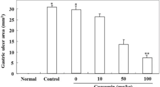
Fig. 1. Curcumin shows the curative effect against in- domethacin-induced gastric mucosal lesions in rats.
Gastric mucosal lesions were induced by oral admin- istration with 25 mg/kg of indomethacin, and then vari- ous doses (10, 50, and 100 mg/kg) of curcumin were treated by oral gavage for 3 days. Curcumin sig- nificantly decreased the gastric ulcer area in the mucosa of stomach in a dose dependent manner, compared with control group. Values are expressed as means ± S.E.M.
*
p
<0.05, significantly different from the untreated nor- mal group.**p
<0.01, significantly different from the con- trol group.Fig. 2. Curcumin inhibited lipid peroxidation induced by in- domethacin in gastric mucosa. MDA production was es- timated by using a thiobarbituric acid reaction. curcu- min distinctly reduced the level of MDA in a dose de- pendent manner in comparison to control group. Values are expressed as means ± S.E.M. *
p
<0.05, significantly different from the untreated normal group.**p
<0.01, sig- nificantly different from the control group.of data obtained using 30 mM tert-butyl hydroperoxide and 80% cumene hydroperoxide. The level of glutathione was expressed in terms of μM/min/mg of protein.
Statistical analysis
All values were represented as means ± S.E.M. Data were analyzed by ANOVA according to General Linear Model procedure. The means were compared by Tukey’s Studentized Range (HSD) test to detect significant differences at p<0.05.
Results and Discussion
Indomethacin is a non-steroidal anti-inflammatory drug (NSAID) that is known to cause gastric mucosal lesions.
Indomethacin comprises polar lipids that have a high affin- ity for the lipophilic areas of cell membranes, where their polar groups trigger membrane disruption, with loss of structural phospholipids and membrane proteins. In addi- tion, this leads to reduced hydrophobicity of the mucosal coat adherent to the mucosal cell surface. Such loss of hydro- phobicity facilitates the entry of water-soluble agents of in- jury, e.g. acid, pepsin, bile salts, etc., which cause lipid per- oxidation and also alter membrane fluidity [15]. Especially, generation of oxygen free radicals and lipid peroxidation play a important role in the development of the gastric mu- cosal lesions induced by indomethacin [4, 19, 22-25].
In this study, the optimal concentration of indomethacin for induction of gastric mucosal lesions was determined on the basis of the previous our study [11]. The concentrations of curcumin were selected on the basis of the preliminary results obtained from cytotoxicity studies using a broad con- centration range for this reagent. Gastric mucosal lesions were caused by oral administration with 25 mg/kg of in- domethacin, and then various doses (10, 50, and 100 mg/kg) of curcumin were treated for 3 days by oral gavage. Gastric lesions were judged macroscopically by clear depth of pene- tration into the gastric mucosal surface in all test groups.
In Fig. 1, indomethacin significantly caused the increase of the gastric ulcer area in the mucosa of stomach, compared with normal group (
*p<0.05). However, curcumin markedly decreased the gastric ulcer area in a dose dependent manner, compared with control group (
**p<0.01).
The development of the gastric mucosal lesions induced by indomethacin is mainly mediated through generation of oxygen free radicals and lipid peroxidation [4, 22, 23, 24].
Therefore, the curative effect of curcumin against in-
domethacin-induced gastric mucosal lesions in rats was
evaluated by measuring the amount of lipid peroxidation
and by comparing the activities of enzymatic scavengers
such as SOD, catalase, and glutathione peroxidase. Fig. 2
shows the effect of curcumin on lipid peroxidation induced
by indomethacin in the gastric mucosa. Indomethacin con-
siderably increased the level of MDA in gastric tissue in
comparison to normal group (
*p<0.05), whereas curcumin
decreased the level of MDA in a dose dependent manner
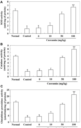
A
B
C
Fig. 3. Curcumin activated radical scavenging enzymes in in- domethacin-induced gastric mucosal lesions. Curcumin markedly increased activities of SOD (A), catalase (B), and glutathione peroxidase (C) in a dose dependent manner in comparison to control group. Values are ex- pressed as means ± S.E.M.*
p
<0.05, significantly differ- ent from the untreated normal group. **p
<0.01, sig- nificantly different from the control group.Table 1. The activities of radical scavenging enzymes at different concentrations of curcumin
Curcumin (mg/kg) SOD (units/mg-protein) Catalase (units/mg-protein) Glutathione peroxidase (μmol/min/mg-protein) Normal
Control 0 1050 100
5.82±0.38 1.12±0.46* 1.38±0.42* 2.04±0.71 4.80±0.68 6.58±0.34**
6.02±0.24 1.54±0.22* 1.70±0.20* 2.02±0.58 5.68±0.54 7.86±0.61**
15.63±0.62 3.78±0.66* 4.45±0.58* 7.02±0.92 14.08±0.42 21.24±1.14**
The normal group received only 5% sodium bicarbonate orally in a volume of 5 ml/kg. The control group received only 25 mg/kg indomethacin.*
p
<0.05, significantly different from the untreated normal group.**p
<0.01, significantly different from the control group.in comparison to control group (
**p<0.01).
In Fig. 3, the effect of curcumin on activities of radical
scavenging enzymes such as SOD, catalase, and glutathione peroxidase was tested. Indomethacin distinctly attenuated activities of SOD, catalase, and glutathione peroxidase in comparison to normal group (
*p<0.05). These results suggest that the inhibition of these enzymatic activities is, at least in part, responsible for oxidative tissue damage of gastric mucosa occurring after indomethacin treatment. In contrast, curcumin increased activities of these enzymes in a dose de- pendent manner in comparison to control group (
**p<0.01).
At the highest dose (100 mg/kg) group of curcumin, the enzyme activities were enhanced by 4.6 fold over 0 dose group (Table 1).
In conclusion, curcumin induce recovery from in- domethacin-induced gastric ulcers through prevention of lipid peroxidation and activation of radical scavenging en- zymes, and such effect is directly involving its antioxidant property. Therefore, we suggest that curcumin is a powerful free radical quencher, and its use may offer an attractive strategy for healing gastric mucosal lesions in humans.
Acknowledgement
This work was supported by Dong-Eui University Grant (2012AA193).
References
1. Aebi, H. 1974. In methods of enzymatic analysis. pp.
674-678, In: Bergmeyer, H. U. (ed.), Academic Press, New York.
2. Ajaikumar, B. K., Sushovan, G., Sunil, K., Parmeswaran, D., Juri, G. and Bharat, B. A. 2007. Curcumin potentiates anti- tumor activity of gemcitabine in an orthotopic model of pancreatic cancer through suppression of proliferation, an- giogenesis, and inhibition of nuclear factor-κB-regulated gene products.
Cancer Res
67, 3853-3861.3. Bradford, M. M. 1976. A rapid and sensitive method for the quantitation of microgram quantities of protein utilizing
the principle of protein-dye binding.
Anal Biochem
72, 248-254.4. Del Soldato, P., Foschi, D., Benoni, G. and Scarpignato, C.
1985. Oxygen free radicals interact with indomethacin to cause gastrointestinal injury.
Agents Actions
17, 484-488.5. Dhandapani, K. M., Mahesh, V. B. and Brann, D. W. 2007.
Curcumin suppresses growth and chemoresistance of hu- man glioblastoma cells via AP-1 and NF-κB transcription factors.
J Neurochem
102, 522-538.6. Djahanguiri, B. 1969. The production of acute gastric ulcer- ation by indomethacin in the rat.
Scand J Gastroenterol
4, 265-267.7. Enyu, L., Jing, W., Weidong, C., Jianning, Z., Weiping, L., Xiaofan, J. and Xiang, Z. 2007. Curcumin induces G2/M cell cycle arrest in a p53-dependent manner and upregulates ING4 expression in human glioma.
J Neurooncol
85, 263-270.8. Hart, F. D. and Boardman, P. L. 1963. Indomethacin: a new non-steroid anti-inflammatory agent.
Brit Med J
2, 965-970.9. Hogeboom, G. H. 1955. In methods in enzymology. pp.
16-19, In: Colowick, S. P. and Kaplan, N. O. (eds.),
Academic Press
, New York.10. Huang, M. T., Lysz, T., Ferraro, T., Abidi, T. F., Laskin, J.
D. and Conney, A. H. 1991. Inhibitory effects of curcumin on
in vitro
lipoxygenase and cyclooxygenase activities in mouse epidermis.Cancer Res
51, 813-819.11. Kim, J. H., Choi, S. K., Lim, W. J. and Chang, H. I. 2004.
Protective effect of astaxanthin produced by
Xanthophyllo- myces dendrorhous
mutant on indomethacin-induced gastric mucosal injury in rats.J Microbiol Biotechnol
14, 996-1003.12. Lawrence, R. A. and Burk, R. F. 1976. Glutathione perox- idase activity in selenium-deficient rat liver.
Biochem Biophys Res Commun
71, 952-958.13. Lin, Y. G., Kunnumakkara, A. B., Nair, A., Merritt, W. M., Han, L. Y., Armaiz-Pena, G. N., Kamat, A. A., Spannuth, W. A., Gershenson, D. M., Lutgendorf, S. K., Aggarwal, B.
B. and Sood, A. K. 2007. Curcumin inhibits tumor growth and angiogenesis in ovarian carcinoma by targeting the nu-
clear factor-κB pathway.
Clin Cancer Res
13, 3423-3430.14. Lodha, R. and Bagga, A. 2000. Traditional Indian systems of medicine.
Ann Acad Med Singapore
29, 37-41.15. McCarthy, D. M. 1995. Mechanisms of mucosal injury and healing: the role of nonsteroidal anti-inflammatory drugs.
Scand J Gastroenterol Suppl
208, 24-29.16. McCord, J. M. and Fridovich, I. 1967. Superoxide dismutase, an enzymatic function for erythrocuprein (hemocuprein).
J Biol Chem
244, 6049-6055.17. Mihara, M. and Uchiyama, M. 1978. Determination of malo- naldehyde precursor in tissues by thiobarbituric acid test.
Anal Biochem
86, 271-278.18. Ohkawa, H., Ohishi, N. and Yagi, K. 1979. Assay for lipid peroxides in animal tissues by thiobarbituric acid reaction.
Anal Biochem
95, 351-358.19. Parks, D. A. 1989. Oxygen radicals: mediators of gastro- intestinal pathophysiology.
Gut
30, 293-298.20. Ruby, A. J., Kuttan, G., Babu, K. D., Rajasekharan, K. N.
and Kuttan, R. 1995. Anti-tumour and antioxidant activity of natural curcuminoids.
Cancer Lett
94, 79-83.21. Shishodia, S., Sethi, G. and Aggarwal, B. B. 2005. Curcumin:
getting back to the roots.
Ann N Y Acad Sci
1056, 206-217.22. Takeuchi, K., Ueshima, K., Hironaka, Y., Fujioka, Y., Matsumoto, J. and Okabe, S. 1991. Oxygen free radicals and lipid peroxidation in the pathogenesis of gastric mucosal lesions induced by indomethacin in rats.
Digestion
49, 175-184.23. Tanaka, J. and Yuda, Y. 1996. Lipid peroxidation in gastric mucosal lesions induced by indomethacin in rats.
Biol Pharm Bull
19, 716-720.24. Vaananen, P. M., Meddings, J. B. and Wallace, J. L. 1991.
Role of oxygen-derived free radicals in indomethacin-in- duced gastric injury.
Am J Physiol
261, G470-475.25. Yoshikawa, T., Naito, Y., Ueda, S., Oyamada, H., Takemura, T., Yoshida, N., Sugino, S. and Kondo, M. 1990. Role of oxy- gen-derived free radicals in the pathogenesis of gastric mu- cosal lesions in rats.