Protective Effects of BK-1202 on the Indomethacin-induced Gastric Ulcer in Rats
Hae-Won Kwon, Dae-Jun Kim
Dept. of Internal Medicine, College of Oriental Medicine, Daegu Hanny University Original Article
⋅Received:14 December 2015 ⋅Revised:18 December 2015 ⋅Accepted:29 December 2015
⋅Correspondence to:Dae-Jun Kim
#907-8 Daejam-dong, Nam-gu, Pohang, Gyungsangbuk-do, South Korea Tel:+82-54-281-0055, Fax:+82-54-281-7463, E-mail:[email protected]
Purpose: The object of this study is to observe the anti-ulcerative effects of BK-1202 (IGM), a mixed herbal formula consisting of 9 herbal drugs, which have been traditional Korean medicine for treating various digestive diseases, on indomethacin-induced gastric ulcer in rat.
Methods: Three different doses of IGM extract (200, 100 and 50 mg/kg) were orally administered once 30 min before indomethacin treatment. Six hours after indomethacin treatment, changes in the gross lesion scores, fundic histopathology, MPO activity and antioxidant activities were observed. The results were compared with two reference groups treated with omeprazole (10 mg/kg), antioxidant and proton pump inhibitor, and DA-9601 (100 mg/kg), a standardized extract of the herb Artemisiaasiatica.
Results: In all three doses of IGM extract, significantly decreased gastric damages were observed in the indomethacin-induced gastric ulcer rats, when compared with the indomethacin-treated control rats. IGM extracts also strengthened the antioxidative defense systems, decreasing the level of lipid peroxidation and catalase activity while increasing the superoxide dismutase and glutathione contents. IGM extracts showed similar anti-ulcerative effects to those shown by equal dose of DA-9601, and the effects of 50 mg/kg IGM extracts were comparable to those of 10 mg/kg omeprazole.
Conclusion: The results obtained in this study suggest that IGM extract has favorable effects on the indomethacin -induced gastric damages by strengthening the antioxidative defense systems and enhancing anti-inflammatory effects.
Key Words : BK-1202, Indomethacin, Gastric ulcer
Introduction
Gastritis and gastric ulcer are defined as pathological conditions developed by exposure of the gastric mucosa to endogenous and/or exogenous aggressive factors and subsequent disturbances in gastric mucosal defenses
1). Gastric ulcers are ulcers occurring in the stomach that are manifested by acute erosive and superficial ulcerative lesions in the regions exposed to gastric juices. In the Korean herbal medicine, gastric ulcer is known to be associated with the symptoms related to epigastric
pain, heartburn, acid regurgitation, and dyspepsia
2).
Since the recognition of the finding of a study that
identified nonsteroidal anti-inflammatory drugs
(NSAIDs) as one of the major causes of gastric
ulcer
3), many anti-ulcerative drugs have been
developed to address the related symptoms. However,
such drugs have a wide range of side-effects, such as
constipation, diarrhea, itching, spotty skin, inhibition
of antifungal agents’ metabolism mediated by proton
pump inhibitor (PPI), headaches, anti-androgenic
effects, dizziness, and misoprostol-induced stillbirth
or discharge of blood in pregnant women
4).
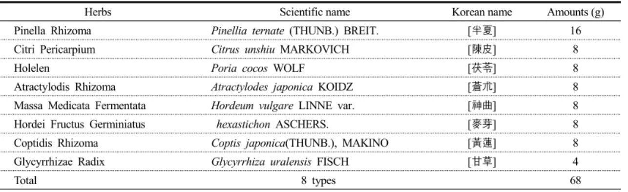
Table 1. Composition of Ijintanggamibang (IG) used in this study
Herbs Scientific name Korean name Amounts (g)
Pinella Rhizoma Pinellia ternate (THUNB.) BREIT. [半夏] 16
Citri Pericarpium Citrus unshiu MARKOVICH [陳皮] 8
Holelen Poria cocos WOLF [茯苓] 8
Atractylodis Rhizoma Atractylodes japonica KOIDZ [蒼朮] 8
Massa Medicata Fermentata Hordeum vulgare LINNE var. [神曲] 8
Hordei Fructus Germiniatus hexastichon ASCHERS. [麥芽] 8
Coptidis Rhizoma Coptis japonica(THUNB.), MAKINO [黃蓮] 8
Glycyrrhizae Radix Glycyrrhiza uralensis FISCH [甘草] 4
Total 8 types 68
Ijintang-Gamibang (IG) recipe prescribes the herbal formula of Ijintang extract added with
Atractylodis rhizoma, Massa medicata fermentata, Hordei fructus germiniatus, and Coptidis rhizoma5). BK-1202 is a new IG formula modified for the purpose of this study by adding Ostrea gigas
6)whose acid-removing effect was verified. To determine the protective effects of BK-1202 against indometacin -induced gastric ulcer, BK-1202 at the doses of 200, 100, and 50 mg/kg was orally administered to rats 30 min prior to administering them indomethacin (25 mg/kg). Six hours after the medication, all animals were sacrificed and postmortem observations were made to check the changes in the surface area of a hemorrhagic gastric ulcerative lesion (lesion score;
mm
2/gastric mucosa), myeloperoxidase (MPO) content in the gastric mucosal tissue, antioxidant defense system, lipid peroxidation, glutathione (GSH), catalase, and superoxide dismutase (SOD) along with histomorphometric alterations. The experimental results were then compared with the results of the reference groups treated with 10mg/kg omeprazole and 100 mg/kg DA-9601, respectively.
Materials and Methods
1. Experimental drugs
The IG used in this study was purchased from a
local pharmaceutical company (Hyosung Pharmaceutical Co., Daegu, Korea) and screened for use through microscopic inspection. Table 1 presents the composition of a sachet of IG. The ingredients contained in a selected sachet (68 g) were dissolved in 2000 ml distilled water and heat-extracted, followed by suction filtration of the extracted liquid and the depressurization and concentration of the filtered extract using a rotary vacuum evaporator (N-N type; LAB Camp, Daejeon, Korea). The extract with viscous consistency thus acquired underwent freeze drying in a programmable freeze dryer (PVTFD10A; Ilshin Lab., Seoul, Korea), which yielded a total of 17.00 g (yield rate: 25.01%) dark brown fluid extract. BK-1202 was then produced by adding 4 g Ostrea gigas to the IG extract, which was them pulverized and stored in a freezer (-20ºC) to be used for the experiments. The prepared BK-1202 powder dissolved well in distilled water up to the concentration of 40 mg/ml. Drugs for the experimental and reference groups, i.e. indomethacin, omeprazole, and DA-9601 were purchased from Sigma (MO, USA) and Dong-A Pharmaceutical Co.
(Yongin, Korea).
2. Lab animals and their management
A total of 56 male Sprague-Dawley rats (6-week
old upon receipt, SLC, Japan) were used for the
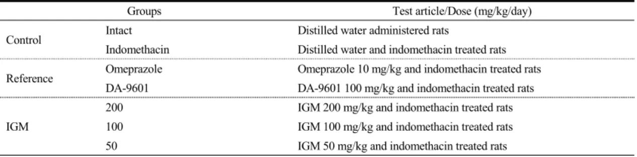
Table 2. Experimental design of this study
Groups Test article/Dose (mg/kg/day)
Control Intact Distilled water administered rats
Indomethacin Distilled water and indomethacin treated rats Reference Omeprazole Omeprazole 10 mg/kg and indomethacin treated rats
DA-9601 DA-9601 100 mg/kg and indomethacin treated rats IGM
200 IGM 200 mg/kg and indomethacin treated rats 100 IGM 100 mg/kg and indomethacin treated rats 50 IGM 50 mg/kg and indomethacin treated rats
experiments after a 7-day acclimation. Throughout the duration of acclimation and experiments, they were kept in polycarbonate boxes designed for rats, 5 rats per box, in constant temperature (20-25℃) and humidity (30-35%) conditions in a 12-h light:dark cycle, and were given ad libitum access to feed (Samyang, Korea) and water. Of 56 rats, 48 were used for developing indomethacin-induced gastric mucosal damage, and the remaining 8 were used as an intact control group. All animals were administered their respective drugs (except for the two control groups, which were administered sterilized distilled water) after 24-h feed deprivation (with free access to water), and gastric mucosal damage was induced by administering indomethacin 30 min after the initial treatment (except for the intact control group, which was administered sterilized distilled water). Individuals were identified by picric acid. All animals were treated in compliance with the “Guide for the Care and Use of Laboratory Animals”
7).
3. Experimental group formation and drug administration
Lab animals were divided in 7 groups, 8 individuals per group, as listed in Table 2: 1) Intact control group treated with sterilized distilled water after the initial administration of sterilized distilled water used as medium, 2) Indomethacin control group with indomethacin-induced gastric mucosal
damage after the administration of the medium sterilized distilled water, 3–7) Experimental groups with indomethacin-induced gastric mucosal damage after being administered omeprazole, DA-9601, and three different doses (200, 100, and 50 mg/kg) BK-1202, respectively (Table 2). BK-1202, omeprazole, and DA-9601 were dissolved in sterilized distilled water and orally administered to the animals using a 3-ml syringe with metallic probe at the ratio of 5 ml per 1 kg animal weight. The omeprazole and DA-9601 doses were set at 10 mg/kg
8)and 100 mg/kg
9), respectively, doses known to be have sure efficacy in reducing indomethacin-induced gastric mucosal damage.
4. Induction of gastric mucosal damage
After 24-h feed deprivation of all animals, the two
reference groups were administered with omeprazole
and DA-9601, respectively, and the three experimental
groups were administered with three different doses
of BK-1202. After 30 min, a single dosage of 25
mg/kg indomethacin dissolved in sterilized distilled
water was orally administered in accordance with the
Guidobono method
10)to induce gastric mucosal
damage. The two control groups were administered
sterilized distilled water in the initial treatment. In
the second treatment, the intact and indomethacin
control groups were administered sterilized distilled
water and 25 mg/kg indomethacin, respectively.
5. Measurement of visually inspected lesions In accordance with the method proposed by Süleyman et al.
11), all animals were sacrificed with cervical dislocation 6 h after being treated with indomethacin. From each animal, the stomach was harvested and, with the greater curvature cut open, fixed in 10% neutral buffered formalin for 24 h. The total surface area of the gastric mucosal lesions with hemorrhagic foci was estimated by the unit of mm
2by overlapping a grid (area: 1 mm
2) on the area of gastric mucosal damage.
6. Measurement of MPO activity
In accordance with the method proposed by Morai et al
l2), the gastric mucus layer was separated and homogenized in an ice-cold solution of 10 mL KCl (100 g/L; pH 7.4; Sigma, MO, USA), and the MPO content within the stomach tissue homogenate was measured using the method proposed by Bradley et al
13)with a light modification as follows. The homogenate underwent 3 freeze-thaw cycles and centrifuged at 1500×g for 10 min at 4°C. Of its supernatant, 100 ml was harvested and added with 1 ml of 1.5 mM/L o-dianisidinehydrochloride (Sigma, MO, USA) containing 1.9 ml of 10 mM/l phosphate buffer (pH 6.0) and 0.0005% (w/v) hydrogen peroxide (Merk, CA, USA). Finally, PMO activity was estimated at the unit of μM/minute/mg tissue by measuring absorbance at 450 nm with a UV-vis spectrophotometer (UV-3600, Shimadzu Scientific Instruments, CO, USA).
7. Measurement of lipid peroxidation Lipid peroxidation was measured by the method of Ohkawa et al
14), by measuring the content of malondialdehyde (MDA) in the prepared stomach tissue homogenate using thiobarbituric acid (Sigma, MO, USA). First, 0.5 ml homogenate was mixed with 0.2 ml solution containing 80 g/l sodium lauryl sulfate (Sigma, MO, USA), 1.5 ml 200 g/l acetic acid
(Merck, CA, USA), 1.5 ml 8 g/l 2-thiobarbiturate, and 0.3 ml sterilized distilled water. The mixed solution was heated to 98°C for 1 h and added with 5 ml n-butanol:pyridine (15:1) (Merck, CA, USA), which was mixed by shaking for 1 min. The solution was then centrifuged at 4000 rpm for 30 min, and the absorbance of the supernatant was measured at 532 nm. Finally, MDA was estimated at the unit of nM/g tissue by comparing the measured absorbance values with the standard curve predetermined using 1,1,3,3-tetramethoxypropane (Sigma, MO, USA).
8. Measurement of glutathione (GSH) content Tissue GSH content was measured by the method of Sedlak and Lindsay
15). The harvested stomach was homogenized in 2 ml of 50 mM Tris–HCl buffer (pH 7.5) containing 20 mM EDTA (Sigma, MO, USA) and 0.2 mM sucrose (Merck, CA, USA) and immersed in 0.1 ml 25% trichloroacetic acid (Merck, CA, USA). Precipitates were then removed by centrifuging at 4200 rpm for 40 min at 4℃. After taking its supernatant, absorbance was measured at 412 nm using 5,5’-dithiobis(2-nitrobenzoic acid) (Sigma, MO, USA). Finally, GSH content was measured at the unit of nM/mg tissue.
9. Measurement of catalase activity
Catalase activity was measured by measuring absorbance of hydrogen peroxide (H
2O
2) decomposition at 240 nm in the presence of catalase
16). Catalase activity was defined as the amount of enzyme necessary for decomposing 1 nM H
2O
2in one min at pH 7.8 and 25℃, and catalase was estimated the unit of mM/min/mg tissue.
10. Measurement of superoxide dismutase (SOD) activity
SOD activity was estimated at the unit of
mM/min/mg tissue using the method proposed by
Sun et al
17). The amount of superoxide radicals
produced was estimated by measuring absorbance at 560 nm with respect to the formazan dye formation from xanthine and xanthine oxidase reacted with nitrotetrazolium blue (Sigma, MO, USA).
11. Histomorphometric analysis
Gastric fundus tissue was harvested, cut into cell layers, fixed in 10% neutral-buffered formalin over 18 h, dehydrated, and embedded in paraffin wax.
The paraffin block was then sliced into 4 μm sections. These tissue sections were stained with hematoxylin & eosin (H&E). The tissue samples thus prepared were analyzed under an optical microscope.
In the histomorphometry, tissue damage such as gastric mucosal damage, edema, and hyperemia was estimated by means of semiquantitative scoring on a 4-grade scale (0 = normal, 1 = slight: mucosal surface injury, 2 = moderate: moderate-to-severe mucosal injury and edema, 3= severe: total mucosal damage). Additionally, using a conventional method used by Ku et al
18), the invasion rate of gastric mucosal damage [(thickness of the damaged gastric mucosa / thickness of the total gastric mucosa) ×100,
%] and the average mucosa thickness around the lesion were measured using CCD image analyzer (DMI-300, DMI, Korea).
Invasive Percentages of Lesions (%)
= (Length of lesions on the crossly trimmed fundic walls / total thickness of crossly trimmed fundic walls) × 100
12. Statistical analysis
All numerical values were expressed as mean ± standard deviation. Statistical analysis was performed using multiple comparison testing, and the equality of variance was tested with Levene’s test. In case of equal variance, one way ANOVA test was used, followed by post-hoc testing with the least-significant differences (LSD) test, to estimate the minimum
difference required for the significance of between groups. When variances were unequal, Kruskal -Wallis non-parametric H test was used, and if significance was established, Mann-Whitney U (MW) test was performed to test the significance between groups. All statistical analysis was performed using SPSS for Windows (Release 14.0K, SPSS Inc., USA), and significance was considered to be present at the 5% level (p≤0.05). Moreover, the degree of indomethacin-induced gastric mucosal damage was estimated by a percent change between the intact control group and indomethacin control group using Eq. [2]. For a more concrete demonstration of the anti-ulcerative drugs, a percent change was performed between treatment group and indomethacin control group using Eq. [3].
= ((Data of indomethacin control – Data of intact control) / Data of intact control) × 100
= ((Data of administered groups – Data of indomethacin control)/Data of indomethacin control) × 100
Results
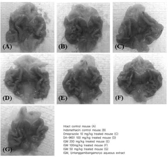
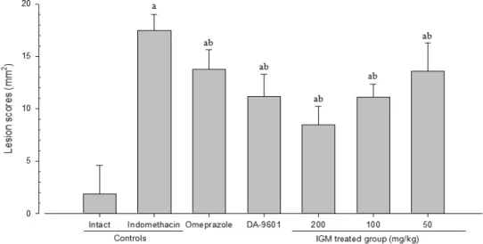
1. Changes in visually inspected lesions In all of the 7 indomethacin-induced gastric mucosal damage groups, hemorrhagic gastric ulcers were observed throughout the gastric mucus layer in broad distribution. While indomethacin control group showed significantly greater (p<0.01) increase in visually inspected lesions than the intact control group, the two reference groups (omeprazole- and DA-9601-treated groups) and three experimental groups (BK-1202-treated groups of three different doses) demonstrated significant decrease (p<0.01) of visually inspected lesions compared with the indomethacin control group (Figs. 1, 2).
The indomethacin control group exhibited changes
in the area affected by gastric mucosal damage as
high as 833.71% with respect to the intact control
Fig. 1. Representative gross observations of the fundic mucosa Intact control mouse (A)
Indomethacin control mouse (B) Omeprazole 10 mg/kg treated mouse (C) DA-9601 100 mg/kg treated mouse (D) IGM 200 mg/kg treated mouse (E) IGM 100mg/kg treated mouse (F) IGM 50 mg/kg treated mouse (G)
IGM, Ijintanggamibangamoryo aqueous extract
group. However, the groups treated with omeprazole, DA-9601, and 200, 100, and 50 mg/kg BK-1202 showed changes of -21.16, -35.88, -51.57, -36.40, and -22.09% with respect to the indomethacin control group.
2. Changes in MPO content
The indomethacin control group showed significantly greater (p<0.01) increase in gastric MPO activity compared to the intact control group, with the amount of change of 302.09% with respect to the intact control group. In contrast, the two reference
groups (omeprazole- and DA-9601-treated groups) and three experimental groups (200, 100, and 50 mg/kg BK-1202-treated groups) demonstrated significant decrease (p<0.01) in gastric MPO activity, -22.55, -31.06, -50.85, -33.02, and -33.91%, respectively, when compared with the indomethacin control group (Fig. 3).
3. Changes in lipid peroxidation
The indomethacin control group showed significantly
greater (p<0.01) increase in gastric MDA content,
i.e. increase in lipid peroxidation, compared to the
Fig. 2. Changes on the gastric lesion score Values are expressed Mean±SD of eight rats a p<0.01 as compared with intact control by LSD test b p<0.01 as compared with indomethacin control by LSD test
intact control group, with the magnitude of change amounting to 570.04% with respect to the intact control group, whereas the omeprazole- and DA-9601-treated reference groups and the 200, 100, and 50 mg/kg BK-1202-treated experimental groups demonstrated significant decrease (p<0.01) in gastric MDA content with changes of -21.51, -37.43, -48.98, -38.66, and -23.11%, respectively, when compared with the indomethacin control group (Table 3).
4. Changes in glutathione (GSH) content The indomethacin control group showed significantly greater (p<0.01) decrease in gastric GSH content compared to the intact control group, exhibiting a change of -59.88% with respect to the intact control group. In contrast, the omeprazole- and DA-9601 -treated reference groups and the 200, 100, and 50 mg/kg BK-1202-treated experimental groups demonstrated significant increase (p<0.01 or p<0.05) in gastric GSH content, marking changes of 20.86, 33.98, 51.40, 34.70, and 22.15%, respectively, when compared with the indomethacin control group
(Table 3).
5. Changes in catalase activity
The indomethacin control group showed significantly greater (p<0.01) increase in gastric catalase activity compared to the intact control group, exhibiting 122.76% increase with respect to the intact control group. In contrast, the omeprazole- and DA-9601 -treated reference groups and the 200, 100, and 50 mg/kg BK-1202-treated experimental groups demonstrated significant decrease (p<0.01) in gastric catalase activity, exhibiting changes of -20.65, -32.49, -45.88, -34.04, and -22.27%, respectively, with respect to the indomethacin control group (Table 3).
6. Changes in superoxide dismutase (SOD) activity
The indomethacin control group showed significantly
greater (p<0.01) decrease in gastric SOD activity
compared to the intact control group, changing
-42.73% with respect to the intact control group. In
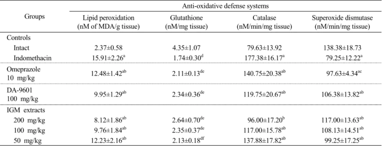
Table 3. Changes on the anti-oxidative defense systems Groups
Anti-oxidative defense systems Lipid peroxidation
(nM of MDA/g tissue)
Glutathione (nM/mg tissue)
Catalase (nM/min/mg tissue)
Superoxide dismutase (nM/min/mg tissue)
Controls
Intact 2.37±0.58 4.35±1.07 79.63±13.92 138.38±18.73
Indomethacin 15.91±2.26a 1.74±0.30d 177.38±16.17a 79.25±12.22a Omeprazole
10 mg/kg 12.48±1.42ab 2.11±0.13de 140.75±20.38ab 97.63±4.34ac DA-9601
100 mg/kg 9.95±1.29ab 2.34±0.36de 119.75±20.67ab 106.38±13.82ab
IGM extracts
200 mg/kg 8.12±1.86ab 2.64±0.70de 96.00±17.20b 117.00±13.63ab 100 mg/kg 9.76±1.84ab 2.35±0.37de 117.00±15.78ab 108.13±14.51ab 50 mg/kg 12.23±2.16ab 2.13±0.18df 137.88±17.82ab 99.25±17.25ab Values are expressed as mean±S.D. of eight rats
IGM, Ijintanggamibangamoryo aqueous extract
a p<0.01 as compared with intact control by LSD test
b p<0.01 and c p<0.05 as compared with indomethacin control by LSD test
d p<0.01 as compared with intact control by MW test
e p<0.01 and f p<0.05 as compared with indomethacin controlby MW test
contrast, the omeprazole- and DA-9601-treated reference groups and the 200, 100, and 50 mg/kg BK-1202-treated experimental groups demonstrated significant increase (p<0.01 or p<0.05) in gastric SOD activity, marking changes of 23.19, 34.23, 47.63, 36.44, and 25.24%, respectively, when compared with the indomethacin control group (Table 3).
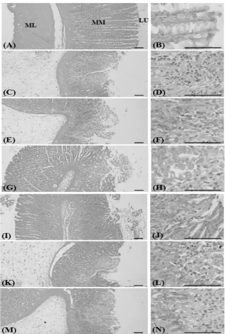
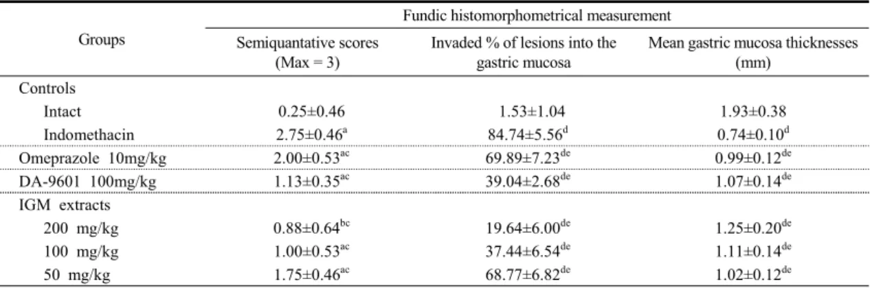
7. Histomorphometric changes
While the indomethacin control group exhibited typical manifestations of gastric ulcer, such as superficial desquamation, necrosis, and inflammatory cell infiltration, such symptoms were considerably suppressed in omeprazole-, DA-9601- and all BK-1202-treated groups (Fig 4). Moreover, while significant increases (p<0.01) were confirmed in the indomethacin control group in terms of the invasion rate of gastric mucosal damage, mean thickness of the gastric mucus layer around the lesion, and semiquantative score, significant decrease (p<0.01) in these pathological conditions were confirmed in the omeprazole-, DA-9601-, and all BK-1202-treated
groups compared to the indomethacin control group (Table 4).
The ratio of gastric mucosal damage in the indomethacin control group to that in the intact control group was as high as 5452.01%, whereas the same for the omeprazole- and DA-9601-treated reference groups and the 200, 100, and 50 mg/kg BK-1202-treated experimental groups were -17.52, -53.92, -76.82, -55.81, and -18.85% with respect to the indomethacin control group.
The change in the gastric mucosa thickness in the indomethacin control group was 61.45% with respect the intact control group, whereas the same for the omeprazole- and DA-9601-treated reference groups and the 200, 100, and 50 mg/kg BK-1202-treated experimental groups were 33.33, 43.94, 67.68, 48.99, and 37.37% with respect to the indomethacin control group.
The change in the gastric mucosa thickness in the
indomethacin control group was 61.45% with respect
the intact control group, whereas the same for the
omeprazole- and DA-9601-treated reference groups
Fig 4. Microscopic appearance of fundic damages Intact control rat (A, B)
Indomethacin control rat (C, D) Omeprazole 10 mg/kg treated rat (E, F) DA-9601 100 mg/kg treated rat (G, H) IGM 200 mg/kg treated rat (I, J) IGM 10 0mg/kg treated rat (K, L) IGM 50 mg/kg treated rat (M, N)
LU, lumen; ML, mucosa layer; SL, submucosa layer; MM, muscle layer All Hematoxylin-Eosin stain
Scale bars = 200 μm
Table 4. Changes on the fundic histomorphometrical analyses Groups
Fundic histomorphometrical measurement Semiquantative scores
(Max = 3)
Invaded % of lesions into the gastric mucosa
Mean gastric mucosa thicknesses (mm)
Controls
Intact 0.25±0.46 1.53±1.04 1.93±0.38
Indomethacin 2.75±0.46a 84.74±5.56d 0.74±0.10d
Omeprazole 10mg/kg 2.00±0.53ac 69.89±7.23de 0.99±0.12de
DA-9601 100mg/kg 1.13±0.35ac 39.04±2.68de 1.07±0.14de
IGM extracts
200 mg/kg 0.88±0.64bc 19.64±6.00de 1.25±0.20de
100 mg/kg 1.00±0.53ac 37.44±6.54de 1.11±0.14de
50 mg/kg 1.75±0.46ac 68.77±6.82de 1.02±0.12de
Values are expressed as mean±S.D. of eight rat IGM, Ijintanggamibangamoryoaqueousextract
a p<0.01 and b p<0.05 as compared with intact control by LSD test
c p<0.01 as compared with indomethacin control by LSD test
d p<0.01 as compared with intact control by MW test
e p<0.01 as compared with indomethacin control by MW test