Received: September 11, 2018 Accepted: October 17, 2018 Trauma and InJury
Correspondence to Hyung Min Kim, M.D.
Department of Emergency Medicine, St.
Vincent’s Hospital, College of Medicine, The Catholic University of Korea, 93 Jungbu-daero, Paldal-gu, Suwon 16247, Korea
Tel: +82-31-249-8377 Fax: +82-31-253-4126
E-mail: [email protected]
utility of Spinal Injury diagnosis using C-Spine lateral X-ray and
Chest, abdomen and Pelvis Computed Tomography in major Trauma
Patients with Impaired Consciousness
Yoon Soo Jang, M.D., Byung Hak So, M.D., Won Jung Jeong, M.D., Kyung Man Cha, M.D., Hyung Min Kim, M.D.
Department of Emergency Medicine, St. Vincent’s Hospital, College of Medicine, The Catholic University of Korea, Suwon, Korea
Purpose: The regional emergency medical centers manage the patients with major
blunt trauma according to the process appropriate to each hospital rather than stan- dardized protocol of the major trauma centers. The primary purpose of this study is to evaluate the effectiveness and influence on prognosis of additional cervical-thorac- ic-lumbar-spine computed tomography (CTL-spine CT) scan in diagnosis of spinal injury from the victim of major blunt trauma with impaired consciousness.
methods: The study included patients visited the urban emergency medical center with
major blunt trauma who were over 18 years of age from January 2013 to December 2016.
Data were collected from retrospective review of medical records. Sensitivity, specifici- ty, positive predictive value, and negative predictive value were measured for evaluation of the performance of diagnostic methods.
results: One hundred patients with Glasgow coma scale ≤13 underwent additional
CTL-spine CT scan. Mechanism of injury was in the following order: driver, pedestri- an traffic accident, fall and passenger accident. Thirty-one patients were diagnosed of spinal injury, six of them underwent surgical management. The sensitivity of chest, ab- domen and pelvis CT (CAP CT) was 72%, specificity 97%, false positive rate 3%, false negative rate 28% and diagnostic accuracy 87%. Eleven patients were not diagnosed of spinal injury with CAP CT and C-spine lateral view, but all of them were diagnosed of stable fractures.
Conclusions: C-spine CT scan be actively considered in the initial examination pro-
cess. When CAP CT scan is performed in major blunt trauma patients with impaired consciousness, CTL-spine CT scan or simple spinal radiography has no significant ef- fect on the prognosis of the patient and can be performed if necessary.
Keywords: Spinal injuries; Tomography; X-rays
INTRODUCTION
Although major trauma centers have been designated nationwide and are in operation, limitations exist in ac- commodating all trauma patients in these centers. There- fore, major trauma patients are often admitted to regional emergency medical centers, and the reality is that each hospital is treating trauma patients with different imaging test and treatment processes, rather than complying with a specialist trauma system. Studies report various multiple organ damages in patients with spinal injury caused by blunt trauma, and spinal fracture occurs in 13% of blunt trauma patients [1]. Neurological complications have also been found in 15-30% of spinal injury patients [2]. Hence, most emergency centers perform brain computed tomog- raphy (CT), as well as chest, abdomen and pelvis CT (CAP CT) when major trauma patients are admitted, and the cervical-thoracic-lumbar-spine CT (CTL-spine CT) scan is also conducted at the discretion of the clinical doctor.
It is relatively easy to determine the necessity of imaging tests if the patient has clear consciousness or if the details of the trauma are known, but in the case of major trauma, it is difficult to completely exclude the possibility of tho- racic-lumbar-spine fracture with history taking, physical examination and neurological tests, even if the patient is conscious [3]. If there is no information about the trauma or if the patient is admitted with impaired consciousness and history taking is not possible, it is more difficult to judge whether there is a spinal injury, making it inevitable to depend on imaging tests, especially CT scans. Vari- ous studies have shown that diagnosis of thoracic-lum- bar-spine injury is possible through reconstructed CAP CT. In addition, advanced trauma life support recom- mends CT without general X-ray for C-spine injuries to reduce exposure to radiation and testing time, but this hasn’t been complied with so far. This study seeks to eval- uate how accurately spinal injury of trauma patients can be diagnosed using C-spine lateral X-ray and CAP CT.
METHODS
Subject patients
This study was conducted with the approval of the Clin-
ical Trial Committee of St. Vincent's Hospital (approval No. VC18ROSI0140). Data was collected for 210 major trauma patients who were admitted to a regional emer- gency medical center during a period of 4 years between January 2013 and December 2016. This emergency center is located in the city center, near two expressways, and the annual average of the number of patients during the study period was approximately 60,000. About 60 to 70 major trauma patients are visited in one year. The criteria for the initial examining doctor in the emergency depart- ment to call the major trauma team included low blood pressure, bicycle or motorcycle injury of more than 30 km per hour, fall from two storey or higher, pedestrian traffic accident hit by a moving car, neurological abnormality, intra-abdominal hemorrhage suspected by focused as- sessment with sonography for trauma, and penetrating wound in the head, cervix-thorax-abdomen at the time of admission. From a total of 210 patients, 100 patients were selected who were over the age of 18, had impaired con- sciousness with Glasgow coma scale (GCS) of less than 13, and conducted C-spine X-ray lateral view, CAP CT and CTL-spine CT at the same time. Patients were excluded if the injury was not by blunt trauma, if the CTL-spine CT wasn’t performed simultaneously at the beginning of ad- mission but conducted additionally during hospitalization after the CAP CT scan was performed, or if the C-spine X-ray or CTL-spine CT was not performed.
Imaging test protocol
The team was activated according to the major trauma team call protocol by emergency department specialists and residents of third-year or higher who examined the major trauma patient initially. Simple radiography, CT scans and FAST were conducted, and the CAP CT was read on the day by the radiologist on duty. After the day of admission, the CTL-spine CT was read by the muscu- loskeletal radiology specialist.
The CT used in this hospital was Discovery CT750HD
(GE healthcare, Chicago, IL, USA), 128 slices, with the
chest using helical thickness 3.75 mm, pitch 0.984:1.000,
the abdomen using helical thickness 5.0 mm, pitch
1.375:1.000, and the spine using helical thickness 2.5 mm,
pitch 0.984:1.000. The CAP-CT scan was reformatted into
the sagittal plane and the coronal plane.
Evaluation and analysis
Based on the results of CTL-spine CT read by the muscu- loskeletal radiology specialist after admission, the records of CAP CT reading by the radiologist on duty and the re- cords of C-spine X-ray reading by the resident or special- ist in radiology department were analysed retrospectively.
R Studio (version 1.1; Rstudio.Inc., Boston, MA, USA) for Windows was used for statistics. In the case of normal distribution of continuous variables, the mean and stan- dard deviation was displayed and the t-test was used for analysis. For non-normal distribution, the median and quartile range was displayed and the Mann-Whitney U test was used for analysis. In the case of nominal variables, frequency and percentage was displayed and the Chi- square test was used, and when the expected frequency of less than five was observed, the Fisher’s exact test was conducted. Sensitivity, specificity, false positive rate and false negative rate were measured for the evaluation of di- agnosis method, and the statistical significance was set to
p<0.005.RESULTS
General characteristics of subjects (Table 1)
Out of the 210 patients who activated the major trauma system during the study period, 100 patients underwent both CAP CT and CTL-spine CT, and had impaired con- sciousness of less than GCS 13. Eighty-four were male, the median GCS was 6.0, and injury severity score (ISS) was 21.0. In the impaired consciousness group, the serum lactate level and ISS were significantly higher. The cause of admission was driver traffic accident, pedestrian traffic accident, fall and passenger traffic accident, in the order of frequency, and the mortality rate was 40%, survival rate 37%, and transfer rate was 23%. In the impaired con- sciousness group, there were 40 patients with spinal inju- ries (40%), who showed no significant differences when compared to the non-spinal injury group in age, gender, initial hemodynamic indicator, and injury score (Table 2).
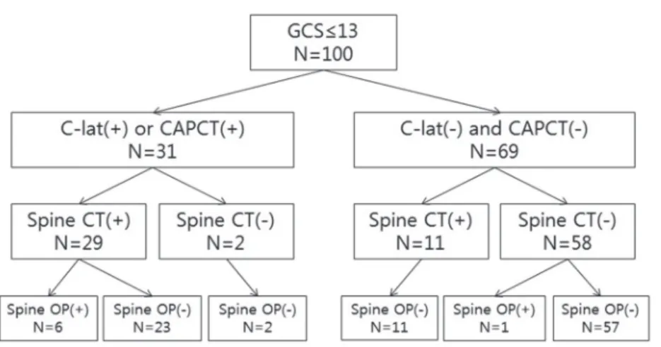
Diagnostic results of the total subject patient group (Fig. 1) Out of the total 100 subject patients, 31 patients were di- agnosed with spinal injury through C-spine lateral view
or CAP CT, and of these 31 patients, only 29 were able to confirm through CTL-spine CT, while 23 patients un- derwent conservative treatment without spinal surgery and six patients underwent surgery. For two patients, it was suspected during C-lateral and CAP CT, but was unconfirmed during the actual CTL-spine CT. Both were C-spine injury patients, and were misdiagnosed in the simple radiography test. Of the 69 patients who were not diagnosed with spinal injury in these tests, 11 were diag- nosed with spinal injury in the CTL-spine CT but recov- ered without surgery. One patient with spinal injury that wasn’t diagnosed even through CTL-spine CT had C3-4, C5-6 cervical myelopathy, which was diagnosed through the patient’s symptoms and the magnetic resonance im- aging test that was conducted later on, and this patient underwent surgery during hospitalization. From spinal injury patients diagnosed through CTL-CT, all six pa- tients who underwent spine surgery were also diagnosed through the C-spine lateral view and CAP CT of the trau- ma series.
C-spine lateral view, CAP CT results (Table 3)
In the total trauma patients, the sensitivity was 77%, spec- ificity was 98%, false positive rate was 2%, false negative rate was 23%, and the diagnostic accuracy was 89%. In patients with impaired consciousness with GCS less than 13, the sensitivity was 72%, specificity was 97%, false positive rate was 3%, false negative rate was 28%, and the diagnostic accuracy was 87%.
Six surgical management patients diagnosed with spi- nal injury using C-spine lateral view, CAP CT (Table 4) Six patients underwent spinal surgery, and they all had C-spine injuries. The average ISS was 17.67.
Eleven patients with spinal injuries not diagnosed using C-spine lateral view, CAP CT (Table 4)
Out of the 11 patients, undiagnosed C-spine injury was
found in seven patients with avulsion fractures of trans-
verse process, spinous process, lamina and spinal body. In
addition, T-spine and L-spine were not diagnosed in three
patients respectively, and they too had avulsion fractures
of transverse process and spinal body.
DISCUSSION
Although there are major trauma centers in operation in each region of Korea, major trauma patients are still admitted to other medical institutions including regional emergency medical centers. Of these medical institutions,
most do not have trauma surgeons on staff or do not have standardized treatment protocols for major trauma patients, so most examinations and treatments are per- formed by initial medical staff in the emergency center.
In the case of imaging tests after initial resuscitation, there are some medical institutions where whole body X-ray
Table 1. Baseline characteristics of study groupVariable All GCS ≤13 GCS >13 p-value
Total 210 (100) 100 (47.6) 110 (52.4)
Age 47.8±18.0 49.3±18.8 46.4±17.6 0.259
Gender (male) 167 (79.5) 84 (84.0) 83 (75.5) 0.173
GCS 14.5 (6.0-15.0) 6.0 (3.0-10.0) 15.0 (15.0-15.0) <0.001
SBP 100.0 (80.0-140.0) 110.0 (80.0-140.0) 100.0 (80.0-130.0) 0.294
DBP 70.0 (50.0-80.0) 70.0 (55.0-90.0) 60.0 (50.0-80.0) 0.257
HR 88.0 (78.0-100.0) 90.0 (78.0-108.0) 88.0 (78.0-99.0) 0.495
Lactate 3.3 (1.9-5.5) 4.2 (2.4-6.7) 2.4 (1.5-4.0) <0.001
BE -7.0±4.6 -8.4±5.2 -5.6±3.3 <0.001
ISS 18.0 (14.5-22.0) 21.0 (17.0-26.5) 16.0 (12.5-20.0) <0.001
RTS 6.9 (5.4-7.8) 5.2 (4.1-6.9) 7.8 (7.1-7.8) <0.001
ELOS 449.0 (225.2-733.8) 318.5 (185.5-647.5) 553.0 (324.0-903.0) <0.001
Trauma mechanism 0.427
Fall down 57 (27.1) 26 (26.0) 31 (28.2)
Driver’s TA 62 (29.5) 30 (30.0) 32 (29.1)
Passenger’s TA 13 (6.2) 7 (7.0) 6 (5.5)
Pedestrian’s TA 60 (28.6) 29 (29.0) 31 (28.2)
Contusion 9 (4.3) 2 (2.0) 7 (6.4)
Penetrating 1 (0.5) 0 (0.0) 1 (0.9)
Unknown 8 (3.8) 6 (6.0) 2 (1.8)
EFAST 0.953
No 113 (53.8) 54 (54.0) 59 (53.6)
Yes, negative 66 (31.4) 32 (32.0) 34 (30.9)
Yes, positive 31 (14.8) 14 (14.0) 17 (15.5)
Spine OP 27 (12.9) 7 (7.0) 20 (18.2) 0.027
Discharge <0.001
Survival 108 (51.4) 37 (37.0) 71 (64.5)
Expire 47 (22.4) 40 (40.0) 7 (6.4)
Transfer 55 (26.2) 23 (23.0) 32 (29.1)
Values are presented as mean±standard deviation, median (interquartile range), or number (%) unless otherwise indicated.
GCS: Glasgow coma scale, SBP: systolic blood pressure, DBP: diastolic blood pressure, HR: heart rate, BE: base excess, ISS: injury severity score, RTS: revised trauma score, ELOS: emergency department length of stay, TA: traffic accident, EFAST: extended focused assessment with sonography in trauma, OP: op- eration.
and CT can be performed in a short time, thanks to the recent development of medical equipment, but most hospitals will prescribe a combination of existing tests by parts. After the initial test, patients may be re-transferred to the CT room depending on the circumstance, to check for spinal injury found in the C-spine lateral view and CAP CT, or at the request of doctors in other depart-
ments. When ordering imaging tests, it is helpful to know the details of trauma, as well as the physical examinations of the patients, results of the FAST, and the symptoms made known by conscious patients, but in the case of pa- tients with impaired consciousness, especially when the details of trauma are unknown, it can be difficult for the initial treating doctor to determine. Therefore, there have
Table 2. Baseline characteristics of study group with GCS ≤13All Spine fracture (-) Spine fracture (+) p-value
Total 100 (100) 60 (60) 40 (40)
Age 49.3±18.8 47.6±19.2 51.8±18.2 0.272
Gender (male) 84 (84.0) 52 (86.7) 32 (80.0) 0.540
GCS 6.0 (3.0-10.0) 6.0 (3.0-9.0) 6.0 (3.0-10.0) 0.963
SBP 110.4±43.3 117.0±37.6 100.0±49.8 0.063
DBP 70.0 (55.0-90.0) 70.0 (60.0-90.0) 60.0 (40.0-80.0) 0.075
HR 91.6±23.7 93.5±21.8 88.6±26.4 0.322
Lactate 4.2 (2.4-6.7) 4.3 (2.4-6.1) 4.2 (2.5-7.5) 0.685
BE -8.4±5.2 -7.7±4.7 -9.4±5.9 0.123
ISS 21.7±7.6 20.4±6.6 23.8±8.7 0.059
RTS 5.2 (4.1-6.9) 5.1 (4.1-6.9) 5.4 (3.7-6.6) 0.614
ELOS 318.5 (185.8-643.2) 248.0 (147.0-617.5) 363.0 (203.5-778.5) 0.106
Trauma mechanism 0.666
Fall down 26 (26.0) 14 (23.3) 12 (30.0)
Driver's TA 30 (30.0) 20 (33.3) 10 (25.0)
Passenger's TA 7 (7.0) 4 (6.7) 3 (7.5)
Pedestrian's TA 29 (29.0) 19 (31.7) 10 (25.0)
Contusion 2 (2.0) 1 (1.7) 1 (2.5)
Unknown 6 (3.0) 2 (3.3) 4 (10.0)
EFAST 0.524
No 54 (54.0) 35 (58.3) 19 (47.5)
Yes, negative 32 (32.0) 18 (30.0) 14 (35.0)
Yes, positive 14 (14.0) 7 (11.7) 7 (17.5)
Spine OP 7 (7.0) 1 (1.7) 6 (15.0) 0.016
Discharge 0.269
Survival 37 (37.0) 26 (43.3) 11 (27.5)
Expire 40 (40.0) 22 (36.7) 18 (45.0)
Transfer 23 (23.0) 12 (20.0) 11 (27.5)
Values are presented as mean±standard deviation, median (interquartile range), or number (%) unless otherwise indicated.
GCS: Glasgow coma scale, SBP: systolic blood pressure, DBP: diastolic blood pressure, HR: heart rate, BE: base excess, ISS: injury severity score, RTS: revised trauma score, ELOS: emergency department length of stay, TA: traffic accident, EFAST: extended focused assessment with sonography in trauma, OP: op- eration.
been various studies on the possibility of excluding spinal injury with only CAP CT, which is performed to check for intra-thoracic or -abdominal injuries in major trauma patients. However, the reality is that many emergency centers still perform additional simple radiography tests or CTL spine tests.
For the evaluation of thoracolumbar injuries, the 5 mm collimation axial CAP CT scan reformatted on the sagittal plan and the coronal plane can diagnose most major frac- tures, and studies report that unless there is a clinical sus-
picion or a need for preoperative planning, CT scan that is not reformatted is sufficient for evaluation, but only in CT with more than 16-slice scanner [4]. Of course, if the distance between the CT sections is thinner, the accuracy of the diagnosis will also increase, but the radiation ex- posure will increase that much more [5]. Other studies have also reported that reformatted CAP CT has better sensitivity in diagnosing T-spine and L-spine fractures than simple radiography [6-8]. In addition, Roos et al.
[9] reported that multi-detected computed tomography
Fig. 1. Diagnostic results for all target groups. GCS:Glasgow coma scale, C-lat: C-spine lateral X-ray, CAPCT: chest abdominopelvic computed tomogra- phy, CT: computed tomography, OP: operation.
Table 3. Sensitivity and specificity of CAPCT+C-spine lateral view
Sensitivity Specificity FP rate FN rate Diagnostic accuracy
All patients 0.77 (0.69-0.86) 0.98 (0.96-1.01) 0.02 (-0.01-0.04) 0.23 (0.14-0.31) 0.89 (0.85-0.93) GCS ≤13 0.72 (0.59-0.86) 0.97 (0.92-1.01) 0.03 (-0.01-0.08) 0.28 (0.14-0.41) 0.87 (0.80-0.94) CAPCT: chest abdominopelvic computed tomography, FP: false positive, FN: false negative, GCS: Glasgow coma scale.
Table 4. Patient operated during spinal injury diagnosed in C-spine lateral view+CAPCT
Patient Age/sex GCS SBP DBP HR Lactate Trauma mechanism ISS Diagnosis
1 35/M 3 60 40 70 2.4 Passenger’s TA 35 Fx. body, C5
2 52/M 4 170 90 80 1.3 Driver’s TA 10 Fx. pedicle, C2, Lt.
Fx. lamina, C7, Lt.
3 55/M 10 100 60 90 1.3 Driver’s TA 15 Fx. body, C3
4 59/M 9 100 70 88 1.7 Pedestrian’s TA 16 Fx. pedicle & transverse process, C2, both
Fx. pedicle & transverse process, C3, Rt.
5 80/M 10 60 40 80 4.1 Unknown 16 Fx. & dislocation, C5
6 26/M 7 130 70 117 7.3 Passenger’s TA 14 Fx. pedicle, C4, Lt.
Fx. lamina, C5, Lt.
CAPCT: chest abdominopelvic computed tomography, GCS: Glasgow coma scale, SBP: systolic blood pressure, DBP: diastolic blood pressure, HR: heart rate, ISS: injury severity score, M: male, TA: traffic accident, Fx.: fracture, Lt.: left, Rt.: right.
according to the trauma protocol of an emergency center is useful in diagnosing thoracolumbar injuries. With the recent development and distribution of advanced CT equipment, is it now possible to obtain information on thoracolumbar fractures without additional exposure to radiation, through the CAP CT with reformatting on the sagittal plane and coronal plane, as well as the picture ar- chiving and communicating system function. However, the available imaging tests will differ depending on the types of CT used in each medical institution.
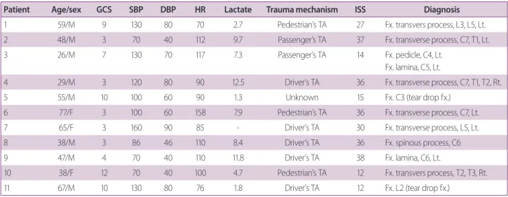
In the results of this study, out of 31 spinal injury pa- tients diagnosed through C-spine lateral X-ray and CAP CT, 29 were identifiable through CTL-spine CT, and of these six patients underwent spinal surgery and they all had C-spine injuries (Table 4). The two patients who were not confirmed in the CTL-spine CT were also C-spine in- jury patients. Similar to existing studies, this result implies that the accuracy of simple radiography test in diagnosing C-spine injury is poor, and also suggests the possibility of diagnosing thoracolumbar injury through CAP CT. There were 11 patients who were diagnosed with spine injury in CTL-spine CT, but not diagnosed in C-spine lateral view and CAP CT, accounting for 26.2% of the total 42 spine injury patients, and as shown in Table 5, most were patients with stable fractures like avulsion fractures in transverse process, spinous process and spinal body, who
do not need emergency surgery. Of these patients, seven were C-spine injury patients, and the diagnosis rate ap- pears to be lower than diagnosing the thoracolumbar area using CAP CT because they were diagnosed with simple radiography of C-spine lateral view. This is consistent with reports from recent studies that CT is more useful than simple radiography in excluding C-spine injury in patients with impaired consciousness caused by blunt trauma [10,11]. For trauma caused by high energy in par- ticular, 37.5% of C-spine injury patients were found to be normal in a simple radiography test but were diagnosed in CT scan [12].
This research institution reformatted the chest CT and was able to identify injury of the 7th C-spine (C7). As shown in studies that report the usefulness of CT scan in evaluating C-spine injuries, it will be helpful to perform C-spine CT to evaluate C-spine injuries in patients in im- paired consciousness in actual emergency centers, or to specify in each hospital’s protocol to include as much of the C-spine as possible when performing an abdominal CT of a trauma patient. Additional thoracolumbar CT or simple radiography of the spine seems unnecessary for patients undergoing CAP CT. This will reduce initial problems for major trauma patients, such as radiation ex- posure, patient monitoring during testing, delayed treat- ment and increased medical costs, etc.
Table 5. Patient with spinal injury not diagnosed in C-spine lateral view+CAPCT
Patient Age/sex GCS SBP DBP HR Lactate Trauma mechanism ISS Diagnosis
1 59/M 9 130 80 70 2.7 Pedestrian’s TA 27 Fx. transvers process, L3, L5, Lt.
2 48/M 3 70 40 112 9.7 Passenger’s TA 37 Fx. transverse process, C7, T1, Lt.
3 26/M 7 130 70 117 7.3 Passenger’s TA 14 Fx. pedicle, C4, Lt.
Fx. lamina, C5, Lt.
4 29/M 3 120 80 90 12.5 Driver’s TA 36 Fx. transverse process, C7, T1, T2, Rt.
5 55/M 10 100 60 90 1.3 Unknown 15 Fx. C3 (tear drop fx.)
6 77/F 3 100 60 158 7.9 Pedestrian’s TA 36 Fx. transverse process, C7, Lt.
7 65/F 3 160 90 85 - Driver’s TA 30 Fx. transverse process, L5, Lt.
8 38/M 3 86 46 110 8.4 Driver’s TA 36 Fx. spinous process, C6
9 47/M 4 70 40 110 11.8 Driver’s TA 38 Fx. lamina, C6, Lt.
10 38/F 12 70 40 100 4.7 Pedestrian’s TA 12 Fx. transvers process, T2, T3, Rt.
11 67/M 10 130 80 76 1.8 Driver’s TA 12 Fx. L2 (tear drop fx.)
CAPCT: chest abdominopelvic computed tomography, GCS: Glasgow coma scale, SBP: systolic blood pressure, DBP: diastolic blood pressure, HR: heart rate, ISS: injury severity score, M: male, TA: traffic accident, Fx.: fracture, Lt.: left, Rt.: right, F: female.
The limitations of this study include the fact that the results were extracted from a small sample of patients at a single regional emergency medical center, and that al- though test reading results that were compared were read by two or more different radiology specialists, they were read a day later than the actual admission of the patient.
This study was also not a blind study. Future improved studies are needed.
CONCLUSION
C-spine lateral X-ray and thoracoabdominal CT showed an accuracy of 87% in diagnosing spine injuries in major trauma patients with impaired consciousness admitted in the emergency center. As the omitted spinal injuries were limited to the cervical spine, it is recommended that C-spine CT scan be actively considered in the initial examination process. Performing spine CT or simple spi- nal radiography test to check for thoracic-lumbar-spine injury in the initial stage of trauma, does not have a significant effect on diagnosing major spinal injury that requires emergency surgery. Therefore, in the case of CAP CT, they are expected to be performed once the patient is stable, for preoperative planning or as required by the surgeon.
REFERENCES
1. Nelson DW, Martin MJ, Martin ND, Beekley A. Evaluation of the risk of noncontiguous fractures of the spine in blunt trau- ma. J Trauma Acute Care Surg 2013;75:135-9.
2. Charles YP, Steib JP. Management of thoracolumbar spine fractures with neurologic disorder. Orthop Traumatol Surg Res 2015;101(1 Suppl):S31-40.
3. Inaba K, DuBose JJ, Barmparas G, Barbarino R, Reddy S, Talv- ing P, et al. Clinical examination is insufficient to rule out tho- racolumbar spine injuries. J Trauma 2011;70:174-9.
4. Smith MW, Reed JD, Facco R, Hlaing T, McGee A, Hicks BM, et al. The reliability of nonreconstructed computerized tomo- graphic scans of the abdomen and pelvis in detecting thora- columbar spine injuries in blunt trauma patients with altered mental status. J Bone Joint Surg Am 2009;91:2342-9.
5. Herzog C, Ahle H, Mack MG, Maier B, Schwarz W, Zangos S, et al. Traumatic injuries of the pelvis and thoracic and lumbar spine: does thin-slice multidetector-row CT increase diagnostic accuracy? Eur Radiol 2004;14:1751-60.
6. Sheridan R, Peralta R, Rhea J, Ptak T, Novelline R. Reformatted visceral protocol helical computed tomographic scanning al- lows conventional radiographs of the thoracic and lumbar spine to be eliminated in the evaluation of blunt trauma patients. J Trauma 2003;55:665-9.
7. Hauser CJ, Visvikis G, Hinrichs C, Eber CD, Cho K, Lavery RF, et al. Prospective validation of computed tomographic screening of the thoracolumbar spine in trauma. J Trauma 2003;55:228- 34; discussion 234-5.
8. Griffey RT, Ledbetter S, Khorasani R. Changes in thoraco- lumbar computed tomography and radiography utilization among trauma patients after deployment of multidetector computed tomography in the emergency department. J Trauma 2007;62:1153-6.
9. Roos JE, Hilfiker P, Platz A, Desbiolles L, Boehm T, Marincek B, et al. MDCT in emergency radiology: is a standardized chest or abdominal protocol sufficient for evaluation of thoracic and lumbar spine trauma? AJR Am J Roentgenol 2004;183:959-68.
10. Widder S, Doig C, Burrowes P, Larsen G, Hurlbert RJ, Kortbeek JB. Prospective evaluation of computed tomographic scanning for the spinal clearance of obtunded trauma patients: prelimi- nary results. J Trauma 2004;56:1179-84.
11. Badhiwala JH, Lai CK, Alhazzani W, Farrokhyar F, Nassiri F, Meade M, et al. Cervical spine clearance in obtunded patients after blunt traumatic injury: a systematic review. Ann Intern Med 2015;162:429-37.
12. Takami M, Nohda K, Sakanaka J, Nakamura M, Yoshida M.
Usefulness of full spine computed tomography in cases of high-energy trauma: a prospective study. Eur J Orthop Surg Traumatol 2014;24 Suppl 1:S167-71.