Introduction
Genus Rhynchocypris belonging to family Cyprinidae (subfamily Leuciscinae) is small fresh- water fishes and comprises 5 species in Korea (Kim and Park, 2002; Kim et al., 2005). Among the Rhynchocypris, R. oxycephalus and R. kum- gangensis coexisted here. Studies on these spe- cies were mostly focused on the early life history, ecology and physiology (Song and Choi, 1997;
Han et al., 1999; Kang and Min, 1999; Song, 2000; Baek et al., 2002; Park et al., 2004; Choi et al., 2005). However, no egg surface structure has
been known until now in Rynchocypris.
Modifications in the egg surface of various tele- osts are closely related to eggs properties: the attachment of the eggs to the substratum in stre- am bed, and sticks or debris; the retention of water or pressure; the protection of embryo; the chorionic respiratory system; the process of water hardening (Blaxter, 1969; Wourms, 1976; Laale, 1980; Groot and Alderdice, 1985; Erickson and Pikitch, 1993; Riehl and Greven, 1993; Thiaw and Mattei, 1996; Riehl and Patzner, 1998). Also, it has been noted that the morphological differ- ences of the adhesive material and the associated inner layers could be a useful aid in the identifi- cation of eggs from different taxa of fish (Johnson and Werner, 1986) and as well as assist in deter-
Structure of Oocyte Surface in Two Korean
Minnow Species, Rhynchocypris kumgangensis and R. oxycephalus (Pisces: Cyprinidae)
Jin-Young Gwak and Jong-Young Park
1,*
PurunKum High School, MuJu-gun, 865, Korea,1Faculty of Biological Sciences, Chonbuk National University and Institute for Biodiversity Research, Chonbuk National University, Jeonju 561-756, Korea
Oocyte surface in two Korean minnows, Rhynchocypris oxycephalus and R. kum- gangensis was examined by light and electron microscope. In two species, the devel- opment of the oocyte was similar, but the follicular layer surrounding full-grown oocyte showed an evident difference. In R. oxycephalus, the follicular layer at the yolk vesicle stage became bilaminar with the retention of its outer squamous cell layer and the acquisition of an inner cuboidal or round cell layer just over the zona radiata. As the oocyte grows, the cuboidal cells of the inner follicular layer began to be replaced by columnar cells. At the yolk granule stage, the columnar cells secreted mucin to their cytoplasm (adhesive materials) and then surround the entire oocyte, as bundles of fence-shaped structures. Whereas, although the follicular layer of R.
kumgangensis had an outer squamous layer and an inner cuboidal or round cell layer at the yolk vesicles as in R. oxycephalus, no inner cells were more changed with the retention of its cuboidal or round cells. Finally, in R. kumgangensis, the adhesive materials did not occur. In Korean two minnows, the structural difference in the oocyte surface seems to be related to their habitats and spawning characteristics as well as taxonomic characters.
Key words : Oocyte structure, Rhynchocypris, adhesive material, zona radiata
*Corresponding author: [email protected]
─
─ 16 ──
mining the phylogenetic relationships between species (Laale, 1980; Groot and Alderdice, 1985;
Riehl and Greven, 1993; Park et al., 2001; Park and Kim, 2001, 2003).
Therefore, we are going to describe the struc- ture of the egg surface and discuss the relation- ship between their habitats and spawning charac- teristics in the coexisted two species, R. oxy- cephalus and R. kumgangensis.
Materials and Methods
10 Females of two species, Rynchocypris oxy- cephalus and R. kumgangensis, were collected from Muju-gucheon dong valley, Muju-gun, Chol- labuk-do, Korea, during the spawning season, May to June, 2006. Muju-Gucheon dong valley where is the upperest stream of the Keum River is keeping cold condition in water, compared to other rivers or streams.
After anaesthetizing with MS222 gravid speci- mens, their full-grown ovaries were excised and
fixed in 10% neutral buffered formaldehyde. We dehydrated these sections through a standard ethanol series to 100%, cleared in xylene and then embedded in wax (Paraplast, Oxford). We deparaffinized 5
µm sections and stained them with Harris hematoxylin, Ehrlich hematoxylin, counter-stained with eosin, and Masson trich- rome stain (Gurr, 1956) for general histology.
Mucin of gland was demonstrated by alcian blue solution (AB) at pH 1.0 and 2.5 (Steedman, 1950;
Lev and Spicer, 1964), and periodic acid-Schiff (PAS) method (Lilllie and Greco, 1947).
For scanning electron microscopy (SEM), the extracted ovaries were prefixed in 2.5% glutaral- dehyde in 0.1 M phosphate buffer at pH 7.2.
Postfixation was performed in 1% osmium tetro- xide in the same buffer. The samples were de- hydrated in a graded alcohol series and critical point dried in CO
2. The dried samples were coat- ed with gold-palladium. Normal eggs and eggs with the follicle-removed were observed with a JEOL JSM-T330A scanning electron microscope.
For photographs and evaluations of the egg sur-
Fig. 1. Developmental stage of the oocyte of Rhynchocypris kumgangensis with Ehrlich haematoxylin and eosin. A, Peri- nucleolus stage (Bar==10µm); B, Yolk vesicle stage (Bar==20µm); C, Middle yolk granule stage (Bar==20µm); D, Mature stage (Bar==20µm). Abbreviations : Arrows, follicular layer; CP, cytoplasm; GV, germinal vesicle; PN, peri- nucleous; YG, yolk granules; YV, yolk vesicle.
face, it was used Carl Zeiss vision (LE REL. 4.4, Germany).
Results
1. General morphology and development
The oocyte development of Rhynchocypris oxy- cephalus and R. kumgangensis was very similar, except for some modifications of inner follicular layer during vitellogenesis in R. kumgangensis.
Their oocytes became developed from peri-nu- cleolus stage which the oocyte has nucleoli locat- ed at the periphery of the germinal vesicle (large nucleus) and was surrounded by a thin, single layer of squamous epithelial cells (Fig. 1A and Fig. 2A). As the oocyte grow, the oocyte increased
in size and its cytoplasm was occupied with yolk materials (vitellogenesis stage) (Fig. 1A to 1D).
At the beginning of the vitellogenesis (early yolk vesicle stage), the follicular layer became two layers (Fig. 1B and Fig. 2B and 2E). By this time, a zona radiata was formed between the follicular layer and ooplasm. As the oocyte grow, the yolk vesicles increased in size and number, and moved to the periphery of the oocyte (late yolk vesicle stage) (Fig. 1C). At this stage, AB-PAS demon- strated that the zona radiata had two distinct layers; an outer thin zone staining strongly and an inner thicker and paler zone which stained weakly (Fig. 3D). As the vitellogenesis proceeds, the cytoplasm became occupied with many dense yolk granules which are a limiting membrane (early yolk granule stage). During later yolk gran-
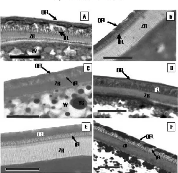
Fig. 2. Developmental stage of the oocyte surface of Rhynchocypris with Ehrlich haematoxylin and eosin (Bar==20µm). A, Peri-nucleolus stage of Rhynchocypris kumgangensis; B, Yolk vesicle stage of R. kumgangensis; C, Yolk granule stage of R. kumgangensis; D, Mature stage of R. kumgangensis; E, Yolk vesicle stage of R. oxycephalus; F, Yolk granule stage of R. oxycephalus. Abbreviations : IFL, inner follicular layer; OFL, outer follicular layer; PN, peri- nucleolus; YG, yolk granules; YV, yolk vesicle; ZR, zona radiata.
ule stage, the yolk granules became fused each other to form several yolk masses (Fig. 1D).
2. Structure of the egg surface
1) Observation by light microscope
R. kumgangensis
By the early yolk vesicle stage, the follicular layer had an outer squamous layer and an inner cuboidal or round cell layer at the yolk vesicles.
But no inner follicular cells were more changed with the retention of its cuboidal or round cells (Fig. 2A to 2D). As the vitellogenesis proceeded, its ooplasm was displaced to many dense yolk granules. By the late yolk granule stage, the oocyte was characterized by the fusion of numer-
ous masses of yolk granules to form a single mass of yolk. At this stage, the zona raidiata was 3.0~
7.0
µm thin.
R. oxycephalus
At early yolk vesicle stage, the inner follicular layer which consisted of a single cuboidal cell layer (inner follicular layer) immediately below the outer squamous layer (outer follicular layer) showed dramatic changes (Fig. 2E to 2F). At early yolk granule stage the follicular bilayer, an outer squamous cell layer and an inner cuboidal cell layer, increased in height. Some of inner cuboidal cells became columnar cell (Fig. 2E). As the oocyte grow, the follicular cells and the zona radiata increased greatly in size, thickness, hei- ght, or number. By this time, the oocyte became
Fig. 3. Special staining reactions on the oocyte surface of Rhynchocypris oxycephalus at the yolk granule stage (Bar==20 µm). A, AB (pH 1.0) reaction; B, AB (pH 2.5) reaction; C, PAS reaction; D, AB-PAS reaction; E, Toluidin blue reac- tion; F, Masson trichrome reaction. Abbreviations: IFL, inner follicular layer; OFL, outer follicular layer; YV, yolk vesicle; YG, yolk granules; ZR, zona radiata.
larger and thicker, and also its inner follicular layer became thicker than the opposition part toward ooplasm (Fig. 2F). At later yolk granule stage, most cuboidal cells of the inner follicular layer was replaced by columnar cells filled with secretions (Fig. 3A to 3F). The inner follicular layer became thicker. By the end of this stage, subsequentially the cytoplasm of the columnar cells lost their cellular integrity with top-situated nucleus and remained as fence-shaped structures, called adhesive material. These secretions were positive to AB (pH 1.0 and 2.5), AB-PAS and Toluidine blue (Fig. 3A to 3F). The thickness of the zona raidata was thicker, 7.0~8.0
µm.
2) Observation by scanning electron microscope
R. kumgangenesis
Without adhesive structure, the full-grown oocyte of R. kumgangensis had a thin zona radia- ta, mean 3.0~7.0
µm, and follicular layer, mean 2~5
µm (Fig. 4A and 4B). The zona radiata show- ing striated appearances in light microscope had
plenty of pore canals and microvilli (Fig. 4A). The microvilli projected from the oocyte toward the follicle cell through the pore canals that traverse the zona radiata (Fig. 4B).
R. oxycephalus
With adhesive structures, the full-grown oocyte had a thicker zona radiata and follicular layer (Fig. 4C and 4D): in the thickness, the zona radi- ata was mean 7.0~8.0
µm and the follicular layer comprising of inner layer (adhesive material) and outer layer mean 4.0~8.0
µm (Fig. 4D). Therewere numerous pore canals in the zona radiata as in R. kumgangensis (Fig. 4D).
Discussion
Two Korean minnows, Rynchocypris oxycepha- lus and R. kumgangensis are sympatric in some regions as Muju Gucheon-dong, the upperest stream of Keum River. The two species have sim- ilar habitats which inhabit valley or the upperest
Fig. 4. Photographs of scanning electron micrograph on the oocyte surface of Rhynchocypris (Bar==20µm). A, Surface structure of oocyte of Rhynchocypris kumgangensis; B, Fine structure of oocyte of R. kumgangensis; C, Surface structure of oocyte of R. oxycephalus; D, Fine structure of oocyte of R. oxycephalus. Abbreviations: IFL, inner folli- cular layer; OFL, outer follicular layer; MV, microvilli; PC, pore canal; YG, yolk granules; ZR, zona radiata.
streams of the river, and spawn in sands or on the surface of gravels during mostly April to June. Also, their eggs were known as a demersal and adhesive type (Song and Choi, 1997; Han et al., 1999; Kim and Park, 2002).
However, through observation on the surface structure of the full-grown oocytes in two min- nows, we could find out a new fact. The adhesive materials to attach the eggs to the substratum or form of egg clumps was not existed in R. kum- gangensis, but occurred in R. oxycephalus. During vitellogenesis, the inner follicular layer of R.
kumgangensis kept the retention of its cuboidal or round cells, but that of R. oxycephalus under- went a dramatic change to secrete adhesive mate- rials. Due to this developmental difference in two species, the entire egg surface of R. oxycephalus became covered with bundles of fence-shaped structure. Based on this histological observation, it could be considered that the egg of R. Kum- gangensis is demersal but non-adhesive, whereas that of R. oxycephalus is demersal and adhesive.
As regards ecological aspect, R. oxycephalus with adhesive materials may be related to more turbid habitat as cool and fast current freshwater sys- tems, compared to R. kumgangensis. Although closely related two species are sympatric, they must have unique environmental factors as egg deposit site, spawning habitat, or behavioral strategy by species. These adhesive structures resembled bullet-shaped structure of Microper- cops swinhonis in appearance and development (Park et al., 1998 and 2001).
In other teleost egg, these morphological cha- racters of egg surface have been used for taxo- nomic perspective (Laale, 1980; Groot and Alder- dice, 1985; Hirai, 1993; Britz et al., 1995; Thiaw and Mattei, 1996; Kim and Park, 1996; Park and Kim, 2001, 2003; Rizzo et al., 2002). In this study, the structure of the oocyte surface revealed inter- specfic difference in two minnows.
By histochemical tests on the adhesive materi- als, the fence-shaped materials of R. oxycephalus was positive to AB, AB-PAS, and PAS reaction, meaning mucoprotein in nature. The nature of adhesive structure in other teleost eggs has been known as neutral mucins of primarily of muco- proteins and mucopolysaccharides, or gelatin (Yorke and McMillan, 1979; Laale, 1980; Abra- ham et al., 1993; Riehl and Bless, 1995; Thiaw and Mattei, 1996; Park et al., 1996, 2001).
Formation of various adhesive structures in teleost eggs may be produced by the following
materials: the follicular epithelium in the goby, Pomatoschistus minutus and some Silurus spe- cies (Kobayakawa, 1985; Abraham et al., 1993);
additional layers produced by the follicular epi- thelium in some Clupea (Gillis et al., 1990); the ovarian wall in the stickleback, Puntungia tymen- sis (Riehl and Greven, 1993); a special follicular epithelium in the perch, Perca fluviatilis (Riehl and Greven, 1993); modification of the zona radi- ata in Cobitidae (Riehl and Patzner, 1998; Park and Kim, 2001, 2003). Based on our results, the adhesive materials in R. oxycephalus may be originated from the follicular epithelium as in Pomatoschistus minutus and some Silurus spe- cies (Kobayakawa, 1985; Abraham et al., 1993).
From our histological and ecological approaches on two minnow species, it seems that the struc- ture of the oocyte surface may be closely related to the systematic relationships as well as the evaluation of environmental factors that deter- mine their habitats and spawning characteristics.
For improving these results about the genus Rh- ynchocypris, however, broad studies on the oo- cyte of other minnows will be needed in future.
References
Abraham, M., V. Hilge, R. Riehl and Y. Iger. 1993. Muco- follicle cells of the jelly coat in the oocyte envelope of the sheatfish (Silurus glanis L.). J. Morphol., 217 : 34~
43.
Baek, H.M., H.B. Song, H.S. Sim, Y.G. Kim and O.K. Kwon.
2002. Habitat segregation and prey selectivity on coha- bitation fishes, Phoxinus phoxinus and Rhynchocypris kumgangensis. Korean J. Ichthyol., 14 : 121~131 (in Korean).
Blaxter, J.H.S. 1969. Development: eggs and larvae. In:
Hoar, W.S., D.J. Randall and E.M. Donalson, (eds), Fish physiology III. Academic Press, New York, pp. 177~252.
Britz, R., M. Kokoscha and R. Riehl. 1995. The anabantoid genera Ctenops, Luciocephalus, Parasphaerichthys, and Sphaerichthys (Teleostei : Perciformes) as a monophy- letic group : evidence from egg surface structure and reproductive behavior. Japan. J. Ichthyol., 42 : 71~79.
Choi, J.S., K.Y. Lee, Y.S. Jang, J.H. Park and O.K. Kwon.
2006. Feeding habit of Rhynchocypris kumgangensis (Cyprinidae) from the Hongcheon River, Korea. Korean J. Environ. Biol., 24 : 29~37 (in Korean).
Erickson, D.L. and E.K. Pikitch. 1993. A histological des- cription of shortspine thornyhead, Sebastolobus alasca- nus, ovaries: structures associated with the production of gelatinous egg masses. Env. Biol. Fish., 36 : 273~282.
Gillis, D.J., B.A. McKewon and D.E. Hay. 1990. Ultrastruc- tural observations on the ovary and eggs, and the devel- opment of egg adhesion in Pacific herring (Clupea haren-
gus pallassi). Can. J. Fish. Aquat. Sci., 47 : 1495~1504.
Groot, E.P. and D.F. Alderdice. 1985. Fine structure of the external egg membranes of five species of Pacific sal- mon and steelhead trout. Can. J. Zool., 63 : 552~566.
Gurr, G.T. 1956. A Practical of Medical and Biological Staining Techniques. New York: Interscience.
Han, H.Y., B.Y. Noh, S.H. O., J.T. Park, J.K. Cho and K.B.
Seong. 1999. Early life history and spawning behavior of Chinese minnow, Rhynchocypris oxycephalus reared in the laboratory. Korean J. Ichthyol., 11 : 177~183 (in Korean).
Hirai, A. 1993. Fine structure of the egg membrane in four species of Pleuronectinae. Japan. J. Ichthyol., 40 : 227
~235.
Johnson, E.Z. and R.G. Werner. 1986. Scanning electron microscopy of the chorion of selected freshwater fishes.
J. Fish. Bio., 29 : 257~265.
Kang, Y.J. and M.S. Min. 1999. Reproductive cycles of Moroco oxycephalus and M. lagowskii in Korea. Korean J. Ichthyol., 11 : 117~125.
Kim, I.S. and J.Y. Park. 1996. Adhesive membranes of oocyte in four loaches (Pisces : Cobitidae) of Korea. Kor- ean J. Zool., 39 : 198~206.
Kim, I.S. and J.Y. Park. 2002. Freshwater fishes of Korea.
Kyo-Hak Publishing Co. Ltd., 463 pp. (in Korean) Kim, I.S., Y. Choi, C.L. Lee, Y.J. Lee, B.Y. Kim and J.H.
Kim. 2005. Illustrated book of Korean fishes. Kyo-Hak Publishing Co. Ltd., 613 pp. (in Korean)
Kobayakawa, M. 1985. External characteristics of the eggs of Japanese catfishes (Silurus). Japan. J. Ichthyol., 32 : 104~106.
Laale, H.W., 1980. The perivitelline space and egg envel- opes of bony fishes; a review. Copeia, 1980 : 210~226.
Lev, R. and S.S. Spicer. 1964. Specific staining of sulphated groups with alcian blue at low pH. Journal of Histo- chemistry and Cytochemistry, 12 : 209
Lillie, R.D. and J. Greco. 1947. Malt diastase and ptyalin in place of saliva in the identification of glycogen. Stain Technology, 22 : 67~70.
Park, I.S., J.H. Im and S.H. Choi. 2004. Effect of dietry thyroxine and iodide on growth of Rhynchocypris oxy- cephalus (Sauvage and Dabry). Korean J. Ichthyol., 16 : 13~18. (in Korean)
Park, J.Y. and I.S. Kim. 2001.Fine structure of oocyte en-
velopes of the related 3 cobitid species in the genus Ik- sookimia (Pisces, Cobitidae). Ichthyol. Res., 48 : 71~75.
Park, J.Y. and I.S. Kim. 2003. Variability of egg envelopes in Korean spined loaches (Cobitidae). Folia Biol., 51 (suppl.) : 187~192.
Park, J.Y., I.S. Kim and Y.J. Lee. 2001. Follicle layer of oocyte of Micropercops swinhonis (Pisces : perciformes).
Korean J. Ichthyol. 4 : 254~260.
Park, J.Y., K.C. Richardson and I.S. Kim. 1998. Develop- mental changes of the oocyte and its enveloping layers, in Micropercops swinhonis (Pisces : Perciformes). Kor- ean J. Biol. Sci., 2 : 501~506.
Riehl, R. and H. Greven, 1993. Fine structure of egg envel- opes in some viviparous goodeid fishes, with comments on the relation of envelope thinness to viviparity, Can.
J, Zool., 71 : 91~97.
Riehl, R. and R. Bless. 1995. First report on egg deposition and egg morphology of the endangered endemic Roman- ian perch. J. Fish Biol., 46 : 1086~1090.
Riehl, R. and R. Patzner. 1998. Mini review: the modes of egg attachment in teleost fishes. Ital. J. Zool., 65 : 415
~420.
Rizzo, E., Y. Sato, B.P. Barreto and H.P. Goninho. 2002.
Adhesiveness and surface patterns of eggs in neutropi- cal freshwater teleosts. J. Fish Biol., 61 : 615~632.
Song, H.B. 2000. Population ecology of fat minnow, Rhyn- chocypris kumgangensis (Cyprinidae) in Korea. Korean J. Ichthyol., 12 : 101~110. (in Korean)
Song, H.B and S.S. Choi. 1997. Development of eggs and larvae of Korean fatminnow, Moroco kumgangensis (Cyprinidae). Korean J. Limnol., 30 : 67~74. (in Korean) Steedman, H.F. 1950. Alcian blue 8G: a new stain for mu- cin. Quarterly Journal of Microscopical Science, 91 : 477~479.
Thiaw, O.T. and X. Mattei. 1996. Ultrastructure of the secondary egg envelope of cyprinodontidae of the genus Epiplatys Gill, 1862 (Pisces, Teleostei). Acta Zool., 77 : 161~166.
Wourms, J.P. 1976. Annual fish oogenesis. I. Differentia- tion of the mature oocyte and formation of the secon- dary egg envelope. Devel. Biol., 50 : 355~366.
Yorke, M.A. and D.B. McMillan. 1979. Nature and cellular origin of the adhesive coats of the lamprey egg (Petro- myzon marinus). J. Morphol., 162 : 313~326.
Received : January 3, 2007 Accepted : March 2, 2007
금강모치와 버들치 난모세포의 표피 구조 곽 진 영∙박 종 영1
푸른꿈고등학교, 1전북대학교 생물과학부, 전북대부설 생물다양성연구소
한국산 버들치속(Rhynchocypris) 어류인 버들치(Rhynchocypris oxycephalus)와 금강모치 (Rhynchocypris kumgangensis) 난모세포의 난막구조에 대해 광학현미경과 전자현미경으로 조사 하였다. 두 종에 있어서 난형성과정은 비슷했으나 난모세포를 둘러싸는 여포세포층(follicular layer)에 있어서는 차이를 보였다. 버들치는 난황포(yolk vesicle)시기에 있어 여포세포층은 안쪽 에 입방형 또는 둥근모양의 세포층(inner follicular layer)이 난막위에 형성되고 그 바깥쪽으로 편평세포층(outer follicular layer)의 2층으로 이루어져 있었다. 난모세포의 발생이 진행됨에 따라 inner follicular layer의 입방형세포는 원주형세포(columnar cell)로 바뀌게 된다. 난황구(yolk
granule)시기에 원주형세포는 세포질에 부착물질인 mucin을 분비해서 난세포 전체를 둘러싸게
된다. 반면에 금강모치의 경우 버들치와 마찬가지로 난황포시기에 안층의 입방형 또는 둥근모양 의 세포층과 바깥층의 편평세포층을 가지게 되지만 안층의 세포는 더 이상 변화를 보이지 않았 으며, 부착물질 또한 형성되지 않았다. 이처럼 한국산 버들치속에 있어 난막의 구조적 차이는 두 종간에 뚜렷한 분류형질로도 이용될 수 있을 뿐 아니라 그들의 서식처 및 산란습성과도 연관이 있는 것으로 생각된다.