한수지 50(4), 388-395, 2017
388
Copyright © 2017 The Korean Society of Fisheries and Aquatic Science pISSN:0374-8111, eISSN:2287-8815 Korean J Fish Aquat Sci 50(4),388-395,2017
Original Article
서 론
생활하수나산업폐수에서유래되는중금속들은기준치이상 초과되면생물에독성을나타내어생태계에많은영향을초래 한다. 특히, 수서생태계로유입된중금속들은퇴적물과결합해 서축적률이높아수서생태계에서식하는생물들은육상생물 에비해더많은영향을받는다(Bryan et al., 1985). 아연(Zn)은 모든생물체의필수적인미량원소이며철부식방지등산업적 이용도가높은금속원소이나, 기준치이상수중에침투할경우 담치류, 굴과같은패류나갑각류의패각형성시탄산칼슘의침 적을저해하며, 독성효과를나타내고(Chang et al., 1996), 지 속적인아연노출은어류의아가미염세포증식및새판괴사등 을일으킨다(Ibrahim et al., 2000). 아연독성연구로는캘리포 니아연안의산업폐수및생활하수에함유된아연성분이 red
abalone Haliotis rufescens 유생에미치는영향(Hunt and An-
derson, 1989)과대만에서마대오분자기를대상으로아연의급
성독성과독성역학(Liao and Lin, 2001) 그리고성장에미치는 영향(Tsai et al., 2004; Liao and Chou, 2005) 등이있으며, 국 내에서는북방전복유생발생및채묘에미치는아연독성연구 (Seo et al., 1999)등이있으나아직까지아연독성연구는미미 한수준이다. 산화아연등중금속미세입자들은세포내미토콘 드리아의붕괴및사멸에의해활성산소생산을증가시켜대사 활성장애를유발한다(Bossy-Wetzel et al., 2004; Ciacci et al., 2012; Guo et al., 2013; Yu et al., 2013). 최근참굴(Crassostrea
gigas)을대상으로산화아연미세입자의독성실험결과를보면
산화아연미세입자를 24시간과 48시간노출하였을때, 아가미 와소화기관의핵막과미토콘드리아의붕괴가일어나고산화스 트레스를유발하여결국폐사하였다(Trevisan et al., 2014). 따
아연 및 알루미늄 용융도금 처리된 강판이 북방전복(Haliotis discus hannai)의 아가미와 간췌장에 미치는 영향
이치훈·박준영·이영돈*
제주대학교 해양과학연구소
Effects of Zinc and Aluminum Hot-dip Galvanized Sheet Steel
on the Gill and Hepatopancreas of the Abalone Haliotis discus hannai
Chi Hoon Lee, Jun Young Park and Young Don Lee*
Marine Science Institute, Jeju National University, Jeju 63333, Korea
We investigated the toxicity of zinc and aluminum hot-dip galvanized sheet steel to abalone Haliotis discus hannai via changes in the gill and hepatopancreas using histological and transmission electron microscopy analysis. Experi- mental groups were composed of one control and four exposure conditions (direct or indirect exposure to zinc and aluminum hot-dip galvanized sheet steel). In the control group, aluminum exposure groups (direct and indirect), and indirect zinc exposure group, abalone mortality was not observed until the end of the experiment, and no histopatho- logical changes were observed in the gill and hepatopancreas. However, the direct zinc exposure group exhibited 100% mortality. Ultrastructural analysis of the cytoplasm of ciliated and microvilli-bearing epithelial cells from gill filaments revealed electron-dense vesicles near the cell membrane and disruption of the nuclear membrane. We also observed swollen mitochondria and a loss of mitochondrial cristae. The hepatopancreas showed similar changes, and we detected highly electron-dense particles within the vesicles. These results suggest that abalone exposed directly to zinc hot-dip galvanized sheet steel experience acute toxicity, causing damage to cell organelles in the gill and hepa- topancreas and, finally, inducing mortality.
Key words: Zinc, Aluminum, Hot-dip galvanized steel sheet, Toxicity, Abalone Haliotis discus hannai
This is an Open Access article distributed under the terms of the Creative Commons Attribution Non-Commercial Licens (http://creativecommons.org/licenses/by-nc/3.0/) which permits unrestricted non-commercial use, distribution, and reproduction in any medium, provided the original work is properly cited.
https://doi.org/10.5657/KFAS.2017.0388 Korean J Fish Aquat Sci 50(4) 388-395, August 2017
Received 30 May 2016; Revised 13 August 2016; Accepted 4 August 2017
*Corresponding author: Tel: +82. 64. 783. 8281 Fax: +82. 64. 782. 8281 E-mail address: [email protected]
아연 및 알루미늄 용융도금강판이 북방전복에 미치는 영향 389
라서, 이연구는연안에서취배수관으로많이이용되는아연및 알루미늄도관이전복에미치는영향을탐색하기위해아연및 알루미늄용융도금강판으로수조를제작한후노출에따른전 복의생존율을조사하였고, 전복아가미와간췌장의세포소기 관의미세구조변화를관찰하였다.
재료 및 방법
실험동물 및 사육환경
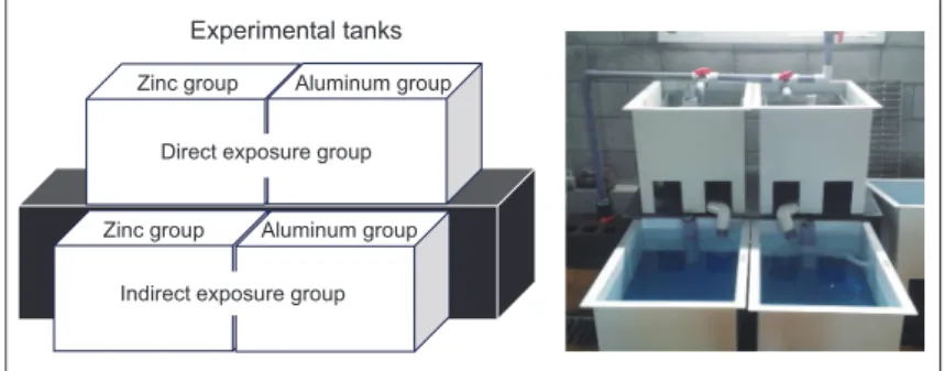
실험에사용된북방전복(Haliotis discus hannai)은각장 7.0- 8.0 cm, 전중량 55-65 g으로외형적이상이없는개체를이용 하였으며, 실험구는대조구, 아연용융도금강판및알루미늄 용융도금 강판 처리구로 나누었고, 실험구별 30마리씩 수용 하였다. 대조구는일반적으로이용되는 ABS수지(acronitrile- butadiene-styren resin)로제작된수조를이용하였고, 처리구는 각각의강판으로제작된수조에직접노출시켜사육한처리구 와강판이해수와접촉후사육수에용해되어있는중금속에영 향을받는지조사하기위해각각의강판으로제작된수조를통 과한해수로사육한간접처리구로구분하였다(Fig. 1). 실험은 총 2회반복하였고, 1차실험은 2014년 8월 6일부터 15일까지, 2차실험은 2014년 22일부터 31일까지각각 10일간실시하였 다. 실험수조는사각수조(50×60×30 cm)를이용하여유수식 으로사육하였고, 각수조당환수량은 4-5 L/min이었으며, 실험 기간중의수온은 21.8-25.0℃, 용존산소는 5.89-7.25 mg/L, 염 분은 31.4-33.4 ppt, pH는 8.22-8.38이었다.
생존율 및 조직학적 변화 분석
실험개체들의생존은실험기간동안각각의처리구별 24시 간간격으로자극시발과촉수의수축반응여부로폐사개체를 판단한후누적폐사율을구하여생존율로환산하여나타냈다. 폐사된개체는조직학적분석을위해아가미, 간췌장을적출하 여 Bouin’s fluid에 24시간고정하였고, 상법인파라핀절편법 으로 5-6 μm의조직절편을제작한후, heamatoxylin과 0.5%
eosin (H-E 염색)으로비교염색하였다. 전자현미경적관찰은 아가미와간췌장의소편을적출하여 2.5% paraformaldehyde-
glutaraldehyde (phosphate buffer, pH 7.4) 용액에 2시간전고 정을한후 1% OsO4 (phosphate buffer, pH 7.4)에 2시간후 고정을하여농도상승순에탄올로탈수하였다. 주사전자현미 경(SEM) 관찰용표본은 amyl acetate로 20분씩 3회치환하고, CO2가스로임계건조한다음 1분동안금이온증착하여 SEM (JSM-7500F, Hitachi, Japan)으로관찰하였다. 투과전자현미 경(TEM)으로관찰하기위해표본은 Epon 812에포매하였고, 이후 LKB-V ultramicrotome을이용하여 1 μm두께로절편을 만들어 toluidine blue로단일염색한후광학현미경하에서관
찰부위를확인한후 80 nm의초박절편을만들었다. 초박절편
은 uranyl acetate와 lead citrate로이중염색을한후 JEOL JEM 1200 EX-II 투과형전자현미경(60 Kv)으로검경하였다.
결 과 전복의 생존율
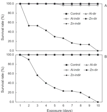
대조구와아연및알루미늄용융도금강판으로제작된사육수 조별노출에따른전복의생존율은다음과같다. 1차실험의전
복생존율은대조구의경우실험종료시 96.7%였다. 처리구별
전복의생존율은알루미늄강판의간접처리구와직접처리구 는실험종료시모두 96.7%, 아연강판의간접처리구는 96.7%
를보였다. 그러나아연강판의직접처리구생존율은처리후 2일경과 53.3%, 5일경과 30.0%, 7일경과 16.7%의누적생 존율을보였으며, 노출후 10일째모두폐사하였다(Fig. 2A).
2차실험기간의 전복생존율은 대조구의경우실험종료시 100%의생존율이었다. 처리구별전복의생존율은알루미늄강
판의간접처리구와직접처리구는실험종료시모두 100%었
고, 아연강판의간접처리구도 100%였다. 그러나아연강판의 직접처리구생존율은처리후 2일경과 90%, 3일경과 56.7%, 5일경과 30.0%의누적생존율을보였으며, 노출후 10일째모 두폐사하였다(Fig. 2B).
아가미와 간췌장의 조직학적 변화
실험기간중아연용융도금강판의직접처리구에서폐사한 전복의아가미와간체장의세포변화를대조구와알루미늄강
Fig. 1. Sketch of the experimental tank containing aluminum and zinc hot-dip galvanized steel sheet.
200μm Gf
Gf Lc
Lc
100μm
Gf
B
10μm C
Sc Sc
A
Hs Hs
El El
40 μm B
Hs El
Hs El
40 μm
C Hs
Hs
El El
40 μm
0.5μm
M M
M B
0.5μm
M
M N
M
A
A
Dg
Dg
100μm B
Dg
Dg
100μm
C
Dg
Dg
100μm
0.5μm N
M
RER
A 0.5μm
M
M
B
0.5μm
N
C 0.5μm
M
M
D
▲ M
0.5μm 0.5μm 0.5μm
ZnP ZnP
ZnP ZnP
ZnP
B C D
ZnP
ZnP ZnP
A
0.5μm
Direct exposure group Experimental tanks
Indirect exposure group Zinc group Aluminum group
Zinc group Aluminum group
0.5μm
N
Hc
A
0.5μmM M
B
0.5μm
N
M
M
C
▲
▲
1.0 μm N
A
0.5μm
0.5μm 1.0μm
N
N
ZnP ZnP
M M
A
B C
이치훈ㆍ박준영ㆍ이영돈 390
판의직접처리구전복과함께광학현미경과전자현미경으로 비교관찰하였다.
아가미
전복아가미는호흡공아래에위치하며, 패각과족부사이에
있는외투막에덮여있다. 아가미는좌·우한쌍으로빗살모양 의새엽이직선상으로뻗어있으며, 새엽의표면은섬모와미세 융모들로덮여있었고, 새엽측면에는다수의분비세포들이관
찰되었다(Fig. 3). 대조구전복과알루미늄및아연용융도금강
판에직접노출된전복의아가미를광학현미경관찰결과, 새 엽은가운데혈림프동을중심으로단층의새엽상피층이둘러 싸고있으며, 새엽상피층은원주형상피세포들로구성되어있 었다. 대조구전복과알루미늄강판직접처리구전복의새엽 은형태적인차이가없었으나(Fig. 4A and B), 아연강판직접 처리구전복의새엽은혈림프동이확장되거나붕괴되는현상 이관찰되었다(Fig. 4C). 대조구와알루미늄강판직접처리구 에서전복의아가미새엽상피층을구성하는섬모와미세융모 의상피세포를투과전자현미경으로관찰한결과, 난형인핵의 핵질은 euchromatin과 heterochromatin이균일하게분포하며
heterochromatin은핵막을따라존재하거나핵질에덩어리형
태로관찰되었고, 크리스테가뚜렷한미토콘드리아가관찰되
었다(Fig. 5). 아연강판직접처리구의전복아가미새엽을투
과전자현미경으로 관찰한결과, 아연미세입자들이존재하였 고, 전자밀도가높은아연미세입자들이세포내유입되는현상 이관찰되었다(Fig. 6). 또한새엽상피층을구성하는섬모와미 세융모상피세포를관찰한결과, 핵들은핵막이붕괴되었으며, 미토콘드리아는크리스테가손실되거나부풀어지고붕괴되는 현상이관찰되었다(Fig. 7).
간췌장
전복간췌장은다수의소화선세관들로이루어져있으며, 소화 선세관은다수의선세포들로구성된다세포선으로관포상형 Fig. 2. Change of survival rate of abalone Haliotis discus hannai
exposed to zinc and aluminum hot-dip galvanized steel sheet (A, 1st experiment; B, 2nd experiment).
0.0 20.0 40.0 60.0 80.0 100.0
Control Al-dir Al-indir Zn-dir Zn-indir
0.0 20.0 40.0 60.0 80.0 100.0
1 2 3 4 5 6 7 8 9 10
Control Al-dir Al-indir Zn-dir Zn-indir
A
B
Survival rate (%)Survival rate (%)
Exposure (days)
Fig. 3. SEM micrographs of the gill of abalone Haliotis discus hannai. A, frontal view of gill; B, gill filament; C, secretory granules in the gill filament. Gf, gill filament; Lc, lateral cilia; Sc, secretory granules.
200μm Gf
Gf Lc
Lc
100μm
Gf
B
10μm C
Sc Sc
A
Hs Hs
El El
40 μm B
Hs El
Hs El
40 μm
C Hs
Hs
El El
40 μm
0.5μm
M M
M B
0.5μm
M
M N
M
A
A
Dg
Dg
100μm B
Dg
Dg
100μm
C
Dg
Dg
100μm
0.5μm N
M
RER
A 0.5μm
M
M
B
0.5μm
N
C 0.5μm
M
M
D
▲ M
0.5μm 0.5μm 0.5μm
ZnP ZnP
ZnP ZnP
ZnP
B C D
ZnP
ZnP ZnP
A
0.5μm
Direct exposure group Experimental tanks
Indirect exposure group Zinc group Aluminum group
Zinc group Aluminum group
0.5μm
N
Hc
A
0.5μmM M
B
0.5μm
N
M
M
C
▲
▲
1.0 μm N
A
0.5 μm
M
M
B 0.5 μm C
M M M
0.5μm
0.5μm 1.0μm
N
N
ZnP ZnP
M M
A
B C
아연 및 알루미늄 용융도금강판이 북방전복에 미치는 영향 391
Fig. 4. Histological change of the gill filament of abalone Haliotis discus hannai in control group (A) and aluminum hot-dip galvanized steel sheet exposure group (B) and zinc hot-dip galvanized steel sheet exposure group (C). A and B, normal morphology of gill filament with he- molymphs sinus and epidermal layer; C, unusual morphology of gill filament with swollen and disrupted hemolymphs sinus. El, epidermal layer; Hs, hemolymph sinus.
200μm Gf
Gf Lc
Lc
100μm
Gf
B
10μm C
Sc Sc
A
Hs Hs
El El
40 μm B
Hs El
Hs El
40 μm
C Hs
Hs
El El
40 μm
0.5μm
M M
M B
0.5μm
M
M N
M
A
A
Dg
Dg
100μm B
Dg
Dg
100μm
C
Dg
Dg
100μm
0.5μm N
M
RER
A 0.5μm
M
M
B
0.5μm
N
C 0.5μm
M
M
D
▲ M
0.5μm 0.5μm 0.5μm
ZnP ZnP
ZnP ZnP
ZnP
B C D
ZnP
ZnP ZnP
A
0.5μm
Direct exposure group Experimental tanks
Indirect exposure group Zinc group Aluminum group
Zinc group Aluminum group
0.5μm
N
Hc
A
0.5μmM M
B
0.5μm
N
M
M
C
▲
▲
1.0 μm N
A
0.5 μm
M
M
B 0.5 μm C
M M M
0.5μm
0.5μm 1.0μm
N
N
ZnP ZnP
M M
A
B C
200μm Gf
Gf Lc
Lc
100μm
Gf
B
10μm C
Sc Sc
A
Hs Hs
El El
40 μm B
Hs El
Hs El
40 μm
C Hs
Hs
El El
40 μm
0.5μm
M M
M B
0.5μm
M M N
M
A
A Dg
Dg
100μm B
Dg
Dg
100μm
C Dg
Dg
100μm
0.5μm N
M
RER
A 0.5μm
M M
B
0.5μm
N
C 0.5μm
M
M
D
▲ M
0.5μm 0.5μm 0.5μm
ZnP ZnP
ZnP ZnP
ZnP
B C D
ZnP
ZnP ZnP
A
0.5μm Direct exposure group
Experimental tanks
Indirect exposure group Zinc group Aluminum group
Zinc group Aluminum group
0.5μm
N Hc
A 0.5μm
M M
B
0.5μm
N
M M
C
▲
▲
1.0 μm N
A
0.5 μm
M
M
B 0.5 μm C
M M M
0.5μm
0.5μm 1.0μm
N
N
ZnP ZnP
M M
A
B C
Fig. 5. TEM micrographs of ciliated and microvilli epithelial cell of gills filaments from abalone Haliotis discus hannai in control (A and B) and aluminum hot-dip galvanized steel sheet exposure group (C). A, normal morphology of nucleus with distinct nuclear envelope (arrowheads) and heterochromatin; B, normal morphol- ogy of mitochondria with distinct cristae (arrows). C, normal morphology of nucleus with intact nuclear envelope (arrowheads) and mitochondrial cristae (arrows). Hc, heterochromatin; M, mito- chondria; N, nucleus.
Fig. 6. TEM micrographs of the gills filaments from abalone Hali- otis discus hannai in zinc hot-dip galvanized steel sheet exposure group. A to C, presence of endocytic vesicles containing electron- dense particles (arrows) and zinc nanoparticles. N, nucleus; ZnP, zinc nanoparticles.
200μm Gf
Gf Lc
Lc
100μm
Gf
B
10μm C
Sc Sc
A Hs
Hs El El
40 μm B
Hs El
Hs El
40 μm
C Hs
Hs
El El
40 μm
0.5μm
M M
M B
0.5μm
M M N
M
A
A Dg
Dg
100μm B
Dg
Dg
100μm
C Dg
Dg
100μm
0.5μm N
M
RER
A 0.5μm
M M
B
0.5μm
N
C 0.5μm
M
M
D
▲ M
0.5μm 0.5μm 0.5μm
ZnP ZnP
ZnP ZnP
ZnP
B C D
ZnP
ZnP ZnP
A
0.5μm Direct exposure group
Experimental tanks
Indirect exposure group Zinc group Aluminum group
Zinc group Aluminum group
0.5μm
N Hc
A 0.5μm
M M
B
0.5μm
N
M M
C
▲
▲
1.0 μm N
A
0.5 μm
M
M
B 0.5 μm C
M M M
0.5μm
0.5μm 1.0μm
N
N
ZnP ZnP
M M
A
B C
태로관찰되었다. 대조구와알루미늄및아연강판직접처리구 간전복간췌장의광학현미경조사결과형태적인차이는관찰 할수가없었다(Fig. 8). 대조구와알루미늄강판직접처리구전
이치훈ㆍ박준영ㆍ이영돈 392
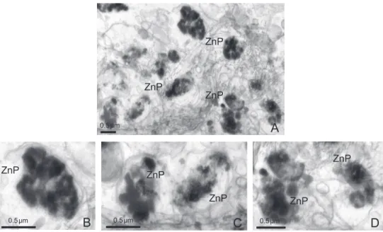
복간췌장의소화선상피세포를투과전자현미경으로관찰한결 과, 난형의핵은염색질이균일하게분포하고있으며뚜렷한핵 막을따라염색질이분포하고있고, 정상적인조면소포체와크 리스테가뚜렷한미토콘드리아가관찰되었다(Fig. 9). 아연강 판직접처리구전복의간췌장을투과전자현미경으로관찰한 결과, 아연미세입자들이존재하였고, 전자밀도가높은아연미 세입자들이세포내유입되는현상이관찰되었다(Fig. 10). 또 한소화선을구성하는상피세포를관찰한결과, 아가미미세융 모상피세포와마찬가지로핵들은핵막이붕괴되었으며, 미토 콘드리아는크리스테가손실되거나부풀어지고붕괴되는현상 이관찰되었다(Fig. 11).
고 찰
산업화및도시화에따른유해독성물질의증가는생태계에
서식하는동·식물의생존및번식, 성장등에위해요인으로작 용한다. 특히해양생태계는외부에서유입된중금속등이쉽게 분해되지않고수서생물의생체내에축적되어형태적, 생리적, 생화학적변화를초래하여수서생물에미치는생태독성에대 한많은연구가진행되고있다(Viarengo, 1985; Dautremepuits et al., 2004; Ju et al., 2006; Son et al., 2015). 미량금속중하나 인아연은생명현상유지에필수적인미량원소로효소의보결 분자단으로서작용하여 면역조절, 성장, 세포보호, 항산화및 항염증작용등각종대사과정을관장한다(Berg, 1990; Maity et al., 2008). 아연은생활용품, 의학관련제품, 산업용제품등 에광범위하게사용되며, 산업용활용은철이나강철의부식을 막기위한용융도금으로해수취배수관및패류종묘생산채묘 기등에많이사용되고있다. 하지만적정농도이상노출하게
되면아연은급성또는만성독성으로작용하게된다(McGeer
et al., 2000; Ho, 2004; Kamaruzzaman et al., 2010; Hwang et Fig. 7. TEM micrographs of ciliated and microvilli epithelial cell of the gills filaments from abalone Haliotis discus hannai in zinc hot-dip galvanized steel sheet exposure group. A, nucleus with disrupted nuclear envelope (arrowheads); B, disrupted mitochondrial cristae (arrows) and showing swollen mitochondria. M, mitochondria; N, nucleus.
200μm Gf
Gf Lc
Lc
100μm
Gf
B
10μm C
Sc Sc
A Hs
Hs El El
40 μm B
Hs El
Hs El
40 μm
C Hs
Hs
El El
40 μm
0.5μm
M M
M B
0.5μm
M
M N
M
A
A
Dg
Dg
100μm B
Dg
Dg
100μm
C
Dg
Dg
100μm
0.5μm N
M
RER
A 0.5μm
M
M
B
0.5μm
N
C 0.5μm
M
M
D
▲ M
0.5μm 0.5μm 0.5μm
ZnP ZnP
ZnP ZnP
ZnP
B C D
ZnP
ZnP ZnP
A
0.5μm
Direct exposure group Experimental tanks
Indirect exposure group Zinc group Aluminum group
Zinc group Aluminum group
0.5μm
N
Hc
A
0.5μmM M
B
0.5μm
N
M
M
C
▲
▲
1.0 μm N
A
0.5 μm
M
M
B 0.5 μm C
M M M
0.5μm
0.5μm 1.0μm
N
N
ZnP ZnP
M M
A
B C
Fig. 8. Histological change of the hepatopancreas of abalone Haliotis discus hannai in control and treatment groups. A, control group; B, aluminum hot-dip galvanized steel sheet exposure group; C, zinc hot-dip galvanized steel sheet exposure group. Dg, digestive gland.
200μm Gf
Gf Lc
Lc
100μm
Gf
B
10μm C
Sc Sc
A
Hs Hs
El El
40 μm B
Hs El
Hs El
40 μm
C Hs
Hs
El El
40 μm
0.5μm
M M
M B
0.5μm
M
M N
M
A
A
Dg
Dg
100μm B
Dg
Dg
100μm
C
Dg
Dg
100μm
0.5μm N
M
RER
A 0.5μm
M
M
B
0.5μm
N
C 0.5μm
M
M
D
▲ M
0.5μm 0.5μm 0.5μm
ZnP ZnP
ZnP ZnP
ZnP
B C D
ZnP
ZnP ZnP
A
0.5μm
Direct exposure group Experimental tanks
Indirect exposure group Zinc group Aluminum group
Zinc group Aluminum group
0.5μm
N
Hc
A
0.5μmM M
B
0.5μm
N
M
M
C
▲
▲
1.0 μm N
A
0.5 μm
M
M
B 0.5 μm C
M M M
0.5μm
0.5μm 1.0μm
N
N
ZnP ZnP
M M
A
B C
아연 및 알루미늄 용융도금강판이 북방전복에 미치는 영향 393
Fig. 9. TEM micrographs of microvilli epithelial cell of the hepatopancreas from abalone Haliotis discus hannai in control (A and B) and aluminum exposure group (C and D). A, normal morphology of nucleus and rough endoplasmic reticulum with ribosomes attached; B, intact mitochondrial cristae (arrows); C and D, normal morphology of nucleus with intact nuclear envelope (arrowheads) and mitochondrial cristae (arrows). M, mitochondria; N, nucleus; RER, rough endoplasmic reticulum.
200μm Gf
Gf Lc
Lc
100μm
Gf
B
10μm C
Sc Sc
A
Hs Hs
El El
40 μm B
Hs El
Hs El
40 μm
C Hs
Hs
El El
40 μm
0.5μm
M M
M B
0.5μm
M
M N
M
A
A
Dg
Dg
100μm B
Dg
Dg
100μm
C
Dg
Dg
100μm
0.5μm N
M
RER
A 0.5μm
M
M
B
0.5μm
N
C 0.5μm
M
M
D
▲ M
0.5μm 0.5μm 0.5μm
ZnP ZnP
ZnP ZnP
ZnP
B C D
ZnP
ZnP ZnP
A
0.5μm
Direct exposure group Experimental tanks
Indirect exposure group Zinc group Aluminum group
Zinc group Aluminum group
0.5μm
N
Hc
A
0.5μmM M
B
0.5μm
N
M
M
C
▲
▲
1.0 μm N
A
0.5 μm
M
M
B 0.5 μm C
M M M
0.5μm
0.5μm 1.0μm
N
N
ZnP ZnP
M M
A
B C
Fig. 10. TEM micrographs of the hepatopancreas from abalone Haliotis discus hannai of zinc hot-dip galvanized steel sheet exposure group.
A to D, presence of endocytic vesicles containing electron-dense zinc nanoparticles. ZnP, zinc nanoparticles.
200μm Gf
Gf Lc
Lc
100μm
Gf
B
10μm C
Sc Sc
A
Hs Hs
El El
40 μm B
Hs El
Hs El
40 μm
C Hs
Hs
El El
40 μm
0.5μm
M M
M B
0.5μm
M
M N
M
A
A
Dg
Dg
100μm B
Dg
Dg
100μm
C
Dg
Dg
100μm
0.5μm N
M
RER
A 0.5μm
M
M
B
0.5μm
N
C 0.5μm
M
M
D
▲ M
0.5μm 0.5μm 0.5μm
ZnP ZnP
ZnP ZnP
ZnP
B C D
ZnP
ZnP ZnP
A
0.5μm
Direct exposure group Experimental tanks
Indirect exposure group Zinc group Aluminum group
Zinc group Aluminum group
0.5μm
N
Hc
A
0.5μmM M
B
0.5μm
N
M
M
C
▲
▲
1.0 μm N
A
0.5 μm
M
M
B 0.5 μm C
M M M
0.5μm
0.5μm 1.0μm
N
N
ZnP ZnP
M M
A
B C