북방전복 (
Haliotis discus hannai
) 의
대식세포이동저해인자 (MIF, Macrophage migration
inhibitory factor) 유전자 동정 및 발현분석
박은희, 신은하, 김영옥, 김동균, 공희정, 김우진, 안철민, 남보혜
국립수산과학원Cloning, characterization, and expression of the macrophage
migration inhibitory factor gene from the pacific abalone (Haliotis
discus hannai)
Eun Hee Park, Eun-Ha Shin, Young-Ok Kim, Dong-Gyun Kim, HeeJeong Kong, Woo-Jin Kim,
Chul Min An and Bo-Hye Nam
Biotechnology Research Division, NIFS, Busan 46083, Korea
ABSTRACT
Macrophage Migration Inhibitory Factor (MIF) are well-defined role as unique cytokine and critical mediator in acute and chronic inflammatory diseases, autoimmune diseases. In this study, we isolated and characterized a full-length of MIF cDNA from the abalone (Haliotis discus hannai). The full-length cDNA of abMIF was of 1264 bp, consisting of a 5’-terminal UTR of 143 bp, an open reading frame of 360 bp and a 3-terminal UTR of 761 bp. The abalone MIF cDNA encodes a 119-amino acid polypeptide with a calculated molecular mass of 13.4 kDa and isoelectric point of 9.07. Multiple alignments and phylogenetic analysis with the deduced abalone MIF protein and showed strong homology with disk abalone (Haliotis discusdiscus). The deduced amino acid sequence of abMIF exhibited homology with other reported MIFs, such as 80%, with that of other disk abalone H. discus discus MIF gene. Quantitative real-time PCR (qRT-PCR) analysis indicated that abMIF was highly expression observed in hapatopacreas, intestine, foot, and gonad of normal conditioned abalone. Even though AbMIF mRNA level in hemocytes was low under the normal condition, it was sharply up-regulated and reached the maximum at 6 h post-infection with Vibrio parahaemolyticus, and then decreased at 24 h post-infection. This result indicates that abMIF plays an important role in responding in the innate immune system.
Keywords: Abalone, Haliotis discus hannai, Macrophage migration inhibitory factor, innate immune system,
Vibrio paraheamolyticus.
서 론
대식세포이동저해인자 (Macrophage Migration Inhibitory
Factor, MIF) 는 활성화된 T림프구의 상층액에서 발견된 최 초의 T세포 유래 사이토카인 (cytokine) 으로 대식세포의 무 작위적인 이동을 억제하는 활성을 가지는 것으로 알려져 왔다 (David, 1966; Bloom and Bennett, 1966).
이후 MIF는 광범위한 포유류 내에서 매우 잘 보존된 단백 질로 많은 연구가 진행되어져 왔는데, 선천성 및 후천성 면역 반응 (Wu et al., 2009; Wang et al., 2010; Li et al., 2010; Kim et al., 2010) 과 각종 염증반응의 초기 단계에서 핵심적 인 역할을 하며, 또한 세포 증식 및 분화 (Calandra et al., 1995), 대식세포 기능 조절 (Onodera et al., 1997), 림프구 면역 (Abe et al., 2001), 종양혈관 신생 (Lue et al., 2002; Received: , 2016; Revised: , 2016; Accepted: , 2016
Corresponding author : Bo-Hye Nam
Tel: +82 (51) 720-2452, e-mail: [email protected] 1225-3480/24633
This is an Open Access article distributed under the terms of the Creative Commons Attribution Non-Commercial License with permits unrestricted non-commercial use, distribution, and reproducibility in any medium, provided the original work is properly cited.
Kamir et al., 2008).
연체동물에서는 소형 전복류인 오분자기 (Haliotis disversicolor supertexta) 에서 처음으로 cDNA와 유전자가 보고되었으며 병원균 감염과 TBT (tributyltin) 노출에 의한 MIF유전자의 발현 양상 변화를 전사체 수준에서 조사되었다 (Wang et al., 2009). 그 후 달팽이 (Acusta despecta sieboldiana) (Baeza Garcia et al., 2010), 진주조개 (Mytilus edulis) (Cui et al., 2011) 와 지중해담치 (Mytilus galloprovincialis) (Parisi et al., 2012) 등에서 유전자와 감 염에 의한 전사체 수준에서의 발현양상 변화 등이 보고되었다. 가장 최근에는 mud crab (Scylla paramamosain) 과 Black tiger shrimp (Penaeus monodon) (Xie et al., 2016) 의 MIF 유전자가 동정, 보고되었다.
본 연구에서는 북방전복 (H. discus hannai) 의 MIF 유전 자의 단편을 대용량 염기서열 분석을 통하여 확보한 후, 유전 자의 전체 염기서열을 확인하고 아미노산 서열을 분석하였다. 정상 전복조직에서 MIF mRNA의 발현과 비브리오균으로 인 위감염 후 시간에 따른 MIF의 발현변화를 전복의 혈구세포에 서 관찰하였다. 본 연구를 통하여 미생물 감염에 대한 북방전 복의 선천성면역 작용에 대한 기초 자료로 활용하고자 하며 향 후 병원균 감염에 대한 바이오마커로서의 활용 가능성에 대해 알아보고자 한다.
재료 및 방법
1. 실험동물 본 연구에 사용된 북방전복 (H. discus hannai) 은 국립수 산과학원 육종연구센터에서 공급받아, 40리터 수조 내에서 18-20℃의 흐르는 해수에 일주일간 순치시킨 후, 외관상 건강 한 개체 (평균 중량; 50 g, 평균 각장; 62 mm, 평균 각폭; 45 mm, 평균 각고; 15 mm) 를 선별하여 사용하였다. 2. 미생물 배양 및 감염 분리를 위해 - 80℃에 보관하였다. 3. 북방전복 MIF 유전자의 전장 cDNA클로닝 북방전복 조직별 (외투막, 아가미, 근육, 간췌장, 혈구) RNAseq 및 De-novo assembly후 BLAST를 이용하여 대용 량 전사체를 분석한 결과, 다른 생물의 MIF 유전자의 5’- 말단 과 높은 상동성을 가지는 약 360 bp의 전복 MIF 전사체 단편 을 찾았다 (data not shown). 전장 전복 MIF (abMIF) cDNA 클로닝을 위해 360 bp 단편의 염기서열을 기초로 하여 abMIF 유전자 특이 프라이머를 디자인하였고 (abMIF ORF-F1: 5’-ACC ATG CCT GTT TTT CTT CTG TTC-3’, abMIF ORF-F2: 5’-ATG CCT GTT TTT CTT CTG TTC AC-3’, abMIFORF-R: 5’-TCA TTT CCA CAG TAC TTC AAA CG-3’, abMIF-5’RACE: 5’-CGT GAA CAG AAG AAA AAC AGG CAT-3’, abMIF-3’RACE: 5’-GGG GTT TCT GTT TGA AAC CAC-3’), SMART RACE cDNA amplification kit (Clontech) 를 이용하여 5’- 및 3’- 말단의 염기서열을 확보하였다. 확보된 5’- 및 3’- 말단의 유전자 단편 을 pGEM-T easy vector (Promega) 에 클로닝하여, E. coli strain XL1-blue로 형질전환 하였다. Plasmid purification kit (Intron) 로 plasmid를 정제한 후에 자동염기 서열분석기 (AB-3730XL, Applied Biosystems) 로 염기서열을 결정하 였다.4. RNA 분리 및RT-PCR 과qPCR을 이용한 발현분석
- 80℃에 보관한 각각의 샘플은 TRIzol reagent (Invitrogen) 를 사용하여 RNA를 분리하였다. 1 μg의 RNA로부터 transcriptor first strand cDNA synthesis kit (Roche Diagnostics) 와 oligo (dT) primer를 이용하여 cDNA를 합성하였다. 북방전 복의 조직에서 MIF 유전자의 발현은 cDNA 서열을 기초로 제작한 프라이머 (abMIF RT-F: 5’-CAT CAA ATT CAC CAG GAA TC-3’, abMIF RT-R: 5’-TGC GCT TTA ACT TAC ATG CAG-3’) 를 이용하여 RT-PCR 과 quantitative
real time-PCR (qPCR) 로 확인하였다. 내재 표준 유전자로 는 북방전복의 18S rRNA (accession no. AY319433; 18S rRNA F: 5’-CTC ACGGAA AGA GCG CGT TTA-3’, 18S rRNA R: 5’-GACTTG CCC TCC AAT AGA TC-3’) 를 사 용하였다. RT-PCR 조건은 다음과 같다: Pre-denaturation 95°C, 5분; denaturation, annealing, extension 각각 95°C 30 초, 55°C 30 초, 72°C 30 초, 35회; extension 72°C, 10 분. 증폭된 PCR 산물은 ethidiumbromide (100 ng/ml) 이 섞 인 1.5% 아가로즈젤에 전기영동하여 확인하였다. qPCR은 FastStart DNA Master SYBR Green I 을 이용하여 Light Cycler system (Roche Diagnostics) 에서 수행하였다. qPCR 조건은 다음과 같다: Taq activation step, 95°C, 10 분; denaturation, annealing, extension 각각 95°C 10 초, 57°C 5 초, 72°C 30 초; 40회 반복. PCR cycle이 끝난 후, 온도를 단계적으로 증가시켜 melting-curve를 분석하였다. abMIF 유전자 발현양은 내재 표준 유전자인 18S rRNA의 상대적인 발현량을 2-ΔΔCT 방법으로 결정하였다 (Livak and Schmittgen, 2001). 모든 실험은 3반복하였으며 데이터는 mean ± SD로 나타내었다. 5. 바이오인포메틱스 분석 abMIF의 cDNA 염기서열로부터 아미노산 서열을 유추하 는 작업은 GENETYX8.0 (SDS Software Development,
Japan) 프로그램을 이용하였다. abMIF의 아미노산 서열을 이용하여 domain 분석은 SMART (http://smart.embl- heidelberg.de) 에서 분석하였다. MIF의 염기서열과 아미노산서열 비교는 NCBI의 BLAST 프로그램 (http://www.ncbi.nlm.nih.gov/ BLAST/) 과 데이터베이스를 이용하였다. 연체동물문에 속하 는 생물 및 기타 생물들의 유사서열을 찾은 후, MEGA 5.0 프 로그램의 CLUSTALW 모듈을 이용하여 multiple sequence alignment를 수행하였으며, phylogenetic tree 는 아미노산 서열에 기초한 Neighbor-Joining 알고리즘으로 분석하여 제 작하였다.
결과 및 고찰
1. abMIF cDNA 염기서열 및 아미노산 분석 북방전복 (H. discus hannai) 의 대용량 유전체 분석 연구 결과 중 119개의 아미노산을 코딩하는 360 bp의 MIF유전자 의 5’- 말단을 포함한 염기서열 일부를 확보하였다. 단편 MIF 유전자 정보를 이용하여 전장 abMIF cDNA를 증폭하기 위하 여 abMIF유전자 특이 프라이머를 이용하여 761 bp와 143 bp가 각각 3'RACE 및 5'RACE 방법으로 증폭되었다 (data not shown). 두 RACE 서열 및 단편 abMIF정보를 조합하 여, 총 1,264bp의 전장 abMIF cDNA를 완성하였다 (Fig. 1). abMIF 유전자는 143 bp의 5'-untranslated region (UTR) Fig. 1. The full-length cDNA and deduced amino acid sequences of abMIF. The deduced amino acid sequence is shownbelow the nucleotide sequence. The initiation code (ATG) and the germination code (TGA) are underlined. The conserved tautomerase activity sites (P2 and K33) are boxed. The conserved oxidoreductase activity (C57) is marked
과 poly-A tail을 포함한 761 bp의 3'-UTR로 구성되어 있었 고, 360 bp의 open reading frame (ORF) 에 119개의 아미 노산을 코딩하고 있었다. 분자량과 등전점은 각각 13.4 kDa 그리고 9.07로 예측되었다. 신호서열예측 프로그램 분석 결과 abMIF는 다른 MIF와 마찬가지로 신호서열을 가지고 있지 않았으며, 상호변이효소 (tautomerase) 의 촉매활성사이트인 proline 잔기와 lysine 잔기가 각각 2번째와 33번째 아미노산 위치에 잘 보존되어 있었다. 또한 57번째 아미노산 위치에는 산화환원효소 (oxidoreductase) 촉매활성사이트인 cysteine 잔기가 잘 보존되어 있었다 (Fig.1). 2. abMIF의 다중서열비교 및 계통수 분석 abMIF의 아미노산 서열을 BLASTP 프로그램을 이용하여 NCBI데이터베이스와 비교해 보았다. 그 결과 abMIF의 아미 노산 서열은 보고된 다른 둥근전복 (H. discus discus) 등의 다른 MIF유전자와 높은 상동성을 보였다 (Fig. 2). 다양한 종 의 abMIF와의 단백질 서열을 비교분석하기 위해, NCBI 데이 터베이스의 다양한 class에서 총 16개 MIF 참고 서열을 선정 하여 phylogenetic tree 를 분석했다. abMIF는 먼저 둥근전 복 (H. discus discus), 오분자기 (H. diversicolor supertexta) 같은 전복류를 포함한 연체동물과 그룹을 형성하 였으며, MIF의 phylogenetic tree는 명확하게 척추동물과 무 Fig. 2. Multiple alignment of the deduced amino acid sequences of abMIF with those of other species. Identical and
similar sites are shown with asterisks (*) and dots (ㆍ), respectively. The gaps (-) are introduced to maximize alignment. The tautomerase catalytic-sites proline residue (P2) and lysine residue (K33) of abMIF are boxed and
marked by triangle (▼), the oxidoreductase catalytic motif in MIF is also boxed and marked by pentacle (★). Sequences were obtained from GenBank: human (Homo sapiens, CAG46452), mouse (Mus musculus, NP034928), rat (Rattus norvegicus, NP112313), pig (Sus scrofa, NP001070681), cow (Bos Taurus, XP594149), frog (Xenopus
laevis, NP001083650 ; NP001083651), zebrafish (Danio rerio, NP001036786), common carp (Cyprinus carpio,
ABY71027), channel catfish (Ictalurus punctatus, NP001187233), tiger puffer (Takifugu rubripes, NP001027889), Bombyx mori (Bombyx mori, NP001040199), guinea pig (Cavia porcellus, XM003477965), Ampioxus (Branchiostoma belcheri, Q698K1), disk abalone (Haliotis discus discus, ACJ65690), small abalone (Haliotis
diversicolor supertexta, ABX76741), mussel (Mytilus galloprovincialis, AEN25591), pearl oyster (Pinctada fucata,
척추동물 그룹으로 나뉘었다. 무척추동물의 MIF는 곤충 MIF 와 연체동물의 MIF를 포함하고 있었다. MIF는 복족류와 이 매패류가 같은 그룹으로 묶였는데, 복족류 그룹에는 H. discus hannai, H. discus discus, H. diversicolor supertexta의 MIF 서열이, 이매패류 그룹에는 Mytilus galloprovincialis 및 Pinctada funcata를 포함하는 MIF 서 열이 포함되었다. 척추 동물 MIF의 homogeneous cluster는 어류, 양서류, 포유류의 MIF가 포함되었다 (Fig. 3).
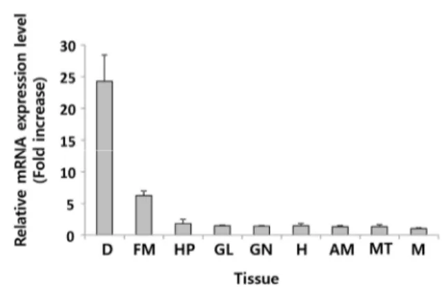
3. abMIF mRNA 의 조직별 발현 분석
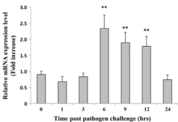
Real-time PCR을 이용하여 abMIF mRNA의 발현을 건 강한 북방전복의 각 조직에서 살펴보았다. 그 결과 abMIF mRNA는 실험에 사용된 9개의 모든 조직에서 다 발현되는 것 이 확인되었으며, 특히 소화관에서 가장 발현이 높았다. 가장 발현량이 낮은 근육과 비교하여 약 25배 이상의 abMIF mRNA가 소화관에서 발현되고 있음이 확인되었다 (Fig. 4). MIF유전자는 건강한 전복에서는 소화관을 제외한 조직에서 기저수준으로 발현하나 소화관에서는 높은 수준으로 발현되는 것으로 보아 전복에 있어서 소화관이 매우 중요한 면역담당 기 관임을 시사한다. 4. 병원균 감염에 의한abMIF mRNA 발현 분석 V. parahaemolyticus로 북방전복을 인위 감염시킨 후, abMIF mRNA의 발현변화를 전복의 혈구세포에서 살펴보았 다. abMIF mRNA의 경우 건강한 전복 혈구세포에서는 기저 수준으로 발현되고 있었으나, V. parahaemolyticus 감염 후 6시간 뒤 발현량이 2.3배 이상 급격히 증가하였으며, 9시간 및 12시간 뒤에는 각각 1.8배와 1.7배로 감소했고, 24시간 뒤에 는 발현이 0시간 수준으로 감소하는 등 급격한 발현변화 양상 을 보였다 (Fig. 5). 오분자기의 경우 간췌장, 근육, 생식소, 아 가미, 외투막, 표피 등 6개의 조직에서 MIF mRNA의 발현양 상을 조사하였으며 간췌장에서 MIF mRNA가 가장 많이 발 현한다고 보고하였으며, 또한 V. parahaemolyticus 감염에 의한 발현양상 변화를 간췌장 조직에서 관찰한 결과 감염 후 24시간째 가장 발현이 많이 유도되었으며, 그 후 96시간째 기 저 수준으로 돌아왔다고 보고하였다 (Wang et al., 2009). 오 분자기 연구에서는 관찰조직에 소화관이 포함되어 있지 않았 으며, 감염에 의한 발현양상 변화도 간췌장에서 관찰하였기에 본 연구결과와 비교가 어려웠다. 그러나 본 연구에서는 abMIF mRNA의 발현량을 살아있는 전복으로부터 채취가 용 Fig. 3. Phylogenetic relationships of abalone GST proteins
with those of other species, calculated using the neighbor-joining method. Sequences were obtained from 15 GenBank: HddMIF (Haliotis discus discus, ACJ65690), Hds (Haliotis diversicolor supertexta, ABX76741), MgMIF (Mytilus galloprovincialis, AEN25591), PfMIF (Pinctada fucata, ADU19847), RnMIF (Rattus norvegicus, NP112313), BtMIF (Bos
Taurus, XP594149), MmMIF (Mus musculus, NP034928), SsMIF (Sus scrofa, NP001070681), HsMIF (Homo sapiens, CAG46452), CcMIF (Cyprinus carpio, ABY71027), IpMIF (Ictalurus punctatus, NP001187233), TrMIF (Takifugu rubripes, NP001027889), BmMIF (Bombyx mori, NP001040199), XlMIF (Xenopus laevis, NP001083650; NP001083651), DrMIF (Daniorerio, NP001036786).
Fig. 4. Tissue-specific mRNA expression of abMIF. Expression
fold-changes of mRNA were detected by qPCR and evaluated by the 2-ΔΔCt method using the abalone 18S
rRNA gene as the internal reference. Data are presented relative to expression in muscle tissue. Error bars represent the SD (n = 3). (D, Digestive duct; FM, Foot muscle; HP, Hepatopancreas, GL, Gills, GN, Gonad, H, Hemocytes; AM, Adductor muscle; MT, Mantle; M, Muscle).
이한 혈구세포에서 관찰하였으며, 결과 또한 감염 후 6시간에 서 9시간이내의 초기에 반응하는 것을 확인하였다. V. parahaemolyticus 감염 시 전복 혈구에서 MIF의 발현이 증 가한다는 것은 MIF가 병원균 감염에 따른 면역 반응에 관여 한다는 것을 의미한다. 이는 병원균 감염에 대한 바이오마커로 의 활용도 가능함을 시사한다. 그러나 abMIF를 병원균 감염 을 예측하는 바이오마커로 활용하기 위해서는 감염 정도에 따 른 발현량의 정량화 연구가 필요하며, 또한 MIF 단백질을 이 용한 간편키트 개발이 필요할 것이다. 또한 어떠한 메커니즘으 로 면역반응에 관여하고 염증반응을 조절하는지에 대한 후속 연구가 필요할 것이다. 지금까지 연구결과를 정리해보면 북방전복 MIF는 아미노산 서열상에 MIF유전자의 특징을 잘 보존하고 있었으며, 발현패 턴을 분석한 결과 병원균 감염에 대한 내재면역반응에도 관여 함을 알 수 있었다. 본 연구를 통해 무척추동물의 면역유전자를 동정하고 병원균 감염에 의한 발현변화를 알아봄으로써 무척추 동물에서 면역시스템을 이해하기 위한 기초자료로 활용하고 병 원균감염에 따른 바이오마커 개발에 활용되리라 기대한다.
결 론
본 연구에서는 북방전복 (H. discus hannai) 의 대용량 염기서열 분석을 통해 MIF 유전자의 전장 cDNA 염기서열을 확보하였다. 북방전복 MIF 유전자의 총 길이는 1,264 bp로 360 bp의 ORF는 총 119개의 아미노산을 코딩하고 있었으며, 등전점은 9.07, 분자량은 13.4 kDa으로 예측되었다. 북방전복사 사
이 논문은 2016년도 국립수산과학원 수산과학연구사업 (R2016024) 의 지원으로 수행된 연구이며 연구비 지원에 감 사 드립니다.REFERENCES
Abe, R., Peng, T., Sailors, J., Bucala, R. and Metz, C.N. (2001) Regulation of the CTL responseby macrophage migration inhibitory factor. The Journal of
immunology, 166: 747-753.
Baeza Garcia, A., Pierce, R.J., Gourbal, B., Werkmeister, E., Colinet, D., Reichhart, J.M., Dissous, C. and Coustau, C. (2010) Involvement of the cytokine MIF in the snail host immune response to the parasite Schistosomamansoni. PLoS pathogens, 6: e1001115. Bacher, M., Meinhardt, A., Lan, H.Y., Mu, W., Metz,
C.N., Chesney, J.A., Calandra, T., Gemsa, D., Donnelly, T., Atkins, R.C. and Bucala, R. (1997) Migration inhibitory factor expression in experimentally induced endotoxemia. The American
journal of pathology, 150: 235-246.
Bernhagen, J., Calandra, T., Mitchell, R.A., Martin, S.B., Tracey, K.J., Voelter, W., Manogue, K.R., Cerami, A. and Bucala, R. (1993) MIF is a pituitary-derived cytokine that potentiates lethal endotoxaemia.
Nature, 365: 756-759.
Bloom, B.R. and Bennett, B. (1966) Mechanism of a reaction in vitro associated with delayed-type hypersensitivity. Science, 153: 80-82.
Calandra, T., Bernhagen, J., Metz, C.N., Spiegel, L.A., Bacher, M., Donnelly, T., Cerami, A. and Bucala, R. (1995) MIF as a glucocorticoid-induced modulator of cytokine production. Nature, 377: 68-71.
Cui, S., Zhang, D., Jiang, S., Pu, H., Hu, Y., Guo, H., Chen, M., Su, T. and Zhu, C. (2011) Amacrophage migration inhibitory factor like oxidoreductase from pearl oyster Pinctadafucata involved in innate immune responses. Fish and shellfish immunology,
Fig. 5. abMIF mRNA expression in hemocytes of abalone
challenged by Vibrio paraheamolyticus. The abMIF mRNA was quantified by its expression relative to the 18S rRNA level. Data are presented relative to expression in muscle tissue. Error bars represent the SD (n = 3). Asterisk (*) represents a significant difference at P < 0.05.
31: 173-181.
David, J.R. (1966) Delayed hypersensitivity in vitro: its mediation by cell-free substances formed by lymphoid cell-antigen interaction. Proceedings of the National
Academy of Sciences of the United States of America,
56: 72-77.
Du, J., Xie, X., Chen, H., Yang, W., Dong, M., Su, J., Wang, Y., Yu, C., Zhang, S. and Xu, A. (2004) Macrophage migration inhibitory factor (MIF) in Chinese amphioxus as a molecular marker of immune evolution during the transition of invertebrate/ vertebrate. Developmental and comparative immunology,
28: 961-971.
Itoa, K., Yoshiura, Y., Ototake, M. and Nakanishi, T. (2008)Macrophage migration inhibitoryfactor (MIF) is essential for development of zebrafish, Daniorerio.
Developmental and comparative immunology, 32:
664-672.
Jaworski, D.C., Jasinskas, A., Metz, C.N., Bucala, R. and Barbour, A.G. (2001) Identification and characterization of a homologue of the pro-inflammatory cytokine Macrophage Migration Inhibitory Factor in the tick, Amblyomma americanum. Insect molecular biology, 10: 323-331. Jin, H.J., Xiang, L.X. and Shao, J.Z. (2007) Molecular
cloning and identification of macrophage migration inhibitory factor (MIF) in teleost fish. Developmental
and comparative immunology, 31: 1131-1144.
Karmir, D., Zierow, S., Leng, L., Cho, Y., Diaz, Y., Griffith, J., McDonald, C., Merk, M., Mitchell, R.A., Trent, J., Chen, Y., Kwong, Y.K., Xiong, H., Vermeire, J., Cappello, M., McMahon-Pratt, D., Walker, J., Bernhagen, J., Lolis, E. and Bucala, R. (2008) A Leishmaniaortholog of macrophage migration inhibitory factor modulates host macrophage responses. The Journal of immunology,
180: 8250-8261.
Kim, S., Miska, K.B., Jenkins, M.C., Fetterer, R.H., Cox, C.M., Stuard, L.H. and Dalloul, R.A. (2010) Molecular cloning and functional characterization of the avian macrophage migrationinhibitory factor (MIF). Developmental and comparative immunology,
34: 1021-1032.
Li, Y., Messina, C., Bendaoud, M., Fine, D.H., Schreiner, H. and Tsiagbe, V.K. (2010) Adaptive immune response in osteoclastic bone resorption induced by orally administered Aggregatibacter actinomycetemcomitans in a rat model of periodontal disease. Molecular oral
microbiology, 25: 275-292.
Livak, K.J. and Schmittgen, T.D. (2001) Analysis of relative gene expression data using real-time quantitative PCR and the 2-△△CT method. Methods,
25: 402-408.
Lue, H., Kleemann, R., Calandra, T., Roger, T. and Bernhagen, J. (2002) Macrophage migration
inhibitory factor (MIF): mechanisms of action and role in diseas. Microbes and infection, 4: 449-460. Marson, A.L., Tarr, D.E. and Scott, A.L. (2001)
Macrophage migration inhibitory factor (mif) transcription is significantly elevated in
Caenorhabditis elegans dauer larvae. Gene, 278: 53-62.
Miska, K.B., Fetterer, R.H., Lillehoj, H.S., Jenkins, M.C., Allen, P.C. and Harper, S.B. (2007) Characterisation of macrophage migration inhibitory factor from Eimeria species infectious to chickens. Molecular and
biochemical parasitology, 151: 173-183.
Onodera, S., Suzuki, K., Matsuno, T., Kaneda, K., Takagi, M. and Nishihira, J. (1997) Macrophage migration inhibitory factor induces phagocytosis of foreign particles by macrophages in autocrine and paracrine fashion. Immunology, 92: 131-137.
Parisi, M.G., Toubiana, M., Mangano, V., Parrinello, N., Cammarata, M. and Roch, P. (2012) MIF frommussel: coding sequence, phylogeny, polymorphism, 3D model and regulation of expression. Developmental and
comparative immunology, 36: 688-696.
Suzuki, M., Takamura, Y., Maeno, M., Tochinai, S., Iyaguchi, D., Tanaka, I., Nishihira, J. and Ishibashi, T. (2004) Xenopuslaevis macrophage migration inhibitory factor is essential for axis formation and neural development. The Journal of biological
chemistry, 279: 21406-21414.
Tan, T.H., Edgerton, S.A., Kumari, R., McAlister, M.S., Roe, S.M., Nagl, S., Pearl, L.H., Selkirk, M.E., Bianco, A.E., Totty, N.F., Engwerda, C., Gray, C.A. and Meyer, D.J. (2001) Macrophage migration inhibitory factor of the parasitic nematode
Trichinellaspiralis. The Biochemical journal, 357:
373-383.
Wang, B., Zhang, Z., Wang, Y., Zou, Z., Wang, G., Wang, S., Jia, X. and Lin, P. (2009) Molecular cloning and characterization of macrophage migration inhibitory factor from small abalone Haliotis diversicolor
supertexta. Fish and shellfish immunology, 27: 57-64.
Wang, F., Shen, X., Guo, X., Peng, Y., Liu, Y., Xu, S. and Yang, J. (2010) Spinal macrophage migration inhibitory factor contributes to the pathogenesis of inflammatory hyperalgesia in rats. Pain, 148: 275-283. Xie, B., Fu, M., Zhao, C., Shi, J., Shi, G., Jiao, Z. and Qiu, L. (2016). Cloning, characterization, and expression of the macrophage migration inhibitory factor gene from the black tiger shrimp (Penaeusmonodon). Fish and shellfish immunology,
56: 489-495.
Zang, X., Taylor, P., Wang, J.M., Meyer, D.J., Scott, A.L., Walkinshaw, M.D. and Maizels, R.M. (2002) Homologues of Human Macrophage Migration Inhibitory Factor from a Parasitic Nematode. The