• 검색 결과가 없습니다.

좌심실 보조장치 이식 환자의 심장눌림증 수술 마취 1례

N/A
N/A
Protected

Academic year: 2021

Share "좌심실 보조장치 이식 환자의 심장눌림증 수술 마취 1례"

Copied!
5
0
0

로드 중.... (전체 텍스트 보기)

전체 글

(1)

© Copyright

Keimyung University School of Medicine 2019

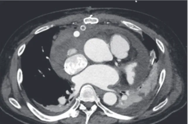
When pericardial tamponade occurs to the left ventricular assist device (LVAD) implanted patients, typical hemodynamic signs of tamponade such as tachycardia and pulsus paradoxus may be masked by LVAD action. For those with normal heart, anesthetic management during pericardial tamponade operation before drainage is to restrict fluid administration and maintain perfusion pressure with vasopressor are recommended. But the things to concern are different in cases of patient with LVAD. Here, we describe a case of performing anesthesia with LVAD implanted patient for pericardial tamponade operation. A 58-year-old male with HeartWare™

(Medtronic, Framingham, MA, USA) LVAD implant was referred for cardiac tamponade surgery. After the induction of general anesthesia, his mean arterial pressure (MAP) decreased to 38 mmHg with device flow 1.8 L/min and device power 2.4 Watts at pump speed 2,400 RPM. Norepinephrine and Epinephrine infusion were initiated. MAP recovered to 70mmHg with device flow 3.7 L/min and power 3.0 Watts after the drainage of 1,200 cc of pericardial fluid. Cardiac tamponade with LVAD implanted patient present with decreased peak flow, mean flow and decreased pulsatility.

LVAD flow depends on pump rotation, preload and afterload. In order to maintain flow in these patients, prevention of preload reduction is important. Since LVAD implantation becoming more popular as Bridge to transplantation and destination therapy, it is important for anesthesiologist to understand the LVAD parameters and factors that affect.

Keywords: Anesthesia, Cardiac tamponade, Heart-assist devices

Received: September 27, 2019 Revised: October 2, 2019 Accepted: October 30, 2019

Corresponding Author: Ji Won Lee, M.D., Department of Anesthesiology and Pain Medicine, Keimyung University School of Medicine, 1095 Dalgubeol-daero, Dalseo-gu, Daegu 42601, Korea

Tel: +82-53-258-7770 E-mail: jiwonlee@dsmc.or.kr

• The authors report no conflict of interest in this work.

Department of Anesthesiology and Pain Medicine, Keimyung University School of Medicine, Daegu, Korea

Sou Hyun Lee, M.D., Ji Won Lee, M.D., Ji Hoon Park, M.D., Ji Seob Kim, M.D.

Anesthetic Management for Cardiac Tamponade in Patient with LVAD

계명대학교 의과대학 마취통증의학교실

이수현 · 이지원 · 박지훈 · 김지섭

좌심실 보조장치 이식 환자의 심장눌림증 수술 마취 1례

(2)

서 론

좌심실 보조 장치 이식 수술은 적절한 약물치료, 심장 재동기화 치료에도 불구하고 말기심부전 증상을 나타내거나 심부전의 급성 악화로 반복적인 입원 치료를 필요로 하는 환자를 대상으로 심장이식 대기 환자 수술(Bridge to transplantation) 또는 심장이식 대체 수술(Destination therapy)을 위해 시행한다. 생체심장이식과 비교했을 때 좌심실 보조 장치는 바로 이식이 가능하며 이식 후 심장 동종 이식 혈관병증, 거부반응, 면역억제제 사용으로 인한 감염과 신독성이 일어나지 않는다는 장점이 있어 많이 시행되고 있다[1]. 현재 미국에서 임상적으로 사용되는 무박동형 좌심실 보조 장치는 애보트 사의 HeartMate-II, HeartMate-III, 메드트로닉 사의 HeartWare 3가지가 있다[2]. 좌심실 보조 장치 이식 수술 후 발생 가능한 합병증은 우심실 부전, 혈전 형성, 출혈, 감염 등이 있으며 출혈로 인한 재수술률은 일반 개흉 수술과 비교하여 4배에 달한다[3]. Slaughter 등[4]이 좌심실 보조 장치 이식 후 1년간 추적관찰을 한 보고에 따르면 심장눌림증 발생률은 5.1%이었다. 심장눌림증이 발생할 경우 혈역학적으로 빈맥, 기이맥이 나타나는데 좌심실 보조 장치 이식 환자는 보조 장치의 활동으로 이러한 혈역학적 변화가 가려지게 된다[5,6]. 따라서 이런 환자들을 대상으로 마취를 할 땐 이식 장치에 대한 이해가 필요하다. 저자들은 HeartWare™ (Medtronic, Framingham, MA, USA) 이식 수술 후 10일째 발생한 심장눌림증으로 응급 혈종 제거 수술을 위한 마취를 경험하였기에 문헌 고찰과 함께 이를 보고하고자 한다.

증 례

환자는 58세, 신장 177.8 cm, 체중 83.5 kg인 남자로 1997년 심한 대동맥판막 협착증으로 금속 대동맥판막 치환술을 받았으며 이후 발생한 확장성 심근병증과 울혈성 심부전의 악화로 2019년 8월 19일 본원에서 HeartWare™이식과 함께 술 후 혈전 발생을 방지하기 위해 생체 인공삽입물을 이용한 대동맥판막 재치환술을 시행하였다. 수술 당일, 수술 후 1일째 출혈로 출혈조절술(Bleeding control)을 시행한 후 좌심실 보조 장치 이식 9일째 평소 70대였던 심박수가 100회 이상으로

상승하였고 HeartWare™기계로부터 반복적인 흡입 경보(Suction alarm)가 발생하였고 박동(Pulsatility)의 감소가 보였으나 평균 동맥압은 70 m m H g대로 유지되었다. 시행한 컴퓨터 단층촬영상 다량의 심장막액, 양측 흉수가 확인되었다(Fig. 1). 좌심실 보조장치 이식 10일째 심장 눌림증으로 인한 응급 혈종 제거 수술을 시행하기 위해 수술실로 입실하였다. 환자는 비강 캐뉼라로 분당 5 L의 산소요법을 받고 있었으며 좌심실 보조 장치 지표는 혈류(Flow) 3.3 L/min, 펌프속도(Pump speed) 2,400 RPM, 파워(Power) 3.0 Watts를 보였다. 마취 전 평균 동맥압은 65 m m H g, 심박수 84회/분 맥박산소포화도는 96% 였다. 좌측 상지의 말초 삽입 중심 정맥관을 통해 측정한 중심 정맥압은 36 mmHg이었다.

안면 마스크로 분당 8 L의 산소를 투여하면서 Midazolam 8 mg을 정주하였고 Remifentanil을 목표농도 조절 주입기(Target controlled infusion)을 사용하여 목표치 농도 1.0 ng/ml로 설정하여 투약하였다. 의식소실 확인 후 마스크를 통한 용수 환기를 시작하였고 Rocuronium 100 mg 정주 1분 뒤 내경 7.5 mm의 기관 내 튜브로 기관 내 삽관을 시행하였다. 삽관 직후 평균 동맥압은 38 mmHg이었으며 혈류 1.8 L/min, 펌프 속도 2,400 RPM, 파워는 2.4 Watts까지 감소하였다(Fig. 2). 즉시 두부 하강 체위를 취한 뒤 Remifentanil 투여를 중지하고 흡입마취제 Sevoflurane 농도는 1.0 Vol%로 낮게 유지하였다.

Phenylephrine 200 mcg, Epinephrine 100 mcg, 3%

염화칼슘 300 mg을 투여하였고 평균 동맥압 49 mmHg 까지 회복되었다. 좌측 목 정맥에 18G 정맥 도관을 삽입해

Fig. 1. Computed Tomography shows large amount of

pericardial effusion with both pleural effusion.

(3)

콜로이드 용액(볼루라이트주 6% 500 ml)을 빠르게 정주하였고 Vasopressin 0.2 unit, Epinephrine 10 mcg을 각각 투약하고 Norepinephrine 0.05 mcg/kg/min, Epinephrine 0.05 mcg/kg/min 속도로 투여하기 시작하였고, 피부 절개 당시 평균 동맥압은 56 mmHg였다. 술 중 경식도 초음파 검사상 우측 심장에 다량의 심장막액이 확인되었고(Fig.

3). 우심방, 우심실 쪽 장액 혈액성 심장막액 1,200 cc를 배액한 후 평균 동맥압은 67 mmHg, 중심 정맥압 24 mmHg, 혈류 3.3 L/min, 펌프속도 2,400 RPM, 파워 3.0 Watts로 회복되었다. 이후 수술 종료 시점까지 Epinephrine 0.04

mcg/kg/min, Norepinephrine 0.02 mcg/kg/min 속도로 유지하였다. 경식도 초음파 검사상 잔여 심장막액이 없는 것을 확인하였고 중등도의 좌측 가슴막 삼출액에 대해 좌측 흉관을 삽입하고 300 cc를 배액한 후 수술을 종료하였다. 총 투여된 교질액은 750 ml, 정질액 900 ml였다. 마취 종료 후 기관삽관 상태로 심장계 중환자실로 이송되었고 이송 직후 평균 동맥압 70 mmHg, 좌심실 보조 장치 지표상 혈류 3.7m/L, 펌프 속도 2,400 RPM, 파워 3.0 Watts 이었다(Fig.

4). 환자는 수술 후 1일째 발관 하였고 Epinephrine, Norepinephrine 지속주입을 중단하였다.

Fig. 2. HeartWare™ monitor parameters during operation. (A) both Power (Watts) and Flow (L/min) decreased soon after the induction of anesthesia (black arrow). (B) Speed (RPM) was constant throughout the operation.

Fig. 3. Intraoperative Transesophageal Echocardiography images immediately after anesthesia induction. (A)

Midesophageal Bicaval View shows pericardial fluid collection adjacent to Right atrium (red arrow heads). (B)

Midesophageal Four Chamber View shows pericardial fluid collection adjacent to Right ventricle (red arrow heads).

(4)

고 찰

HeartWare™좌심실 보조 장치는 좌심실 심첨에서 혈액을 빨아들이는 좌심실 연결로(Inflow cannula), 펌프, 상행 대동맥 근위부로 혈액을 뿜어내는 대동맥 연결로 (Outflow cannula)로 구성되어 있다. HeartWare™이식 환자를 평가할 때는 펌프 속도, 펌프 파워, 혈류, 박동 4가지 지표에 대한 이해가 필요하다. 펌프 속도는 프로펠러의 분당 회전수로 유일하게 조절 가능한 지표이다. 펌프 파워는 프로펠러가 설정된 펌프 속도를 유지하는데 필요한 전력이며 전력의 증가는 혈전으로 인한 펌프의 폐쇄로 인해, 전력의 감소는 혈전이 아닌 이유로 펌프로 유입되는 혈류가 감소할 때 발생한다. 혈류는 펌프 속도, 좌심실 압력(전부하)과 상행대동맥 압력(후부하) 차이인 압력수두(Head pressure)에 의해 결정된다. 펌프 속도를 낮게 하거나 압력수두가 커지면 혈류는 감소하고, 반대의 경우 혈류는 증가한다. 혈류는 심장주기에 따라 변하는데 최대 혈류와 최소 혈류의 차이를 박동이라고 정의한다[2,7-8].

본 증례에서 환자는 이식 수술 후 9일째 흡입 경보가 발생했고 박동이 감소했다. 흡입 경보는 혈류가 기준치의 40% 이상 감소할 때 발생하는데, 좌심실 심근에 의한 좌심실 연결로의 폐쇄가 원인으로 저혈량증, 우심부전, 심장눌림증 시에 나타나며[9] 박동의 감소는 저혈량증, 저혈압,

우심부전, 수축력 저하가 원인으로 나타난다[8]. 흡입 경보가 발생하면 좌심실 연결로로 피를 과도하게 빨아들여 좌심실 연결로의 폐쇄가 일어나지 않도록 펌프 속도를 줄이게 되는데, 본 환자에서 임상 최소속도인 2,400 RPM으로 조절하였으나 효과가 없었다. 본 환자의 경우 저혈량증 의심 하 5% 알부민 200 mL 정주 하였으나 반복해서 흡입 경보가 발생하였고 심장눌림증 의심 하에 CT 촬영 후 환자는 혈종 제거 수술을 위해 수술실로 입실하게 되었다.

HeartWare™이식 환자에서 심장눌림증이 발생할 경우, 심장막공간에 다량의 체액이 축적되어 좌우 심실 충만을 제한하게 되고 이는 좌심실 수축기압을 많이 감소시킨다.

따라서 수축기 압력수두는 증가하고 최대혈류와 박동은

감소하게 된다[8]. 좌심실 보조 장치가 없는 환자에선

심장막액을 배액 하기 전 용적 부하는 배액 후 용적 과부하를

초래할 수 있음으로 전신순환 유지를 위해 혈관 수축제를

주입할 것을 권고하고 있지만[10] 좌심실 보조 장치 이식

기계의 혈류는 전부하 증가와 후부하 감소 시 증가하게 되므로

용적 부하의 중요성도 크다고 볼 수 있다. 본 증례에서

전신마취유도 후 혈류와 파워의 감소, 평균 동맥압 감소는

전부하의 감소로 인한 것으로 보인다. 우심방, 우심실에 다량

존재한 가슴막삼출액 배액 후 평균 동맥압, 혈류, 파워는

수술 전 수준으로 회복되었는데 이는 우심실 박출의

Fig. 4. HeartWare™ monitor after being transferred to Cardiac Intensive Care Unit.

(5)

증가와 이로 인한 좌심실수축기압의 증가로 인한 압력수두의 감소와 전부하의 증가 때문일 것이다.

본 증례 보고에는 몇 가지 제한점이 있다. 마취유도 전, 수술 중 이식장치 지표 중 혈류, 펌프 속도, 파워에 대해선 지속해서 관찰 하였으나, 박동의 변화는 관찰하진 못했다.

HeartWare™이식 환자에서 심장눌림증 발생 시 박동의 감소와 호흡에 따른 박동의 변화를 보이고 회복 시 박동의 증가가 나타나므로[11] 적절히 모니터링을 하였다면 술 중 용적 부하 적정성을 판단하는데 도움이 되었을 것이다. 또한 폐동맥압 감시를 했다면 우심실 기능 및 폐혈관 저항을 고려하여 세심한 마취관리를 시행할 수 있었겠지만 환자 상태가 응급하였기에 폐동맥 카테터를 거치하지 못했다.

요 약

우리나라에서 좌심실 보조 장치 이식 수술이 급여화 되었고 말기 심부전 환자는 점점 증가하고 있으나 이식 심장은 부족하므로 심장이식 대기 환자 수술을 위한 좌심실 보조 장치 이식수술은 점점 늘어날 것으로 보인다. 따라서 앞으로 좌심실 보조 장치를 가진 환자가 수술을 위해 마취를 경험하는 경우가 증가할 것이며 본원에서도 심장이식 및 좌심실 보조 장치 이식 수술이 증가하고 있는 바 이식 장치의 지표와 변동요인에 대해 마취의가 알고 있어야 하겠다.

증례와 같이 좌심실 보조장치 이식을 받은 환자에서 심장눌림증으로 마취를 하게 된다면 전신순환 유지를 위한 혈관수축제 주입 못지않게 압력수두의 증가로 인한 혈류감소를 막기 위해 전부하를 증가시키는 조치도 필요하다.

참 고 문 헌

1. Eisen HJ. Left ventricular assist devices (LVADS):

history, clinical application and complications. Korean

Circ J 2019;49(7):568-85.

2. Tchoukina I, Smallfield MC, Shah KB. Device management and flow optimization on left ventricular assist device support. Crit Care Clin 2018;34:453-63.

3. Miller LW, Guglin M. Patient selection for ventricular assist devices: a moving target. J Am Coll Cardiol 2013;61:1209-21.

4. Slaughter MS, Pagani FD, McGee EC, Birks EJ, Cotts WG, Gregoric I, et al. HeartWare ventricular assist system for bridge to transplant: combined results of the bridge to transplant and continued access protocol trial.

J Heart Lung Transplant 2013;32:675-83.

5. Topilsky Y, Price TN, Atchison FW, Joyce LD. Atypical tamponade hemodynamic in a patient with temporary left ventricular assist device. Interact Cardiovasc thorac Surg 2011;12:832-4.

6. Appleton C, Gillam L, Koulogiannis K. Cardiac tamponade. Cardiol Clin 2017;35:525-37.

7. Birati EY, Rame JE. Left ventricular assist device management and complications. Crit Care Clin 2014;30:607-27.

8. Rich JD, Burkhoff D. HVAD Flow Waveform morphologies: theoretical foundation and implications for clinical practice. ASAIO J 2017;63:526-35.

9. Larose JA, Tamez D, Ashenuga M, Reyes C. Design concepts and principle of operation of the HeartWare ventricular assist system. ASAIO J 2010;56:285-9.

10. O'Connor CJ, Tuman KJ. The intraoperative management of patients with pericardial tamponade.

Anesthesiol Clin 2010;28:87-96.

11. Zalawadiya SK, Lindenfeld J, DiSalvo T. Rapid

diagnosis of cardiac tamponade using pulsatility index

variability in a patient with a HeartWare ventricular

assist device. Circulation 2015;131:e387-8.

수치

Fig. 1. Computed Tomography shows large amount of  pericardial effusion with both pleural effusion.
Fig. 2. HeartWare™ monitor parameters during operation. (A) both Power (Watts) and Flow (L/min) decreased soon  after the induction of anesthesia (black arrow)

참조

관련 문서

To connect multiple External Devices, from [Device-Specific Settings] in the [Device/PLC] window, click [Add Device] to add another External Device.... 

When a process or deviation alarm occurs, the controller switches to the single loop display for the loop with the alarm and displays the alarm code on the screen.. Software

– Head rotation to the left causes extension of left arm and leg and flexion of right arm and leg; head rotation to the right causes extension of right arm and leg and

The “Asset Allocation” portfolio assumes the following weights: 25% in the S&P 500, 10% in the Russell 2000, 15% in the MSCI EAFE, 5% in the MSCI EME, 25% in the

빈칸에 들어갈 말로 가장 적절한 것은?. occurs when a foreign object lodges in the throat, blocking the

1 John Owen, Justification by Faith Alone, in The Works of John Owen, ed. John Bolt, trans. Scott Clark, "Do This and Live: Christ's Active Obedience as the

4,5 However, because of variations in the anatomy of the hard palate between patients, particular care must be taken during donor tissue harvesting not to damage structures

And the State, local governments and the citizen have basic obligation to prevent disasters and, if any disaster occurs, to minimize damages from such a disaster, and to