서론
치아 교합면의 점진적인 마모는 나이가 들면서 진행되는 정상 적인 과정이다.1 생리적 치아 마모는 치조골 및 치아 맹출로 치아 마모를 보상하여 수직 고경이 일정하게 유지 될 수도 있으나 보 상 기전보다 더 빠르게 마모를 보이는 경우는 수직고경이 감소 할 수 있다.2 심한 마모 환자에서 수직고경의 감소 여부는 논란 이 되어왔다. Briggs와 Bishop,3 Hemmings 등,4 Sato 등5은 수직 고경의 감소가 발생될 경우 수직고경의 거상을 통해 보철적으로 회복해야 한다고 주장하였으며, Dahl과 Krogstad,6 Ramfjord와 Blankenship,7 Dawson 등은 교모에 의한 백악질의 성장, 치아의 정출, 치조골의 보상성장으로 인하여 수직고경이 유지된다고 주 장하였다.
치아 상실 및 치아 마모로 교합이 붕괴된 환자는 저작기능의 감소, 심미성 감소, 전방유도 기능의 상실 등 다양한 문제가 발생 할 수 있다.8 마모된 치아를 회복 시키기 위해서는 수직 고경 변 경을 통한 전악 수복을 고려할 수 있다.9 적절한 수복을 위해서 는 치료 전 정확한 진단 과정이 필수적이다. 수직고경 상실 여부 의 평가, 상실된 부분의 수복방법의 결정, 교합 양식의 선택 등에 대해 평가 해야 하며, 임상적인 검사를 하여 잔존치 발치 여부 및 무치악 부위에 대해 전반적인 평가를 시행한다.
본 증례의 환자는 새로운 수직 고경을 정한 후 임시 국소의치 와 임시 보철물을 장착하여 환자의 적응도를 평가 함으로써 변 경된 수직고경의 적절성 여부를 판단하였다. 적응한 수직고경을 참고하여 최종적으로 상악은 고정성 보철물과 가철성 국소의치, 하악은 고정성 보철물로 수복하였다. 이에 기능적, 심미적으로
치아 마모로 인한 수직고경감소와 과개교합을 가진 환자에서 전악 수복 증례
서성용 이나영* 강정경
중앙보훈병원 치과보철과
A case of full mouth rehabilitation in patient with loss of vertical dimension and deep bite due to tooth wear
Seong-Yong Seo, Na-Young Lee*, Jeong-Kyung Kang
Department of Prosthodontics, Veterans Health Service Medical Center, Seoul, Republic of Korea
The collapse of the posterior occlusion destroys the normal occlusal plane and causes excessive wear reducing the vertical dimension. Reduced vertical dimension of occlu- sion causes not only aesthetic and functional problems but also overloading on the temporomandibular joints and abnormalities of muscle nerve system. In order to improve the collapsed occlusal relationship, it is necessary to consider the change of the vertical dimension. It is necessary to make a precise diagnosis and analysis before the treat- ment and to evaluate the adaption of patient to the new vertical dimension of occlusion. A patient with excessive overbite often has occlusal problems of tooth wear and tooth eruption. Considering these considerations, overall prosthodontic restoration is required to solve the problem. A patient of 68 year old man in this case who suffered major tooth wear and maxillary posterior teeth loss was treated with elevation of vertical dimension of occlusion by maxillary removable dental prosthesis and mandibular fixed prosthesis. (J Korean Acad Prosthodont 2018;56:31-9)
Keywords: Deep bite; Partially edentulous patient (partial edentulism); Removable dental prostheses; Full mouth rehabilitation
*Corresponding Author: Na-Young Lee
Department of Prosthodontics, Veterans Health Service Medical Center 53 Jinhwangdo-ro 61-gil, Gangdong-gu, Seoul 05368, Republic of Korea +82 (0)2 2225 1658: e-mail, [email protected]
Article history: Received July 10, 2017 / Last Revision September 4, 2017 / Accepted September 5, 2017
2018 The Korean Academy of Prosthodontics
This is an Open Access article distributed under the terms of the Creative Commons Attribution Non-Commercial License (http://creativecommons.org/
licenses/by-nc/3.0) which permits unrestricted non-commercial use, distribution, and reproduction in any medium, provided the original work is properly cited.
c cc
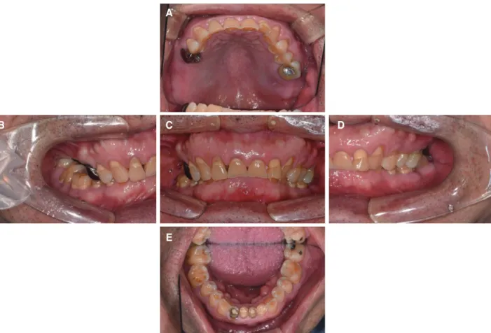
본 환자는 68세 남성으로 상악 좌, 우측 어금니를 상실하여 전 반적인 치료를 받고 싶다는 주소로 내원하였다. 의과적 병력은 고혈압 약을 복용 중 이었으며 턱관절 및 기타 저작근 장애, 비기 능적 악습관은 없었다. 구강내 소견은 상악은 상악 우측 제1, 2 대구치, 상악 좌측 제2대구치가 상실되었으며 상악 우측 제2소
근을 가지는 사각형의 안모 형태로 보아 강한 교합력을 의심할 수 있었으며, 측면에서는 상악 전돌양 안모가 관찰되었다. 측두 하악 관절 방사선 사진상 과두의 위치는 정상이었다 (Fig. 1, Fig.
2, Fig. 3, Fig. 4).
Fig. 1. Intraoral photograph before treatment. (A) Upper, (B) Right, (C) Frontal, (D) Left, (E) Lower.
B C
A
E
D
Fig. 2. Panoramic radiograph before treatment.
2. 진단모형 분석 및 보철 수복공간 평가
모형 분석 시 순측 전정 최저점으로부터 전치 절단연 까지 거 리는 상악 20 mm, 하악 18 mm로 한국 성인 유치악 평균인 상악 20.8 mm, 하악 17.3 mm와 크게 차이 나지 않았으며10 상하악 순 측 전정간 거리는 35 mm로 평균인 35 mm와 같았다. 상악 전치 의 길이는 7 mm, 하악 전치의 길이는 6 mm로 정상수치인 상악 10 mm, 하악 8 mm 보다 작았다. Overbite는 7 mm로 깊은 수직 피개량을 보였고, overjet은 2 mm로 측정되었다. Freeway space
는 7 - 8 mm로 평균 2 - 4 mm 보다 크게 측정되었으며11,12 Willis 법13으로 안면 계측시 동공에서 구각까지의 거리는 81 mm, 코끝 에서 턱끝까지의 거리는 75 mm로 작았다. 우측 구치부는 지지 상실로 인한 하악 우측 대구치 부위의 정출로 보철 수복 공간이 부족한 상태였다. 상악 전치 및 상악 좌우측 소구치의 구개측 마 모로 과도한 수직피개를 가지는 과개 교합이 발생한 것으로 생 각되며 구치부 상실로 인한 대합치의 정출과 심한 마모로 인해 역스피만곡(reverse curve of spee)과 역윌슨만곡(reverse curve of willson)이 관찰되었다. 다양한 수직고경 평가방법으로 평가 한 결과 상기 환자는 과도한 치아 마모가 있으며 수직 고경의 상 실이 있어 수복을 위한 악간 공간이 부족한 Turner의 분류 I으로 진단하였다 (Fig. 5, Fig. 6).
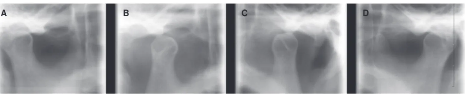
Fig. 3. TMJ series before treatment. (A) Rt. close, (B) Rt. opening, (C) Lt. opening, (D) Lt. close.
A B C D
Fig. 4. Facial photographs before treatment. (A) Frontal view, (B) Lateral view.
A B
Fig. 5. Diagnostic wax-up model. (A) Right, (B) Frontal, (C) Left.
A B C
Fig. 6. Vertical dimension of occlusion evaluation.
81 mm
75 mm
치료 하기로 결정하였다. 환자의 수술에 대한 불안감과 경제적 이 유로 상악 무치악부는 가철성 국소의치로 수복하고 모든 잔존치 는 도재-금속주조관으로 수복하기로 결정하였다. 발치한 하악 우측 제2대구치는 추후 임플란트 치료를 하기로 결정하였다.
4. 치료과정
수직고경 거상량을 결정하기 위해 예비인상을 채득하여 진 단 모형을 제작하였다. 구강 내에서 교합평면을 동공간선과 Camper’s plane에 맞춰서 설정하였다. 양손 수조작술을 이용하 여 중심위로 유도하여 악간 관계 기록을 채득한 후 반조절성 교 합기(Artex, Girrbach Dental, Pforzheim, Germany)를 이용하 여 마운팅을 시행하였다. 보철물 제작에 필요한 공간을 확보하 고 과개교합을 개선하기 위해 상하악 견치를 기준으로 수직고경 을 4 mm 거상하기로 결정하였다.14,15 새로 설정한 수직 고경에 맞추어 진단용 납형제작 후 상악 임시의치를 포함하는 임시 보 철물을 제작하였다 (Fig. 7). 교합양식은 좌, 우측은 군기능 교합
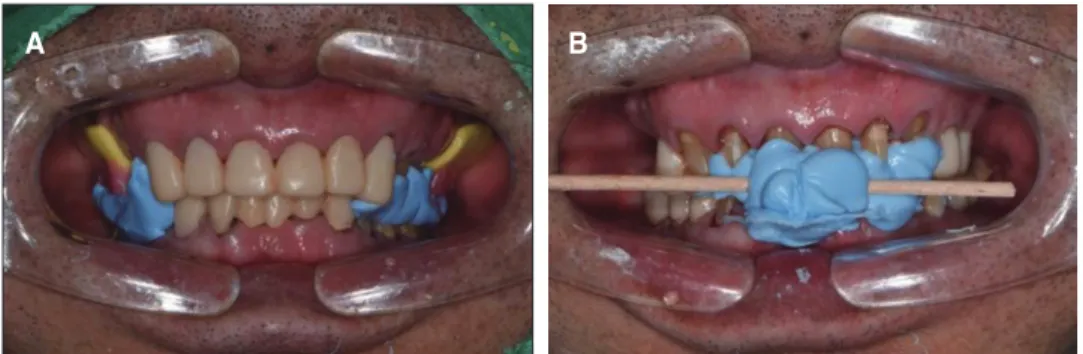
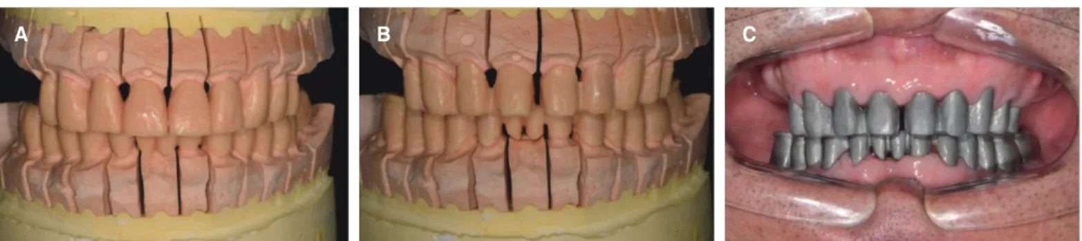
최종 치아 삭제 후 개인트레이와 polyvinyl siloxane (Imprint II Garant. Light Body, 3M ESPE, St. Paul, MN, USA)으로 인 상 채득 후 작업모형과 다이를 제작하였다. 임시 보철 수복 기간 동안 적응된 환자의 수직고경을 그대로 유지하기 위해 임시 수 복물을 부분적으로 제거하여 악간 기록을 채득하고 안궁이전을 하여 마운팅을 시행하였다 (Fig. 8). 임시 보철물상의 전방 및 측 방 유도를 최종보철물에 전달하기 위해서 임시보철물 모형을 마 운팅한 후 자가 중합레진(Pattern resin, GC Corporation, Tokyo, Japan)을 이용하여 전방, 측방운동을 시켜 개인 전방유도판 (customized anterior guide table)을 제작17하였다. 최종 수복물 의 교합양식은 좌우측 군기능 교합으로 납형 형성후 도재 금속 구조물 제작을 위한 되깎기(cut-back)를 시행하였다 (Fig. 9). 환 자의 안정적 교합 및 도재 파절방지를 위해 교합면은 금속으로 수복하였다. 가철성 국소의치의 지대치인 상악 우측 견치는 설 측결절 레스트(cingulum rest), 상악 우측 제1소구치와 좌측 제 1, 2소구치는 근심 레스트를 위한 surveyed crown을 설계하였다 (Fig. 10).
Fig. 7. Provisional restoration. (A) Occlusal view (maxillary), (B) Frontal view, (C) Occlusal view (mandible).
A B C
Fig. 8. Interocclusal relationship registrations using provisional restorations. (A) Registration of posterior restoration, (B) Registration of anterior restoration.
A B
최종 고정성 보철물을 구강 내 임시합착(RelyX TempNE, 3M ESPE, Neuss, Germany) 후 가철성 국소의치 제작을 위해 개인 트레이(Vertex Trayplast, Vertex dental B.V., Zeist, Netherlands) 를 이용하여 modeling compound (Pericompound, GC Co., Tokyo, Japan)로 변연 형성하고, polyvinylsiloxane 인상재 (Exadenture, Light bodied, GC Co., Tokyo, Japan)로 인상 채득 하였다 (Fig. 11). 상악 국소의치의 주연결장치는 전후방 구개 판(Anterior-posterior palatal strap)으로 설계하였고, 지대치에 가해지는 부하를 최소화 하기 위해 상악 우측 제1소구치와 상 악 좌측 제2소구치에 RPA로 설계하였다. Framework의 적합도 를 확인하고 상악 교합제를 이용하여 중심위를 다시 채득하였 다. 마운팅한 후 치아배열(Endura teeth, Premiere dental, Kuala
Lumpur,Malaysia)을 시행하고 의치상용 레진을 중합하여 의치 를 제작하였다. 의치가 조직과 균일한 접촉을 갖도록 내면적합 (Fit CheckerII, GC Co., Tokyo, Japan) 확인 후 전방부 고정성 보철물이 균일하게 접촉하도록 의치의 교합 조정을 시행하였다.
중심위와 최대교두 감합위가 일치함을 확인하였으며, 처음 계획 한 임시보철물과 동일한 교합양식을 재현하였다 (Fig. 12, Fig.
13).
최종 수복(Fuji CEM, GC Co., Tokyo, Japan) 후 검진시 방사 선 사진상 특별한 이상이 발견되지 않았으며 측두 하악 관절 방 사선 사진에서 안정적인 과두 위치를 확인하였다 (Fig. 14, Fig.
15). 6개월간의 경과 관찰 기간 동안 심미와 기능이 잘 유지되고 있다.
Fig. 9. Full contour wax-up & cut back & coping try in. (A) Full contour wax-up (B) Cut back (C) Coping try in.
A B C
Fig. 10. Porcelain fused to metal crown try in. (A) Occlusal view (maxilla), (B) Frontal view (C) Occlusal view (mandible).
A B C
Fig. 11. Maxillary removable partial denture fabrication. (A) Impression taking, (B) Working cast of maxilla, (C) Wax denture.
A B C
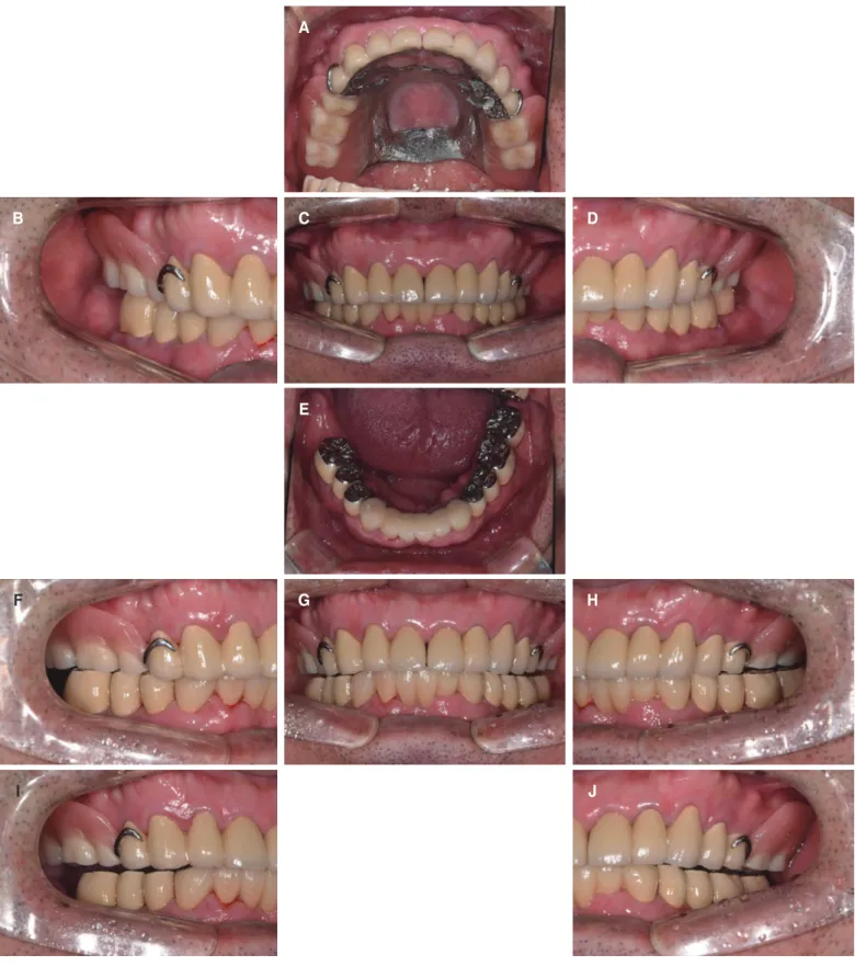
Fig. 12. Definitive prosthesis. (A) Upper, (B) Right, (C) Frontal, (D) Left, (E) Lower, (F) Right working, (G) Protrusive, (H) Left working, (I) Right balancing, (J) Left balancing.
B
F
I
C
E
G
D
H
J
고찰
과개교합은 상악의 절치가 하악절치의 절반이상을 덮고 있 는 것을 말한다. 과개교합 환자는 치은 연조직의 손상, 수복공 간의 부족, 치아마모 등의 문제를 발생시킨다. 본 환자는 Akerly 의 과개교합 분류에 의하면 상,하악 전치의 마모가 발생한 Class IV로 분류되었다.18 과개교합의 치료에서는 안정적인 교합점 과 약한치아를 보호 할 수 있는 적절한 교합 양식이 중요한데19 Torbjörner와 Fransson20은 군기능교합이 견치에 가해지는 측방 력이 줄여준다고 말하였다.
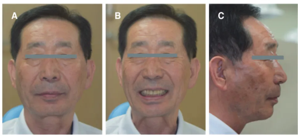
2급 악간 관계를 가지는 환자는 생리적 안정위시 1급 악간 관 계를 가지는 사람보다 하악이 보다 더 전방으로 위치하는 경향 이 있으며 전방으로 이동된 하악은 심미성 향상,입술 봉쇄,근육 기능, 발음, 호흡을 도와준다고 하였다.21 2급 악간 관계를 가지 는 환자는 넓은 하악의 움직임을 보이며 0.5 - 1 mm 이상의 long centric이 필요하다고 하였다.21 Ash와 Ramfjord22는 long centric 이 기능 범위보다 크다면 부작용은 없다고 하였다. 또한 2급 악 간 관계를 가지는 환자의 치료에서 적절한 교합의 형성시 수직 고경의 증가는 가능 하다고 하였으며 Jensen은 3 - 5 mm 증가가 가능하다고 하였다.23,24 Curtis 등25은 2급 악간 관계를 가지는 환 Fig. 13. Facial photographs after treatment. (A) Frontal view, (B) Frontal view smile, (C) Lateral view.
A B C
Fig. 14. Panoramic radiograph after treatment.
Fig. 15. TMJ series after treatment. (A) Rt. close, (B) Rt. opening, (C) Lt. opening, (D) Lt. close.
A B C D
하였지만 5 mm 까지의 수직 고경의 증가는 안전하다고 하였다.
본 증례에서 수직고경을 평가하기 위해 치료전 진단모형상에 서 치관길이 평가, 전치 절단연에서 구강 전정까지의 거리평가, 교합시 악간 거리 측정, Willis 안면 계측법을 참고하여 수직 고 경 감소 여부를 평가 하였다. 새로 설정한 수직 고경은 근신경계 의 적응 여부를 평가하여야 하는데 이는 1 - 3개월간 의 기간을 두고 평가 하여야 한다.16 10주간의 기간 동안 임시 의치의 불편 을 제외한 근육의 피로감, 측두하악관절 증상, 임시치아의 파절 이 관찰되지 않아서 최종 보철물을 제작하였다. 치료 완료 후 6 개월간의 관찰 기간 동안 상악 국소의치 인공치의 마모는 없었 지만 추후 정기검진 시 인공치의 마모가 발생하면 인공치의 금속 수복 필요성에 대해 환자에게 교육을 진행하였다.
결론
본 증례에서는 과도한 마모와 구치부 상실이 있는 과개 교합 환자를 치관 길이 평가, 악간 거리 측정, Willis 안면계측법,진단 납형의 제작을 통해 수직 고경을 평가하여 증가 시켰으며, 임시 수복물을 제작하여 증가된 수직 고경에 대한 환자의 기능적, 심 미적 적응을 확인한 후 최종 보철물을 제작하여 장착하였다. 관 찰 기간 동안 특별한 이상을 보이지 않았으며 만족스러운 결과 를 얻을 수 있었다. 최종 수복 완료 후에도 장기적인 안정성을 위 해서 정기적인 관찰을 통해 교합의 유지를 확인해야 하고 교합 조정, 의치의 이장이 필요 시 시행되어야 할 것으로 생각된다.
ORCID
Seong-Yong Seo https://orcid.org/0000-0003-3596-7687
References
1. Dawson PE. Functional occlusion: From TMJ to smile de- sign. St. Louis; Mosby Elsevier, 2007. p. 430-52.
2. Murphy T. Compensatory mechanisms in facial height adjust- ment to functional tooth attrition. Aust Dent J 1959;4:312-23.
3. Briggs P, Bishop K. Fixed prostheses in the treatment of tooth wear. Eur J Prosthodont Restor Dent 1997;5:175-80.
4. Hemmings KW, Darbar UR, Vaughan S. Tooth wear treated with direct composite restorations at an increased vertical di- mension: results at 30 months. J Prosthet Dent 2000;83:287-93.
5. Sato S, Hotta TH, Pedrazzi V. Removable occlusal overlay splint in the management of tooth wear: a clinical report. J
worn dentition. J Prosthet Dent 1984;52:467-74.
9. Rivera-Morales WC, Mohl ND. Relationship of occlusal vertical dimension to the health of the masticatory system. J Prosthet Dent 1991;65:547-53.
10. Park JH, Jeong CM, Jeon YC, Lim JS. A study on the oc- clusal plane and the vertical dimension in Korean adults with natural dentition. J Korean Acad Prosthodont 2005;43:41-51.
11. Silverman MM. The speaking method in measuring vertical dimension. J Prosthet Dent 1953;3:193-9.
12. Rivera-Morales WC, Mohl ND. Restoration of the vertical dimension of occlusion in the severely worn dentition. Dent Clin North Am 1992;36:651-64.
13. Willis FM. Features of the face involved in full denture pros- thesis. Dent Cosmos 1935:77:851-4.
14. Abduo J, Lyons K. Clinical considerations for increasing oc- clusal vertical dimension: a review. Aust Dent J 2012;57:2- 15. Abduo J. Safety of increasing vertical dimension of occlu-10.
sion: a systematic review. Quintessence Int 2012;43:369-80.
16. Carlsson GE, Ingervall B, Kocak G. Effect of increasing ver- tical dimension on the masticatory system in subjects with natural teeth. J Prosthet Dent 1979;41:284-9.
17. Naylor CK. Fabrication of a custom anterior guide table. J Prosthet Dent 1979;42:466-9.
18. Akerly WB. Prosthodontic treatment of traumatic overlap of the anterior teeth. J Prosthet Dent 1977;38:26-34.
19. Beddis HP, Durey K, Alhilou A, Chan MF. The restorative management of the deep overbite. Br Dent J 2014;217:509- 20. Torbjörner A, Fransson B. Biomechanical aspects of prosthet-15.
ic treatment of structurally compromised teeth. Int J Prostho- dont 2004;17:135-41.
21. Curtis TA, Langer Y, Curtis DA, Carpenter R. Occlusal con- siderations for partially or completely edentulous skeletal class II patients. Part I: Background information. J Prosthet Dent 1988;60:202-11.
22. Ash MM. Occlusion. 4th ed. Saunders (W.B.) Co. Ltd., 1995.
p. 72,3,6.
23. Hellsing G. Functional adaptation to changes in vertical di- mension. J Prosthet Dent 1984;52:867-70.
24. Jensen WO. Occlusion for the Class II jaw relations patient. J Prosthet Dent 1990;64:432-4.
25. Curtis TA, Langer Y, Curtis DA, Carpenter R. Occlusal con- siderations for partially or completely edentulous skeletal Class II patients. Part II: Treatment concepts. J Prosthet Dent 1988;60:334-42.
치아 마모로 인한 수직고경감소와 과개교합을 가진 환자에서 전악 수복 증례
서성용 이나영* 강정경 중앙보훈병원 치과보철과
구치부 교합의 붕괴는 정상적인 교합평면을 소실 시키고, 과도한 마모를 일으켜 수직고경을 감소시킨다. 감소된 수직 고경은 심미적, 기능적 문제를 일
으킬 뿐만 아니라 측두하악관절에 과하중을 야기하고 근신경계 이상을 일으킬 수 있다. 이렇게 붕괴된 교합관계를 개선하기 위해서 수직고경 변경을
고려하여야 하는데 치료 전 정확한 진단과 분석이 필수적이며 새로운 수직 고경에 대한 적응 평가를 함께 하여야 한다. 그리고 과도한 수직피개를 가지 는 과개교합 환자는 치아 마모 및 치아정출의 교합 문제를 가지는 경우가 많다. 이러한 사항을 고려해 보았을 때 치아마모로 수직고경이 감소된 과개 교합 환자의 문제 해결을 위해서는 전반적인 보철수복을 하여야 한다. 본 증례는 68세 남자환자로 다수 치아의 마모와 상악 구치의 결손부위를 치료하 기 위해 수직고경 증가를 동반한 상악 가철성 국소의치 및 상하악 고정성 보철물로 수복한 증례이다. (대한치과보철학회지 2018;56:31-9)
주요단어: 과개교합; 부분무치악; 가철성보철; 전악 수복
*교신저자: 이나영
05368 서울 강동구 진황도로 61길 53 중앙보훈병원 치과보철과 02 2225 1658: e-mail, [email protected]
원고접수일: 2017년 7월 10일 / 원고최종수정일: 2017년 9월 4일 / 원고채택일: 2017년 9월 5일
2018 대한치과보철학회
이 글은 크리에이티브 커먼즈 코리아 저작자표시-비영리 3.0 대한민국 라이선스에 따라 이용하실 수 있습니다.
c cc