Original Articles Korean Circulation J 2000;;;;30((((8))))::::947-957
혈장 섬유소원의 농도 및 섬유소원 유전자 다형성이 관동맥 질환의 발생에 미치는 영향
연세대학교 의과대학 심혈관연구소,
1심장내과
2박현영
1
·오수환2
·권혁문2
·김동수2
·홍범기2
·이남호2
·장양수1,2
The Effects of Plasma Fibrinogen and β β β Fibrinogen Gene β Polymorphisms on the Development of Coronary Artery Disease
Hyun-Young Park
1, Soohwan Oh
2, Hyuck Moon Kwon
2, Dongsoo Kim
2, Bum Kee Hong
2, Nam Ho Lee
2and Yangsoo Jang
1,21
Yonsei Cardiovascular Research Institute,
2Department of Internal Medicine, Yonsei University College of Medicine, Seoul, Korea
ABSTRACT
Background:Elevated plasma fibrinogen level has been shown to be an independent risk factor of ischemic heart disease, cerebrovascular disease, and peripheral vascular disease. The aim of this study is to determine the associations of plasma fibrinogen levels, coronary artery disease (CAD), classical vascular risk factors, and genetic polymorphisms at position -455 and C448 of the β-fibrinogen gene. Methods: : : :We measured the plasma fibrinogen levels and lipid levels in 374 patients with angiographically defined CAD and 290 control patients.
The genotypes of β-fibrinogen in randomly selected patients were determined by restriction fragment length polymorphism and allele-specific oligomer hybridization. Results: : :1) Higher plasma fibrinogen levels were : observed in the patients with CAD, especially who had multiple coronary artery atherosclerosis. 2) The plasma fibrinogen levels were higher in smokers, and positively related with age and total cholesterol levels. 3) The two polymorphisms were in tight linkage disequilibrium and the genotype frequencies were similar in patients and control subjects. The significant association between β-455G/A genotype and plasma fibrinogen was noted in females but not in males. 4) In logistic regression model, the elevated plasma fibrinogen was an independent risk factor of CAD. Conclusion: : :The present study shows that the plasma fibrinogen level is an independent risk : factor for coronary atherosclerosis, and the genetic variants of the β fibrinogen gene are associated with an increased plasma fibrinogen in females. ((((Korean Circulation J 2000;30((((8)))):947-957))))
KEY WORDS:Fibrinogen·Coronary artery disease·Genetics·Polymorphism.
서 론
혈장 섬유소원(fibrinogen)은 혈액 응고인자의 일종
으로 혈소판의 수용체와 작용하여 혈소판 응집 및 혈액 의 viscosity에 영향을 미쳐 혈전 형성에 관여하며, 조 직 손상이나 감염, 염증 등의 외부적 요인에 의해 증가 하는 급성상 반응 물질이다.
1)2)최근 혈장 섬유소원의 농도는 관상동맥질환, 뇌졸중, 경동맥의 동맥경화 진행 정도와 좋은 상관관계가 보고되고 있어,
3-8)혈장 섬유 소원의 농도 상승이 관동맥질환의 위험인자로 인식되 고 있으며, 국내에서도 혈청 섬유소원의 농도는 관동맥
논문접수일:1999년8월16일심사완료일:2000년9월25일
교신저자:120-752, 장양수, 서울 서대문구 신촌동 134 연세대학교 의과대학 심혈관연구소
전화:(02)361-7350/8365・전송:(02)365-1878 E-mail:[email protected]
질환의 위험인자라는 보고가 있어 관심을 끌고 있다.
9)혈장 섬유소원의 농도에 영향을 미치는 인자들로는 인종, 성별, 흡연, 고혈압, 당뇨등이 알려져 있으며,
10-14)최근 유전적인 영향에 대해서도 많은 연구들이 진행되 어 오고 있다. 섬유소원은 간에서 생성되는 당단백으로 Aα, Bβ, γ로 분류된 3종류의 polypeptide chain이 disulfide 결합에 의해 연결되어(Aα-Bβ-γ)
2의 형 태를 이루고 있다.
15)섬유소원을 구성하는 polypeptide 는 α, β, γ 섬유소원 유전자로부터 합성되며 이 유전 자들은 4번 염색체의 long arm에 50 kb 미만의 cluster의 형태로 위치한다. 각 섬유소원 유전자에서의 mRNA합성은 따로 이루어지나, 전사 조절은 상호 밀 접한 관련이 있는것으로알려져있다.
16)17)이중섬유소원 의Bβ-polypeptide chain의 합성은 실험적으로 성숙 된 섬유소원(mature fibrinogen molecule)의 합성의 조절에 중요한 역할을 하며, 따라서 Bβ-polypeptide chain의 합성이 혈장 섬유소원의 농도에 영향을 미칠 것으로 추정된다.
18)혈장 섬유소원의 농도에 미치는 유 전학적 영향은 이미 밝혀져 있는 유전자다형성(genetic polymorphism)을 바탕으로 연구가 진행되어 오고 있 다. 현재까지 섬유소원 유전자에는 10여가지 이상의 유전자다형성이 보고되었고, 이중 β 섬유소원 유전자 에서는 유전자 전사조절부위에 5(β-1420, β-854, β-455, β-249, β-148), coding region에 2(β C345, β C448), intron에 2(β+1689, Intron 16 I/D), 그리고, 3’-untranslated region(UTR)에 BclⅠ pol- ymorphism이 알려져 있다.
19-22)이 중 혈장 섬유소원 의 농도와 관련이 있는 유전적 다형성으로는 β-455, β-148, β C448, 3’-UTR의 BclⅠ polymorphism 등이 있으나, 보고자마다 차이가 있다.
23)24)그리고, 섬 유소원 유전자다형성과 경동맥 경화증, 관상동맥질환, 말초혈관질환등과의 관련성에 대해서도 보고가 있으 나,
20)22)25)26)질환별, 인종등에 따라 차이가 있다.
본 연구에서는 혈관조영술을 시행한 환자들 중 관동 맥질환군과 혈관조영술상 정상 소견을 보인 정상 대조 군을 대상으로 혈장 섬유소원의 농도가 관동맥질환에 미치는 영향 및 혈장 섬유소원과 기존의 위험인자들과 의 관련성에 대해 알아보고, β섬유소원의 유전자다형성 중 유전자 전사조절부위에 존재하는 β-455G/A, 엑손 에 존재하는 β C448G/A이 혈장 섬유소원의 농도 및 관상동맥질환의 발생에 미치는 영향을 분석하였다.
대상 및 방법
대 상
1997년 1월부터 1998년 4월까지 영동 세브란스병 원을 방문하여 관동맥조영술을 시행하였던 환자 중 조 영술상 세 관동맥중 적어도 하나의 관동맥에 50%이상 의 유의한 협착이 관찰되었던 374명을 환자군으로 하 였으며, 세 혈관 모두에서 30%이상의 협착이 관찰되 지 않았던 290명을 대조군으로 하였다. 환자군 및 대 조군에서 혈장 섬유소원의 농도 및 기존의 동맥경화증 관련인자들에 대해 조사하였다. 환자들의 임상진단은 안정성 및 불안정성 협심증이 414명, 심근경색증으로 진단받았던 경우가 135명, 이형협심증 및 판막질환등 을 사유로 관동맥조영술을 시행하였던 경우가 115명이 었다. 혈관병변의 침범정도는 혈관조영술상 50%이상 협착이 있었던 혈관의 수에 따라 단일혈관질환군, 다혈 관질환군으로 나누어 분석하였다.
방 법
섬유소원, 지질 농도의 측정
혈장섬유소원의농도는Fibriprest(Diagnostica Stago, France) 시약으로 자동응고기기인 STA(Diagnostica Stago, France)를 이용하여 측정하였다. 혈장 섬유소 원의 농도는 급성상 반응물질로 심근 경색환자들에 있 어서는 섬유소원의 농도측정은 심근경색 발생 후 적어 도 1개월이 지난 뒤 측정하였다. 총콜레스테롤과 중성 지방은 Hitachi 747(Boehringer Mannheim, Germ- any) 자동생화학분석기를 이용하여 효소법으로 측정하 였으며, 고밀도지단백 콜레스테롤(HDL-cholesterol) 은 직접법으로 Hitachi 747(Boehringer Mannheim, Germany) 자동생화학분석기를 이용하여 측정하였다.
저밀도지단백 콜레스테롤은 Friedwald 공식을 이용하 여 계산하였다. Lp(a)의 농도는 TintElize Lp(a) kit (Biopool, Umea, Sweden)를 이용하여 측정하였다.
유전형분석
환자들로부터 혈액 7 ml을 채취하여 EDTA가 첨가된
시험관에 보관하였고, genomic DNA는 Wizard DNA
purification system (Promega Co., Madison, Wisc-
onsin, U.S.A.)를 사용하여 말초 백혈구로부터 추출하 였다. 분리된 genomic DNA는 Tris-EDTA(TE) 용 액으로 용해하여 분석전까지 4℃에 보관하였다. 섬유소 원의 β-455G/A 및 β C448G/A 유전자형분석을 위 해 이용된 primer의 염기서열은 다음과 같다.
β-455G/A sense:
5’-CTATTCAGCACAAAAAAAGGGTC-3’
antisense:
5’-CTACTCACAAGGCAACCACTAAA-3’
β C448G/A sense:
5’-TTCAGGTTAACATCAGATCCCAG –3’
antisense:
5’-CGTCTGCTTGAGAGTTTTAGAGGA-3’
PCR은 20 μl반응으로 genomic DNA 50 ng, sense 와 antisense primer 각각 10 pmole, 62.5 μM dNTP, 1.5 mM MgCl 2, 10 mM Tris-HCl(pH 8.8), 11 mM ammonium sulfate, 6.7 mM β-mercap-toethanol, 4.5 μM EDTA, 0.4 U Taq polymerase(AmpliTaq Gold , Roche Molecular System Inc., Branchburg, New Jersey, U.S.A.)와 혼합하여 시행하였다. PCR 조건은 먼저 95℃에서 10분간 변성시킨 뒤, 94℃에서 1분간 변성, 60℃에서 1분간 annealing, 72℃에서 1분 간 신장시키는 과정을 35회 반복 후, 72℃에서 5분간 다시 신장시켰다. PCR 산물은 1.5% agarose gel에 전
기영동하여 확인하였다.
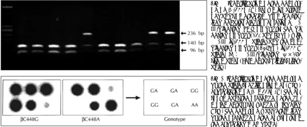
β-455G/A 유전자 다형성은 Hae Ⅲ 제한효소에 의 해 DNA 단편길이 다형성(restriction fragment length polymorphism, RFLP)을 보이는 것으로, PCR 후 증폭 된 산물 15 μl에 Hae Ⅲ 제한효소 5 U 및 buffer를 혼 합 후 37℃에서 6시간이상 반응시켰다. 이를 ethidium bromide를 함유한 2% gel에 전기영동하여 절단부위 유무를 UV transilluminator에서 관찰하여 유전형을 결정하였다(Fig. 1).
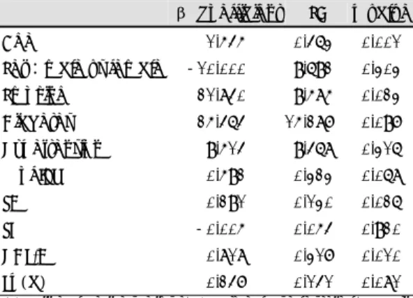
βC448G/A 유전자 다형성은 allele specific oligo- mer(ASO) hybridization 방법으로 유전형을 결정하였 다. PCR 산물을 0.4 N NaOH 200 μl로 변성 후 ny- lon membrane(Hybond-N+, Amersham, UK)에 tr- ansfer후 UV irradiation하여 PCR 산물을 membrane 에 cross-link시켰다.
32P로 표지된 ASO(448G:5’- TCAATGAGAAGATG-3’,448A:5’-TCAATGA AGAAGATG-3’)로 35℃에서 6시간 이상 hybridi- zation 후 실온에서 2×SSC/0.1%SDS로 2회 세척한 다음 448G는 45℃에서, 448A는 40℃에서 2×SSC/
0.1%SDS로 10분간 세척하였다. Hybridized memb- rane은 phosphor-imaging plate reader(Bio-ima- ging analyser system 2500
®, Fuji Film Co., Tokyo, Japan)를 이용하여 autoradiogram을 시행하여 유전형 을 결정하였다(Fig. 2).
통계 및 분석
통계분석은 SPSS for window 7.0 program을 이용하 여 분석하였다. 수치 자료는 평균±표준오차(mean±
Fig. 1. Identification of the genoty- pe of β-455G/A. Shown is pattern of bands on ethidium bromide-sta- ined gel after electrophoresis of Hae
Ⅲ-digested PCR products. The di- gested product (140 and 96 bp) with
HaeⅢ is -455 G allele, and the non-digested product (236bp) is -455 A allele. M:HaeⅢ-digested ψ×174 size marker (Fermentas, Vilnius, Lithu- ania).
Fig. 2. Identification of the genotype
by the allele-specific oligomer (ASO)
hybridization of β C448G/A. The hot
spot means the presence of the co-
mplementary sequence to labeled
ASO. The genotype was determined
by the presence of the hot spots on
the hybridized membrane.
SE)로 표시하였다. 수치척도는 Student t-test, AN- OVA, MANOVA로, 명목척도는 chi-square test로 분석하였다. 변수간의 상관관계는 Pearson 상관분석을 이용하였으며, 관동맥질환의 위험인자분석을 위해 lo- gistic regression analysis를 사용하였다. 두 유전자 다형성간의 linkage는 Hill
27)과 Thompson 등
28)이 기 술한 방법으로 분석하였다. p값은 0.05 이하인 경우 의 미있다고 판정하였다.
결 과
대상군의 혈장 섬유소원의 농도 및 임상적 특징 관동맥조영술상 병변이 있었던 군에서 대조군에 비해 연령, 남성비, 당뇨, 고혈압, 혈장 섬유소원의 농도 및 고밀도지단백콜레스테롤을 제외한 지질농도의 수치가 유의한 차이를 나타내었다(자료생략). 혈관병변의 침범 정도와 혈장섬유소원 및 관련인자들의 관련성을 알아보 기 위하여 환자군을 단일혈관질환군 및 다혈관질환군으 로 나누어 분석한 임상적 소견, 혈장 섬유소원의 농도 및 혈청 지질농도는 Table 1과 같다. 단일혈관질환군에 서는 대조군에 비해 남성 및 흡연의 빈도만이 유의한 차 이를 나타내었으며, 혈장 섬유소원의 농도는 단일혈관질 환군에서 대조군에 비해 증가되어 있었으나, 통계적으로 유의한 차이는 관찰되지 않았다(312.9±6.3 vs. 303.8
±4.6 mg/dL, p>0.05). 다혈관질환군에서는 고밀도지 단백 콜레스테롤을 제외한 모든 위험인자의 빈도 및 수 치가 증가되어 있었으며, 단일혈관군과 비교하였을 때 연령, 고혈압의 빈도, 혈장 섬유소원, Lp(a)의 농도가 유의하게 증가되어 있었다. 각 군에서 혈장 섬유소원의 농도에 영향을 미칠 수 있는 연령, 성별, 당뇨, 혈압 및 혈중 지질농도등을 covariate로 설정하여 분석한 결과 에서도 혈장섬유소원의 농도는 다혈관질환군에서 정상 혈관군 혹은 단일혈관군에 비해 유의하게 증가되어 있었 다(adjusted mean:정상혈관군;305.9±6.8 mg/dL, 단일혈관질환군;310.0±8.0 mg/dL, 다혈관질환군;
345.4±7.3 mg/dL, F value 8.63, p<0.001).
혈장 섬유소원의 농도 및 각 위험인자간의 상관관계 혈중 지질농도 및 비만도, 연령등과의 연관관계를 분 석한 결과 연령, Lp(a) 농도, 총콜레스테롤 및 저밀도지 단백콜레스테롤과 유의한 양의 상관관계를, 고밀도지단
백콜레스테롤과는 음의 상관관계를 보여주었다(Table 2). 남녀 성별로 나누어 혈장 섬유소원의 농도차이를 분석한 결과 남자에서는 319.6±4.8 mg/dL, 여자에서 는 324.0±5.6 mg/dL로 유의한 차이가 없었다(p=
0.90). 현재 흡연여부에 따라 분석한 결과에서는 흡연 자에서 326.1±7.5 mg/dL, 비흡연자 혹은 최소 1년간 금연자에서는 315.4±4.8 mg/dL로 흡연자에서 유의 하게 증가되어 있었다(p=0.01). 전체 대상군에서 혈 장 섬유소원의 농도와 관련이 있는 인자들을 알아보기 위하여 linear regression 분석을 시행한 결과, 연령(p
=0.001), 흡연(p=0.02), 총콜레스테롤(p=0.03), 고 밀도지단백 콜레스테롤(p=0.01)이 혈장 섬유소원의 농도와 유의한 상관관계를 보여주었다(Table 3).
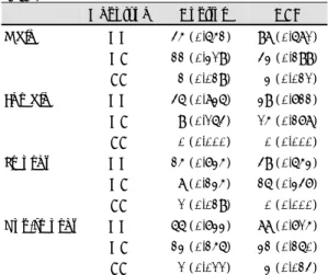
β섬유소원 유전자 다형성과 혈장 섬유소원과의 관계 β섬유소원의 유전형 분석은 전체 664명의 환자 중 게놈 DNA를 얻을 수 있었던 322명의 환자에서 시행 하였다. 유전형 분석이 가능했던 환자들은 정상혈관군 이 124명, 단일혈관 질환군이 78명, 다혈관질환군이 120명이었으며, 임상적 특징은 Table 4에서 보는 바 Table 1. Comparisons of clinical characteristics, fibrin- ogen concentration and the lipid profiles according to degree of coronary stenosis in control subjects and patients
Control Single VD Multiple VD
Number 290 166 208
Age (years) 54.4±0.6 57.7±0.7* 60.4±0.7*
†Sex (% of male) 54.5 62.7 71.2*
Diabetes (%) 3.4 7.2 12.0*
Hypertension (%) 41.4 44.0 56.7*
†Smoking (%) 31.0 43.2* 44.4*
Fibrinogen (mg/dL) 303.8±4.6 312.9±6.3 352.4±7.8*
†TC (mg/dL) 190.0±2.3 192.1±2.8 203.2±3.0*
†TG (mg/dL) 151.9±5.2 158.5±6.4 175.5±6.6*
HDL-C (mg/dL) 39.6±0.7 40.1±1.6 37.8±0.8 LDL-C(mg/dL) 118.4±2.3 120.5±2.7 128.5±2.8*
Lp(a) (mg/dL) 26.1±2.9 24.8±2.6 35.6±3.0*
†Control:Control subjects without angiographically de- fined coronary artery stenosis, VD:vessel disease, Sin- gle VD:significant stenosis at single coronary artery, Multiple VD:stenotic lesions at more than one coron- ary artery, TC:total cholesterol, TG:triglycerides, HDL- C: high-density lipoprotein cholesterol, LDL-C: low- density lipoprotein cholesterol
*p<0.05 compared with control,
†p<0.05 compared
with single VD
와 같이 전체 환자군과 유의한 차이가 없었다. 유전자 분석 대상환자에서 두 유전자다형성에서의 유전자형의 분포는 Hardy-Weinberg equation을 벗어나지 않았 으며, 두 유전자다형성간에는 유의한 상관관계(linkage disequilibrium)가 관찰되었다(|D|=0.97, p<0.05).
전체 대상군에서 β -455G/A와 β C448G/A 유전자 다형성의 각 유전형에 따른 혈장 섬유소원의 농도 차이 는 관찰되지 않았다(Fig. 3). 성별 및 흡연여부로 나누 어 각 유전형에 따른 혈장 섬유소원의 농도를 분석한 결과 여자에서는 β-455 G/A에서 변이 유전형을 가 진 경우 혈장 섬유소원의 농도가 유의하게 증가되어 있 었으나, 남자에서는 유전형에 따른 유의한 차이가 없었 다(Fig. 4a). 흡연여부로 나누어 분석한 결과 흡연자 비흡연자 모두에서 유전형에 따른 혈장 섬유소원의 농 도는 유의한 차이가 없었다(Fig. 4b).
β섬유소원 유전자 다형성과 관동맥질환과의 관계 대조군 및 관동맥질환군간에 유의한 유전형 분포의 차이는 관찰되지 않았으며, 단일혈관질환군과 다혈관질 환군을 비교하였을 때 혈관병변의 침범정도에 따른 차 이 역시 관찰되지 않았다(Table 5). β-455G/A 유전 자 다형성에서 성별에 따라 나누어 유전형의 분포를 분 석한 결과 유전형의 빈도는 관동맥질환군과 대조군 사 이에 유의한 차이가 관찰되지 않았으며, 흡연 여부로 나누어 분석한 결과에서도 유전형의 빈도에는 차이가 없었다(Table 6).
Table 2. Correlations between biochemical parameters in study population
Parameters Fibrinogen Age TC TG HDL-C LDL-C Age (ye ars) 0.17**
TC (mg/dL) 0.08* -0.02
TG (mg/dL) 0.05 -0.07 0.32**
HDL-C (mg/dL) -0.10** 0.06 0.23** -0.15**
LDL-C (mg/dL) 0.10* 0.01 0.84** -0.05 0.23**
Lp(a) (mg/dL) 0.16** 0.06 0.22** -0.20 -0.15 0.26**
TC:total cholesterol, TG:triglyceride, HDL-C:high density lipoprotein cholsterol, LDL-C:low density lipoprotein cholesterol, Lp (a):lipoprotein (a)
*Correlation is significant at the 0.05 level.
**Correlation is significant at the 0.01 level.
Table 3. β-Coefficients and SEs of plasma fibrinogen levels in study population from multiple linear regression analysis
β coefficient SE p value Age 1.545 0.463 0.001 Sex:male vs female -10.000 9.692 0.303 Smoking 21.840 9.585 0.023 Diabetes 25.464 15.287 0.097 Hypertension 9.514 9.468 0.316
% of IBW 0.592 0.323 0.068 TC 0.291 0.130 0.026 TG -0.005 0.054 0.920 HDL-C 0.818 0.317 0.010 Lp(a) 0.247 0.141 0.081 IBD:ideal body weight, TC:total cholesterol, TG:tr- iglyceride, HDL-C:high density lipoprotein cholesterol, Lp(a):lipoprotein(a)
Table 4. Comparisons of clinical characteristics, fibrin- ogen concentration and the lipid profiles in control su- bjects and patients whose genotypes were analyzed
Control single VD Multiple VD Number 124 78 120 Age (years) 54.4±0.9 57.1±1.0* 60.6±0.9*
Sex (% of male) 55.3 66.7 76.7*
Diabetes (%) 4.1 8.3 14.1.0*
Hypertension (%) 49.2 38.5 55.8 Smoking (%) 27.4 30.8* 42.5*
Fibrinogen (mg/dL) 305.5±7.6 309.9±8.3 336.5±11.0*
TC (mg/dL) 185.5±3.5 193.1±4.0 199.7±3.9*
TG (mg/dL) 149.8±8.2 158.4±9.7 177.3±8.0*
HDL-C (mg/dL) 37.9±0.9 38.5±1.4 37.2±0.9 LDL-C (mg/dL) 118.7±3.1 122.9±4.1 125.1±3.7 Lp(a) (mg/dL) 23.8±2.1 24.3±3.7 32.6±3.6 Control:Control subjects without angiographically defi- ned coronary artery stenosis, VD:vessel disease, Single VD:significant stenosis at single coronary artery, Multiple VD:stenotic lesions at more than one coronary artery, TC:total cholesterol, TG:triglycerides, HDL-C:high-de- nsity lipoprotein cholesterol, LDL-C:low-density lipoprot- ein cholesterol
*p<0.05 compared with control,
†p<0.05 compared with
single VD
관동맥질환의 위험인자분석
관동맥질환 발생의 유의한 위험인자를 분석하기 위하 여 multiple stepwise logistic regression analysis를 시행한 결과 연령, 남성당뇨, 혈장섬유소원의 농도, 흡연, 총콜레스테롤 농도가 독립된 위험인자로 분석되었으며 각 요인들의 Odd ratio는 Table 7에서 보는 바와 같다.
고 찰
죽상동맥경화반의 파열로 인한 혈전형성은 급성 심 근경색의 발생 및 동맥경화반의 진행에 중요한 역할을 하는 것으로 알려져 있다.
29)섬유소원은 혈장에 존재하
는 당단백으로 트롬빈에 의해 섬유소로 전환되어 혈액 응고인자 ⅩⅢ의 작용에 의해 안정된 섬유소를 형성함 으로써 지혈작용에 관여한다.
혈장 섬유소원의 상승이 관동맥질환과 같은 동맥경 화성 질환의 발생과 관련이 있음은 이미 여러 연구결과 에서 보고되었는데,
3-8)30)본 연구결과에서도 관동맥질 환군에서 혈장 섬유소원의 농도가 정상혈관을 보인 군 에 비해 증가되어 있었으며, 특히 다혈관질환군에서 유 의하게 증가되어 있었고, multiple logistic regression 분석결과에서도 높은 혈장섬유소원은 관동맥질환의 유 의한 위험인자로 판명되었다. 이는 기존의 보고와도 일 치하며, 한국인에서도 역시 상승된 혈장 섬유소원의 농 Table 5. Fibrinogen β -455G/A and β C448G/A genotype frequencies of control subjects and patients
Subgroup Genotype Control CAD
Single VD Multiple VD β -455G/A GG 93 (0.738) 135 (0.689) 57 (0.731) 78 (0.661) GA 31 (0.246) 58 (0.296) 21 (0.269) 37 (0.314) AA 2 (0.016) 3 (0.015) 0 (0.000) 3 (0.025) β C448G/A GG 89 (0.817) 75 (0.714) 28 (0.737) 47 (0.701) GA 18 (0.165) 28 (0.267) 9 (0.237) 19 (0.284) AA 2 (0.018) 2 (0.019) 1 (0.026) 1 (0.015) CAD:Patient group with angiographically defined coronary artery disease.
The number in parenthesis represents the genotype frequencies.
Fig. 3. Fibrinogen levels by β fibrinogen genotype (a) β-455G/A polymorphism (b) β C448G/A polymorphism.
Fig. 4. Fibrinogen levels by genotype β-455G/A polymorphism in male and females (a) and smokers and nonsmokers (b).
도는 관동맥질환 발생의 유의한 위험인자임을 시사한 다. 상승된 혈장 섬유소원이 어떠한 기전에 의해 관동 맥질환의 발생 위험도를 증가시키는지에 대해서는 아 직 명확하지 않으나, 다음과 같은 기전들이 제시되었다.
첫째, 혈장 섬유소원은 혈액의 viscosity에 관여하는 인자로, 증가된 혈장섬유소원은 viscosity를 증가시켜 혈류(rheology)에 영향을 미침으로써 혈전형성을 용이 하게 할 것이다.
31)둘째, 상승된 혈장 섬유소원의 농도 는 혈소판의 응집에도 관여하여 급성심근경색과 같은 허혈성 질환의 발생에 영향을 미친다.
32)셋째, 섬유소 원은 thrombin의 중요한 substrate로 혈장에서의 농도 가 증가된 경우 fibrin 형성의 촉진뿐만 아니라, 더욱 단단한 clot을 형성하여 허혈성 혈관질환의 발생 및 경 과에 관여할 것이다. 한편, 혈전형성과정뿐 아니라, 초 기 동맥경화반의 발생과정에서도 혈장 섬유소원이 관 여하며 동맥경화반의 진행정도와도 밀접한 관련이 있 는 것으로 보고되고 있으며, 이에 관련된 기전으로는
섬유소와 Lp(a)의 친화성, FDP등에 의한 혈관평활근 세포의 증식등이 거론되고 있다.
33)34)혈장 섬유소원의 농도에 미치는 유전적 영향은 혈장 섬유소원의 농도가 여러 환경적 요인의 영향을 받으므 로 분석이 쉽지가 않다. NHLBI twin study에 의하면 혈중 섬유소원 농도의 maximum likehood heritability 는 약 30% 정도로 알려져 있다.
35)섬유소원을 구성하 는 Aα, Bβ, γ peptide는 각각 분리된 유전자에 의해 합성되며, 섬유소원의 합성에 있어 Bβ-chain의 합성 이 rate-limiting step으로 작용한다.
18)현재까지 β 섬유소원 유전자의 다형성을 이용한 β 섬유소원유전 자와 혈장섬유소원의 농도 및 동맥경화성 질환과의 관 련성에 대해 여러 연구결과가 발표되었으나, 아직 논란 의 여지가 많다. 유전자전사 조절부위에 존재하는 β -455G/A 유전자다형성의 경우 AA 유전형에서 GA 혹은 GG 유전형에 비해 혈장 섬유소원의 농도가 높음 이 보고되기도 하였으나,
22)36)37)그렇지 않다고 보고한 경우도 있다.
38)β -455 G/A 유전형이 혈장 섬유소원 의 농도에 영향을 미치는 기전으로는 β -455G/A 유 전형과 interleukin -6 responsive element와 근접하 여 존재하는 β -148C/T 유전형과 밀접한 연관관계 를 이루고 있기 때문으로 설명되기도 하였으나,
39)β -148 C/T 유전형에 따른 혈장 섬유소원의 농도도 유 의한 차이가 없다고 보고되기도 하였다.
25)엑손내 존재 하는 β C448G/A는 β 섬유소원 유전자의 carboxy terminal로부터 13번째 아미노산이 arginine에서 ly- sine으로 치환된 것으로 Carter 등은 AG 혹은 AA 유 전형을 가지는 경우 GG 유전형을 가진 경우에 비해 남 자, 특히 일반인 남자에서 혈장 섬유소원의 농도가 유 의하게 증가됨을 보고하였다.
40)41)반면 관동맥질환과 의 관계에서는 오히려 여자에서 변이 유전형의 빈도가 높다고 보고하고 있어 β C448G/A 유전자변이는 섬유 Table 6. Fibrinogen β -455G/A genotype frequencies
of control subjects and patients by gender and smoking status
Genotype Control CAD Male GG 45 (0.652) 98 (0.681) GA 22 (0.319) 43 (0.299) AA 2 (0.029) 3 (0.021) Female GG 46 (0.836) 39 (0.722) GA 9 (0.164) 15 (0.278) AA 0 (0.000) 0 (0.000) Smoker GG 25 (0.735) 49 (0.653) GA 8 (0.235) 26 (0.347) AA 1 (0.029) 0 (0.000) Non-smoker GG 66 (0.733) 88 (0.715) GA 23 (0.256) 32 (0.260) AA 1 (0.011) 3 (0.024) CAD:Patient group with angiographically defined coronary artery disease.
Table 7. Multiple logistic regression analysis of risk factors for coronary artery disease
β Wald p OR [95% CI]
Age 0.047 18.6 <0.001 1.048 [1.025-1.071]
Diabetes 0.974 5.64 0.018 2.648 [1.186-5.913]
Fibrinogen 0.003 5.67 0.018 1.003 [1.000-1.005]
Male 0.533 6.07 0.014 1.704 [1.115-2.604]
Smoking 0.793 12.69 <0.001 2.211 [1.429-3.421]
TC 0.007 5.92 0.015 1.007 [1.001-1.012]
TC:total cholesterol
소원의 농도보다는 섬유소원의 기능과 관련지어 관동맥 질환과 관련이 있을 것으로 추정되고 있다. 본 연구에서 는 두 유전자다형성간에는 유의한 연관관계를 보여 두 유전형이 섬유소원의 농도에 미치는 영향은 거의 동일 하다고 할 수 있는데, 전체 대상군에서 β-455G/A 유 전형에 따른 혈장 섬유소원의 농도차이는 관찰되지 않 았으나, 남녀로 나누어 분석한 결과 여자에서는 β- 455A 대립인자를 가지는 경우 혈장 섬유소원의 농도가 유의하게 증가되어 있었다. 이는 Carter 등
40)41)이 유전 자변이를 가지는 경우 남자에서 더욱 뚜렷이 증가되어 있었다는 보고와는 차이가 있다. 혈장 섬유소원의 농도는 연령, 혈중 지질수치, 흡연정도, 비만등에 의해 영향을 받 을 수 있어 유전형에 따라 비교하여 보았으나 남녀 모두 에서 유의한 차이는 없었다. 따라서, 여자에서의 유전형 에 따른 섬유소원의 농도는 유전적 영향과 관련 있으리 라 추측된다. 하지만, 남자에서 유전형에 따른 차이가 나 타나지 않은 이유는 유전적 영향이 크지 않을 가능성과 함께 환자의 흡연강도등의 환경적 요인이 고려되지 못하 여 나타난 결과일 수도 있다. Hunphries 등
42)도 β - 455G/A에서의 유전자다형성이 혈장 섬유소원의 농도 에 기여하는 정도가 2% 정도인데 반해 비만등과 같은 환경적 요인들은 2.6%로 보고하여, 환경적 영향을 많 이 받는 대상군에서는 유전적 영향이 상쇄되어 나타날 수도 있을 것으로 생각된다. 따라서, 유전자 전사 조절 부위의 유전자다형성과 혈장 섬유소원 농도와의 관련 성을 더욱 명확히 밝히기 위해서는 추후 segregation analysis등을 통한 분석이 이루어져야 할 것이다. β섬 유소원 유전자형과 관동맥질환과의 관련성은 기존의 일부보고에서 유의한 상관관계를 보여주었던 것과는
달리,
41)43)관동맥질환여부, 혹은 혈관침범정도에 따른
유전형의 분포에는 차이가 없었다. 한편, 본 연구에서 는 환자군과 대조군 모두에서 β-455에서 GG 유전형 의 빈도가 남자에 비해 여자에서 유의하게 높았는데, 남녀 성별에 따른 연령에 차이가 없어 여자에서 GG 유 전형의 빈도가 높은 이유를 설명하기가 어렵다. 향후 많은 대상군에서의 분석을 통해 남녀의 유전형 빈도에 차이가 있는지 관찰하여야 할 것이다.
혈장 섬유소원에 영향을 미치는 인자들을 분석한 결 과, 본 연구 대상군에서는 연령, 흡연, 총콜레스테롤, 고 밀도지단백 콜레스테롤 등이 유의한 요인으로 분석되 었다. 연령은 decade당 혈장 섬유소원 농도를 약 10~
20 mg/dL정도 증가한다고 알려져 있으며,
11)본 연구 대상군에서도 연령이 50세 이하인 군에서는 평균 291 mg/dL, 50대에서는 328 mg/dL, 60대 이상인 군에서 는 334 mg/dL로 연령에 따라 점차 증가하는 소견을 보여주었다. 성별에 따른 차이로는 여성이 남성에 비해 높다고 보고되었으며,
11)본 연구결과에서도 여성에서 약간 증가되어 있었으나(324.0±5.6 vs. 319.6±4.8 mg/dL), 유의한 차이는 관찰할 수 없었다. 당뇨 환자 및 고혈압 환자에서도 혈장 섬유소원의 농도가 상승되 어 있다고 보고되기도 하였으나,
13)14)이들 질환에 의해 상등된 것인지 아니면, 다른 인자나 이들 질환에서 동 반된 염증반응에 의한 것인지에 대해서 명확히 밝혀진 바는 없는 실정이다. 본 연구결과 당뇨환자에서는 유의 하지 않으나 증가된 소견을 보였으며, 고혈압 유무에 따른 유의한 차이는 관찰할 수 없었다. 혈청 콜레스테 롤 농도와 섬유소원의 농도와의 상관관계에 대해서는 이미 여러 문헌에서 보고된 바가 있으며,
44)45)실험동 물모델에서도 고콜레스테롤 식이가 섬유소원농도를 상 승시킬 수 있음이 보고되어
46)연구자등의 결과와 일치 한다. 그러나, Lp(a)와의 관련성에 대해서는 Song 등
9)이 보고한 바와 같이 단순상관분석에서는 혈장 섬유소 원 농도와 밀접한 상관관계가 관찰되었으나, 총콜레스 테롤 등의 요인들과의 관계를 보정한 결과 유의한 결정 인자는 아닌 것으로 분석되었다.
이상의 결과로 한국인에 있어서도 혈장 섬유소원의 상승은 관동맥질환의 발생에 중요한 역할을 하며, 특히 다혈관질환의 유의한 결정요인이 되나, β 섬유소원의 유전형은 여성에서만이 혈장 섬유소원의 농도와 관련 이 있었으며 관동맥질환의 발생과는 유의한 관련이 없 었다.
본 연구의 제한점으로는 대조군의 선택에 있어 정상
대조군으로 일반인이 아니라 혈관조영술 상 정상 혹은
30% 이하의 미미한 병변을 가진 환자들을 대상으로
하였기 때문에 대조군에서 미세혈관폐쇄나 향후 관동맥
질환의 발생 가능성이 배제할 수 없다는 점이다. 그러나,
일반인을 대상으로 하는 경우 무증상 관동맥질환 환자
들이 포함될 가능성이 있어 환자-대조군 연구의 특성
상 피할 수 없는 문제점이라고 생각된다. 그리고, 혈장
섬유소원의 농도는 여러 환경적인 영향을 받으므로 본
연구에서와 같이 1회 측정한 수치로 비교하는데는 다소
무리가 있다고 생각된다. 급성 심근경색 환자들에서는
1~2개월 후 추적 조사한 검사결과로 비교하였으나, 기 타 염증성 질환이나 영양상태 스트레스 등에도 영향을 받았을 가능성이 있다. 이러한 문제점들을 극복하기 위 해서는 향후 연령이나 성별이 조절된 대상군에서의 비 교 및 수개월 혹은 수년 추적조사한 섬유소원동도와의 관련성에 대한 연구가 이루어져야 할 것이다.
요 약
연구배경:
혈장 섬유소원은 혈액응고 및 혈류의 결정인자로 최 근 심혈관질환의 주요 위험인자로 인식되고 있다. 본 연구에서는 혈관조영술을 시행한 환자들을 대상으로 혈장 섬유소원의 상승이 관동맥질환의 발생 및 진행정 도에 미치는 영향, 섬유소원농도와 기존의 위험요인들 과의 관계, 혈장 섬유소원 유전자다형성이 혈장 섬유소 원 농도 및 관동맥질환의 발생에 미치는 영향을 알아보 고자 하였다.
방 법:
1997년 1월부터 1998년 4월까지 관동맥조영술을 시행한 환자 664명(혈관질환군 374명, 정상혈관군 290명)을 대상으로 혈장섬유소원의 농도를 포함한 임 상 및 생화학적 지표들에 대한 분석을 하였다. 섬유소 원 유전자다형성 중 β섬유소원 유전자에 존재하는 β-455G/A, β C448G/A에 대한 유전형을 분석하였 으며, 분석방법은 restriction fragment length poly- morphism 및 allele-specific oligomer hybridization 을 이용하였다.
결 과:
1) 혈장 섬유소원은 관동맥질환군, 특히 다혈관질환 군에서 유의하게 증가되어 있었다. 2) 혈장 섬유소원 농 도는 연령, 흡연, 콜레스테롤 농도와 유의한 상관관계를 보여주었으나, 성별, 당뇨, 고혈압에 따른 유의한 상관관 계는 관찰되지 않았다. 3) β섬유소원 유전자 다형성과 관동맥질환과의 관련성은 관찰되지 않았으나, 여자에서 β-455G/A에서 AA 혹은 AG 유전형을 가진 경우 혈 장 섬유소원의 농도가 유의하게 증가되어 있었다. 4) 관 동맥질환의 독립적인 위험인자는 연령, 남성당뇨, 혈장 섬유소원의 농도, 흡연, 콜레스테롤 농도등이었다.
결 론:
한국인에 있어서도 혈장 섬유소원의 상승은 관동맥
질환의 독립적 위험인자이며, 특히 다혈관질환의 유의 한 결정요인이었다. 혈장 섬유소원농도는 기존의 위험 인자들과 밀접한 관련성을 보이며, 여자에서 β 섬유소 원의 유전자형에 따라 섬유소원의 농도가 유의한 차이 를 보여주었으나, 남자에서는 관찰되지 않았다. 관동맥 질환의 존재여부에 따른 β섬유소원 유전형의 빈도는 유의한 차이가 없었다.
중심 단어 :섬유소원・관동맥질환・유전자다형성.
본 논문은 1998년도 연세대학교 학술연구비의 지원으로 수행되었음.
REFERENCES
1)
Ernst E, Koenig W, Matrai A. Plasma viscosity and he-moglobin in the presence of cardiovascular risk factors.
Clin Hemorheol 1988;8:507-15.
2)
Fey GH, Fuller GM. Regulation of acute phase gene ex-pression by inflammatory mediators. Mol Biol Med 1987;
4:2-38.
3)
Kannel WB, Wolf PA, Castelli WP, D’agostino RBD.Fibrinogen and risk of cardiovascular disease: The Fr- amingham study. JAMA 1987;258:1183-6.
4)
Yarnell JWG, Baker IA, Sweetnam PM, Bainton D, O’B- rien JR, Whitehead PJ, Elwood PC. Fibrinogen, viscosity,and white cell count are major risk factors for ischemic heart disease: The Caerphilly and Speedwell Collabor- ate Heart Disease Studies. Circulation 1991;83:836-44.
5)
Heinrich J, Balleisen L, Schulte H, Assmann G, van de Loo J. Fibrinogen and factor VII in the prediction ofcoronary risks: Results from the PROCAM study in healthy men. Arterioscler Thromb 1994;14:54-9.
6)
Resch KI, Ernst E, Matrai A, Paulsen HF. Fibrinogen andviscosity as risk factors for subsequent cardiovascular events in stroke survivors. Ann Intern Med 1988;19:
634-6.
7)
Banerjee AK, Pearson J, Gilliland EL, Goss D, Lewis JD, Stirling Y, Meade TW. A six year prospective study of fib-rinogen and other risk factors associate with mortality in stable claudicants. Thromb Haemost 1992;68:261-3.
8)
Smith WCS, Woodward M, Tunstall-Peoe H. In;FowkesFGR, ed. Intermittent claudication in Scotland. Epide- miology of peripheral vascular disease. London, UK:
Springer-Verlag;1992. p.117-23.
9)
Song TH, Kim CJ, Ryu WS, Oh BH, Lee MM, Park YB,et al. Fibrinogen as risk factors for the coronary artery disease. Korean Circulation J 1996;26:1115-21.
10)
Iso H, Folsom AR, Sato S, Wu KK, Shimamoto T, Koike K, Iida M, Komachi Y. Plasma fibrinogen and its corre-lates in Japanese and US population samples. Arterioscl Thromb 1993;13:783-90.
11)
Krobot K, Hense HW, Cremer P, Eberle E, Keil U. Det-erminants of plasma fibrinogen: Relation to body weight,
waist-to-hip ratio, smoking, alcohol, age, and sex: Res-
ults from the second MONICA Augsburg Survey 1989-
90. Arterioscler Thromb 1992;12:780-8.
12)
Meade TW, Chakrabarti R, Haines AP, North WRS, Sti- rling Y. Characteristics affecting fibrinolytic activity andplasma fibrinogen concentrations. BMJ 1979;1:153-6.
13)
Kannel WB, D’Agostino RB, Wilson PW, Belanger AJ, Gagnon DR. Diabetes, fibrinogen, and risk of cardiova-scular disease: The Framingham experience. Am Heart J 1990;120:672-6.
14)
Smith WC, Lowe GD, Lee AJ, Tunstall-Pedoe H. Rheo-logical determinants of blood pressure in a Scottish ad- ult population. J Hypertens 1992;10:467-72.
15)
Doolittle RF. Fibrinogen and fibrin. Sci Am 1981;245:92-101.
16)
Chung DW, Harris JE, Davie EW. Nucleotide sequenceof the 3 genes coding for human ibrinogen. In: Liu CY, Chien S, eds. Thrombosis, coagulation and fibrinolysis.
New York, NY: Plenum Publishing Corporation;1990.
p.29-38.
17)
Henry I, Uzen G, Weil K, Nicolas H, Kaplan JC, Marg- uerie C, Kahn A, Junien C. The gene coding for Aα-, B β-, and γ-chains of fibrinogen map to 4q2. Am J HumGenet 1984;36:760-8.
18)
Roy SN, Mukhopadhyay G, Redman CM. Regulation offibrinogen assembly. J Biol Chem 1990;265:6389-93.
19)
Humphries SE, Cook M, Dubowitz M, Stirling Y, Meade TW. Role of genetic variation at the fibrinogen locus indetermination of plasma fibrinogen concentrations. La- ncet 1987;1:1452-5.
20)
Scarabin PY, Bara L, Ricard S, Poirier O, Cambou JP, Arveiler D, et al. Genetic variation at the β fibrinogenlocus in relation to plasma fibrinogen concentrations and risk of myocardial infarction; The ECTIM study.
Arterioscler Thromb 1993;13:886-91.
21)
Thomas A, Lamlum H, Humphries S, Green F. Linkagedisequilibrium across the fibrinogen locus as shown by five genetic polymorphism, G/A
-455 (HaeⅢ), C/T-148 (HindⅢ/AluⅠ), T/G1689(AvaⅡ) and BclⅠ (βfibrino- gen) and Taq I (α-fibrinogen), and their detection by PCR. Hum Mutat 1994;3:79-81.
22)
Behague I, Poirier O, Nicaud V, Evans A, Arveiler D, Luc G, et al. β Fibrinogen gene polymorphisms areassociated with plasma fibrinogen and coronary artery disease in patients with myocardial infarction: The EC- TIM study. Circulation 1996;93:440-9.
23)
Zito F, Castelnuovo AD, Amore C, D’Orazio A, Donati MB, Iacoviello L. BclⅠ Polymorphism in the fibrinogen β chain gene is associated with the risk of familial my-ocardial infarction by increasing plasma fibrinogen lev- els: A case-control study in a sample of GISSI-2 patients.
Arterioscler Thromb Vasc Biol 1997;17:3489-94.
24)
Thomas AE, Green FR, Kelleher CH, Wilkes HC, Bren- nan PJ, Meade TW, Humphries SE. Variation in thepromoter region of the β fibrinogen gene is associated with plasma fibrinogen levels in smokers and non-smo- kers. Thromb Haemost 1991;65:487-90.
25)
Schmidt H, Schmidt R, Niederkorn K, Horner S, Becsagh P, Reinhart B, et al. β-Fibrinogen gene polymorphism (C148→T) is associated with carotid atherosclerosis:Results of the Austrian Stroke Prevention Study. Arteri- oscler Thromb Vasc Biol 1998;18:487-92.
26)
Yu Q, Safavi F, Roberts R, Marian AJ. A variant of betafibrinogen is a genetic risk factor for coronary artery disease and myocardial infarction. J Investig Med 1996;
44:154-9.
27)
Hill WG. Estimation of linkage disequilibrium in rand-omly mating populations. Heredity 1974;33:229-39.
28)
Thompson EA, Deeb S, Walker D, Motulsky AG. Thedetection of linkage disequilibrium between closely lin- ker markers: RFLPs at the AI-CIII apolipoprotein genes.
Am J Hum Genet 1988;42:113-24.
29)
Davis M. The contribution of thrombosis to the clinicalexpression of coronary atherosclerosis. Thromb Res 1996;
82:1-32.
30)
Ernst E, Resch KL. Fibrinogen as a cardiovascular riskfactor: A meta-analysis and review of the literature. Ann Intern Med 1992;118:956-63.
31)
Lowe GDO. Blood viscosity and cardiovascular disease.Thromb Haemost 1992;67:494-8.
32)
Meade TW, Vickers MV, Thrompson SG, et al. Epidem-iological characteristics of platelet aggregability. BMJ 1985;290:428-32.
33)
Thompson WD, Smith EB, Stirk CM, Wang J. Fibrindegradation products in growth stimulatory extracts of pathological lesions. Blood Coagul Fibrinolysis 1993;4:
113-5.
34)
Smith EB. Fibrinogen, fibrin and the arterial wall. Eu-ropean Heart J 1995;16:11-5.
35)
Reed T, Tracy RP, Fabsitz RR. Minimal genetic influen-ces on plasma fibrinogen level in adult males in the NHLBI study. Clinical Genetics 1994;45:71-7.
36)
Tybjærg-Hansen A, Agerholm-Larsen B, Humphries SE, Abildgaard S, Schnohr P, Nordestgaard BG. A commonmutation (G-455→A) in the β-fibrinogen promoter is an independent predictor of plasma fibrinogen, but not of ischemic heart disease. A study of 9,127 individuals based on the Copenhagen city heart study. J Clin Invest 1997;99:3034-9.
37)
Thomas AE, Green FR, Himphries SE. Association ofgenetic variation at the β-fibrinogen gene locus and pl- asma fibrinogen levels; Interaction between allele freq- uency of the G/A-455 polymorphism, age, smoking. Cli- nical Genetics 1996;50:184-90.
38)
Kessler C, Spitzer C, Stauske D, Mende S, Stadlmller J, Walther R, Rettig R. The apolipoprotein E and β-fibri-nogen G/A-455 gene polymorphisms are associated with ischemic stroke involving large-vessel disease. Arterios- cler Thromb Vasc Biol 1997;17:2880-4.
39)
Anderson GM, Shaw AR, Schafer JA. Functional char-acterization of promoter elements involved in regulation of human B beta-fibrinogen expression; Evidence for binding of novel activator and repressor proteins. J Biol Chem 1993;13:1183-93.
40)
Carter AM, Catto AJ, Bamford JM, Grant PJ. Gender-specific associations of the fibrinogen Bβ448 polymor- phism, fibrinogen levels, and acute cerebrovascular di- sease. Arterioscler Thromb Vasc Biol 1997;17:589-94.
41)
Carter AM, Ossei-Gerning N, Wilson IJ, Grant PJ. Ass-ociation of the platelet Pl
Apolymorphism of glycoprotein
Ⅱb/Ⅲa and the fibrinogen Bβ448 polymorphism with
myocardial infarction and extent of coronary artery dis- ease. Circulation 1997;96:1424-31.
42)
Humphries SE, Ye S, Talmud P, Bara L, Wilhelmsen L,Tiret L. European Atherosclerosis Research Study:Ge-
notype at the fibrinogen locus (G
-455-A β-gene) is asso- ciated with differences in plasma fibrinogen levels in Young Men and women from different regions in Europe.
Arterioscler Thromb Vasc Biol 1995;15:96-104.
43)
de Maat MPM, Kastelein JJP, Jukema WJ, Zwinderman AH, Jansen H, Groenemeier B, Bruschke AVG, Kluft C.-455G/A polymorphism of the β-fibrinogen gene is as- sociated with the progression of coronary atherosclero- sis in symptomatic men. Arterioscler Thromb Vasc Biol 1998;18:265-71.
44)
Lowe GD, Drummond MM, Third JL, Bremner WF,Forbes CD, Prentice CR, Lawrie TD. Increased plasma