518
Received: November 17, 2010 Revision Received: January 3, 2011 Accepted: January 24, 2011
Correspondence: Young Mi Hong, MD, Department of Pediatrics, School of Medicine, Ewha Womans University, 911-1 Mok-dong, Yang- cheon-gu, Seoul 158-710, Korea
Tel: 82-2-2650-2841, Fax: 82-2-2653-3718 E-mail: [email protected]
• The authors have no financial conflicts of interest.
cc This is an Open Access article distributed under the terms of the Cre- ative Commons Attribution Non-Commercial License (http://creativecom- mons.org/licenses/by-nc/3.0) which permits unrestricted non-commer- cial use, distribution, and reproduction in any medium, provided the origi- nal work is properly cited.
Introduction
Pulmonary arterial hypertension includes a group of dis- eases characterized by progressive increase in the pulmo- nary vascular resistance, leading to right ventricular failure and premature death. Pulmonary endothelial dysfunction, characterized by impaired production of vasodilators and
over-expression of vasoconstrictors, has been implicated in the pathogenesis of the disease.1)2) Despite intensive efforts, the molecular mechanism by which endothelial vasomedia- tors contribute to pulmonary hypertension, has not been ful- ly elucidated. Pulmonary vascular remodeling is character- ized by endothelial cell injury, infiltration of smooth muscle cells in the subintima, and thickening of the medial layer in proximal vessels.3) It is usually secondary to the high pulmo- nary blood flow or pressure, typically seen in various con- genital heart disease.4)5) To date, nitric oxide (NO) and endo- thelin-1 (ET-1) have been identified as major endothelium- dependent vasomediators. NO is a potent endogenous vaso- dilator produced in the lung and other tissues by endothelial nitric oxide synthase (NOS3).6)
Monocrotaline (M) has been reported to injure the endo- thelium of pulmonary arteries and to induce progressive pul- monary hypertension in rats, even after a single subcutaneous (sc) injection.7) Simvastatin, is a 3-hydroxyl-3-methylglutaryl Open Access
Changes of Pulmonary Pathology and Gene Expressions After Simvastatin Treatment in the Monocrotaline-Induced Pulmonary Hypertension Rat Model
Yun Hee Lee, MD1, Kwan Chang Kim, MD2, Min-Sun Cho, MD3, and Young Mi Hong, MD1
1Departments of Pediatrics, 2Thoracic and Cardiovascular Surgery and 3Pathology, School of Medicine, Ewha Womans University, Seoul, Korea
ABSTRACT
Background and Objectives: Simvastatin’s properties are suggestive of a potential pathophysiologic role in pulmonary hy- pertension. The objectives of this study were to investigate changes of pulmonary pathology and gene expressions, including en- dothelin (ET)-1, endothelin receptor A (ERA), inducible nitric oxide synthase (NOS2), endothelial nitric oxide synthase (NOS3), matrix metalloproteinase (MMP) 2, tissue inhibitor of matrix metalloproteinases (TIMP) and caspase 3, and to evaluate the effect of simvastatin on monocrotaline (M)-induced pulmonary hypertension. Materials and Methods: Six week old male Sprague-Dawley rats were treated, as follows: control group, subcutaneous (sc) injection of saline; M group, sc injection of M (60 mg/kg); and simvastatin group, sc injection of M (60 mg/kg) plus 10 mg/kg/day simvastatin orally. Results: On day 28, right ventricular hypertrophy (RVH) significantly decreased in the simvastatin group compared to the M group. Similarly, right ventricular pressure significantly decreased in the simvastatin group on day 28. From day 7, the ratio of medial thick- ening of the pulmonary artery was significantly increased in the M group, but there was no significant change in the simvasta- tin group. The number of muscular pulmonary arterioles was significantly reduced in the simvastatin group. On day 5, gene expressions of ET-1, ERA, NOS2, NOS3, MMP and TIMP significantly decreased in the simvastatin group. Conclusion:
Administration of simvastatin exerted weak inhibitory effects on RVH and on the number of muscular pulmonary arterioles, during the development of M-induced pulmonary hypertension in rats. Simvastatin decreased gene expressions on day 5.
(Korean Circ J 2011;41:518-527)
KEY WORDS: Hypertension, pulmonary; Gene expression; Monocrotaline; Simvastatin.
coenzyme A reductase inhibitor (HMG CoA inhibitor), and is efficacious in both experimental and clinical pulmonary hypertension.8-12) However, in this animal model, the effect of simvastatin on M-induced pulmonary hypertension has not been established. It has been suggested that simvastatin causes endothelial cell apoptosis and attenuates severe pul- monary hypertension.13)
The objectives of this study were to investigate the changes produced by simvastatin treatment in pulmonary pathology and gene expressions of ET-1, endothelin receptor A (ERA), inducible nitric oxide synthase (NOS2), NOS3, matrix metal- loproteinase (MMP) 2, tissue inhibitor of matrix metallo- proteinases (TIMP) and caspase-3 genes, in the M-induced pulmonary hypertension rat model.
Materials and Methods
Materials
Six-week-old male Sprague-Dawley rats, weighing approx- imately 250-300 g, were used for this study. All rats were hou- sed in climate-controlled conditions, with a 12 hours light/12 hours dark cycle, and had free access to chow and water.
Pulmonary hypertension was induced by the sc injection of 60 mg/kg monocrotaline (M) (Sigma Chemicals, St. Louis, MO, USA), dissolved in 0.5 N HCl solution. The rats were grouped, as follows: control group (n=18), sc injection of sa- line (0.1 mL/kg); M group (n=36), sc injection of M; simvas- tatin group (n=36), sc injection of M plus 10 mg/kg/day si- mvastatin by gavage, during all experimental days. The rats were sacrificed after 1 day, 5, 7, 14 and 28 days. Lung tissues were removed and immediately frozen at -70°C for enzyme analysis, post-fixed in 10% formalin and processed routinely for paraffin embedding. All protocols were approved by the Institutional Review Board of the School of Medicine of Ewha Womans University.
Methods Organ weights
The rats were weighed and observed for general appear- ance during the study period. At the scheduled time, the an- imals were sacrificed and the hearts and lungs were rapidly removed. The wet weights of the right ventricle (RV), left ventricle and septum (LV+S) were measured, and the ratio of organ weight to body weight was calculated. The RV to LV+S ratio {RV/(LV+S)} was used as an index of RVH.
Estimation of mean right ventricular pressure
The animals were placed in the supine position and instru- mented with an arterial pressure line (Physiological Pressure Transducer, MLT1199; AD Instruments, Oxfordshire, UK).
Hemodynamic parameters were recorded at baseline, and
after 5, 7, 14 and 28 days. The catheter was placed in the RV to estimate the mean right ventricular pressure (RVP).
Morphometric analysis of the pulmonary arteries
Following gentle perfusion through trachea, the lung tissue was fixed with 10% buffered formalin for 24 hours, and then embedded in paraffin. Hematoxylin-eosin and Victoria blue stains were performed on 3 um-thick sections to evaluate the histopathologic changes of pulmonary blood vessels. More than 20 images of pulmonary arterioles (25-100 μm diameter) per tissue section were captured at a magnification of ×400, using the microscopic digital camera, and were analyzed us- ing an image analysis program (analySIS, Olympus Soft Im- aging Solutions, Singapore). The external diameter (D) and medial thickness on either side (M1 and M2) were measur- ed along the shortest diameter. The medial wall thickness was expressed, as follows: % wall thickness={(M1+M2)/2/D}×100.
The dimension of pulmonary arteries (i.e., the distance be- tween both sides of the outer elastic layer) and the thickness of the medial layer (i.e., the distance between the inner and outer elastic layers) were measured. The percent wall thick- ness of the medial layer of arterioles was determined by di- viding the medial wall thickness by the dimension. In addi- tion, the number of pulmonary arterioles accompanied by respiratory bronchioles and present in the alveolar wall was determined. A total number of 20 randomly selected micro- scopic fields per tissue section were assessed at a magnifica- tion ×200.14)
RNA extraction and cDNA synthesis
Total RNA was extracted by using the TRIzol ReagentTM (Invitrogen, Carlsbad, CA, USA), according to the Trizol me- thod protocol, and re-suspended in diethyl pyrocarbonate water. The final RNA amount was spectrophotometrically de- termined at 260/280 nm. Quality was assessed as the absence of smear of 18 S and 28 S bands, analyzed with Bio analyzer 2100 (Agilent). RNA samples were stored at -70°C until used.
cDNAs were synthesized from 1 ug of total RNA, according to the manufacture’s protocol (Hight Capacity RNA-to-cD- NA kit, Appllied Biosystems, USA).
Gene expression analysis by real time reverse transcription-polymerase chain reaction
Real-time quantitative polymerase chain reaction (PCR) was performed in triplicate in 384-well plates. The 384-well high-throughput analysis was performed, using the ABI Pri- sm 7900 Sequence Detection System Software (Applied Bio- systems, CA, USA) and white colored 384-well plates (ABgene, Hamburg, Germany), for intensification of the fluorescent sig- nals by a factor of three. The system operates using a thermal cycler and a laser that is directed via fiber optics to each of the 384 sample wells. The fluorescence emission from each sam-
ple is then collected by a charge-coupled device-camera and the quantitative data were analyzed using the Sequence De- tection System Software (SDS version 2.0, Applied Biosys- tems, Carlsbad, CA, USA). The reaction mixtures contained 10 pmol/uL of each primer and 2X SYBR Green PCR Master Mix (Applied Biosystems, Carlsbad, CA, USA), which in- cludes the HotStarTaqt DNA-Polymerase in an optimized buf- fer, the dNTP mix (with dUTP additive), the SYBRs Green I fluorescent dye, and ROX dye as the passive reference. Each of the 384-well real-time quantitative PCR plates included serial dilutions (1, 1/2 and 1/4) of cDNA, which were used to generate relative standard curves for genes.15)
The resulting first-strand of cDNA was normalized by the glyceraldehyde 3-phosphate dehydrogenase gene. The nor- malized cDNA was the used as a template for the PCR proce- dure. The specific primers for rat ET-1 were 5’-TCTCGGA GAG CAGAGACACA-3’ (forward) and 5’-TGGACTTTG GAGTTTCTCCCT-3’ (reverse). The specific primers for ERA were 5’-CACAGGCTTCAGTGTGCAT T-3’ (forward) and 5’-CAACACAGGCCCTTAGCTTC-3’ (reverse). The specific primers for NOS2 were 5’-GGGCCACCTTTATG TTTGTG-3’ (forward) and 5’-CCTCAACCTGCTCCTC ACTC-3’ (reverse). The specific primers for NOS3 were 5’-CT GCGGTGATGTCACTATGG-3’ (forward) and 5’-AAAT GTCCTCGTGGTAGGGT-3’ (reverse). The specific primers for MMP2 were 5’-AAGAGGCCTGGTTACCCTGT-3’
(forward) and 5’-AAGTAGCACCTGGGAGGGAT-3’ (re- verse). The specific primers for TIMP were 5’-GACCTA TAGTGCTGGCTGTG-3’ (forward) and 5’-GATCGCTCT GGTAGCCCTTCT-3’ (reverse). And finally, the specific primers for caspase 3 were 5’-GAAAGCATCCAGTAGGC-3’
(forward) and 5’-TAAGGAAGCCTGGAGCACAG-3’ (re- verse) (Table 1) (Fig. 1).
All primers were amplified using the same conditions.
The thermal cycling conditions were 50°C for 2 minutes and 95°C for 10 minutes, followed by 40 cycles of 95°C for 30 seconds, 60°C for 30 seconds, and 72°C for 30 seconds. In order to exclude the presence of unspecific products, the melt- ing curve analysis of products was performed routinely after
finishing amplification by high-resolution data collection, during an incremental temperature increase from 60°C to 95°C, with a ramp rate of 0.21°C/second. We then converted, on the basis of the equation, the real-time PCR cycle num- bers to gene amounts (ng). The real-time PCR analysis was performed on an Applied Biosystems Prism 7900 Sequence Detection System (PE Applied Biosystems).16)
Liver enzymes and lipid profiles
Liver enzymes, such as aspartate aminotransferase (AST) and alanine aminotransferase (ALT), and lipid profiles, such as total cholesterol, triglyceride, high density lipoprotein- cholesterol (HDL-C), and low density lipoprotein-cholester- ol (LDL-C), were measured in each group.
Statistical analysis
Results were expressed as the mean±standard deviation.
An unpaired two-tailed t-test and the Mann-Whitney test were used, and a p<0.05 was considered statistically signifi- cant. Statistical Package for the Social Sciences (SPSS) for win- dows version 12.0 (SPSS, Chicago, IL, USA) was used for all statistical analyses.
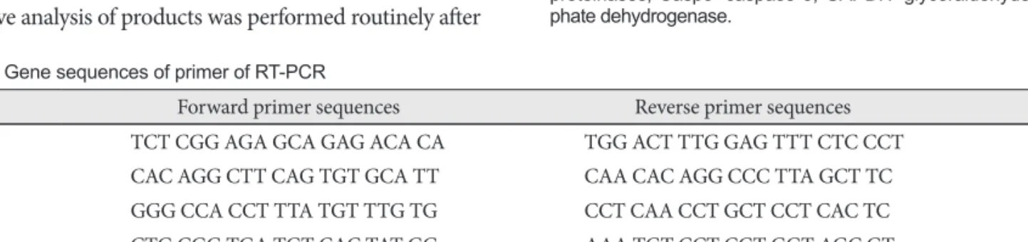
Table 1. Gene sequences of primer of RT-PCR
Gene Forward primer sequences Reverse primer sequences Size
ET-1 TCT CGG AGA GCA GAG ACA CA TGG ACT TTG GAG TTT CTC CCT 156 bp
ERA CAC AGG CTT CAG TGT GCA TT CAA CAC AGG CCC TTA GCT TC 118 bp
NOS2 GGG CCA CCT TTA TGT TTG TG CCT CAA CCT GCT CCT CAC TC 107 bp
NOS3 CTG CGG TGA TGT CAC TAT GG AAA TGT CCT CGT GGT AGC GT 140 bp
MMP2 AAG AGG CCT GGT TAC CCT GT AAG TAG CAC CTG GGA GGGAT 137 bp
TIMP GAC CTA TAG TGC TGG CTG TG GATC GCT CTG GTA GCC CTT CT 133 bp
Casp3 GAA AGC ATC CAG CAA TAG GC TAAGGA AGC CTG GAG CAC AG 100 bp
RT-PCR: reverse transcription-polymerase chain reaction, ET-1: endothelin-1, ERA: endothelin receptor A, NOS2: inducible nitric oxide synthase, NOS3: endothelial nitric oxide synthase, MMP2: matrix metalloproteinase 2, TIMP: tissue inhibitor of matrix metalloproteinases, Casp3: Caspase 3
Fig. 1. Typical example of RT-PCR products are shown for the level of ET-1, ERA, NOS2, NOS3, MMP 2 and TIMP mRNA measured in the lung tissue. The RT-PCR products from the transcripts of ET-1, ERA, NOS2, NOS3, MMP2, TIMP, Casp 3 and GAPDH were 156 bp, 118 bp, 107 bp, 140 bp, 137 bp, 133 bp 100 and 89 bp, respectively.
ET-1: endothelin-1, ERA: endothelin receptor A, NOS 2: inducible ni- tric oxide synthase, NOS 3: endothelial nitric oxide synthase, MMP-2:
matrix metalloproteinase 2, TIMP: tissue inhibitor of matrix metallo- proteinases, Casp3: caspase 3, GAPDH: glyceraldehyde 3-phos- phate dehydrogenase.
501 bp 489 bp 404 bp 331 bp 242 bp 190 bp 147 bp 110 bp
067 bp
ET-1156 ERA
118 NOS2107 NOS3140
MMP2137 TIMP 133 Casp3
100 Rat GAPDH
89
Results
Changes of the total body weight and heart weight From day 5 until day 28, the total weight of the rats decre- ased significantly in the M group compared with the control group. On day 28, the RV weight significantly increased in the M group, and significantly decreased in the simvastatin group compared with the M group. From day 7, the LV and septum weight significantly decreased in the M group and there were no significant changes in the simvastatin group.
On day 28, the RV/LV+S weight ratio significantly decreased in the simvastatin group, compared with the M group (Table 2).
Estimation of the right ventricular pressure
From day 5, the mean RVP significantly increased in the M group, compared with control group. On day 28, the RVP sig- nificantly decreased in the simvastatin group compared with the M group (Table 3).
Histologic study
The basic pulmonary architecture was similar in each gr- oup. The predominant changes in the pulmonary vasculature occurred in the M group on day 7 and included the develop- ment of medial thickening in the pulmonary arterioles, com- pared with both the control group and the simvastatin group (Fig. 2).
Morphometric analysis of the pulmonary arteries The medial wall thickness of the pulmonary artery
In the M group, the ratio of medial thickening to the ex- ternal diameter of the pulmonary artery significantly incre-
ased from day 7, compared to control. In the simvastatin gr- oup, the ratio of medial thickening of the pulmonary artery was not significantly different from the M group (Table 4).
The number of muscular pulmonary arterioles
The number of muscular pulmonary arterioles significant- ly increased in the M group, comapared with the C group.
From day 7, the number of muscular pulmonary arterioles significantly decreased in the simvastatin group, compared to the M group (Table 5).
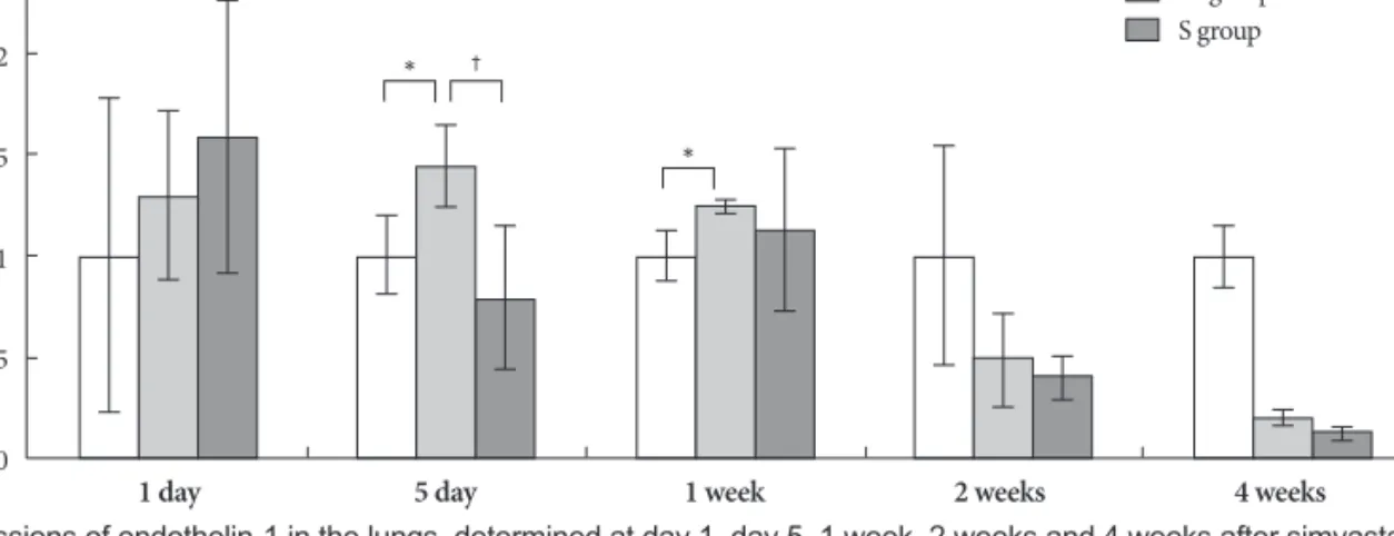
Gene expressions in rat lung tissues Gene expressions of endothelin-1
On days 5 and 7, in the M group, the expression of gene ET-1 was higher than in the control group. On day 5, in the Simvas- tatin group, the same gene was lower expressed than in the M group (Fig. 3).
Gene expressions of endothelin receptor A
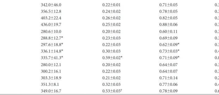
On day 1, the ERA gene was more expressed in the M gr- Table 2. Changes in the whole body weight, right ventricle weight, and left ventricle and septum weight, measured in each group on day 1, 5, 7, 14 and 28
Group/Day Total weight (g) RV (g) LV+S (g) RV/LV+S
C 01 05 07 14 28
284.8±15.2 342.0±46.0 356.5±12.8 403.2±22.4 436.0±19.7
0.18±0.02 0.22±0.01 0.24±0.02 0.26±0.02 0.25±0.02
0.69±0.09 0.71±0.05 0.78±0.05 0.82±0.05 0.88±0.06
0.26±0.06 0.31±0.03 0.30±0.02 0.30±0.02 0.32±0.03 M01
05 07 14 28
280.6±10.0 288.8±12.7*
297.6±18.8*
336.1±14.8*
335.7±41.3*
0.20±0.02 0.23±0.03 0.22±0.03 0.30±0.03 0.59±0.02*
0.60±0.11 0.69±0.09 0.62±0.09*
0.73±0.03*
0.71±0.09*
0.33±0.05 0.33±0.03 0.33±0.03 0.41±0.02 0.83±0.04 S 01
05 07 14 28
280.0±12.1 300.2±16.1 303.3±18.9 351.3±8.1 349.0±16.7
0.20±0.02 0.22±0.03 0.21±0.02 0.32±0.03 0.53±0.03†
0.64±0.07 0.64±0.07 0.71±0.14 0.77±0.06 0.78±0.09
0.31±0.02 0.37±0.04 0.29±0.03 0.41±0.02 0.67±0.03†
*p<0.05 vs. the corresponding value in the C group, †p<0.05 vs. the corresponding value in the M group. C: control, M: monocrotalin, S: sim- vastatin, RV: right ventricle, S; septum, LV: left ventricle
Table 3. Right ventricular pressure in each group (mmHg)
Days C group M group S group
01 12.3±3.5 14.1±2.5 17.0±1.1
05 11.6±2.4 17.3±5.3* 15.0±2.1
07 10.2±3.1 15.4±3.5* 17.0±2.1
14 13.2±3.2 22.6±2.9* 24.0±1.4
28 12.5±1.9 30.3±2.8* 13.7±2.6†
*p<0.05 vs. the corresponding value in the C group, †p<0.05 vs. the corresponding value in the M group. C: control, M: monocrotaline, S: simvastatin
oup than in control group. On day 5, however, expression of the same gene significantly decreased in the simvastatin gr- oup, compared with the M group (Fig. 4).
Gene expressions of inducible nitric oxide synthase On day 7, expression of NOS2 significantly increased in the M group compared to control group. On day 5, NOS2 was less expressed in the simvastatin group than in the M group (Fig. 5).
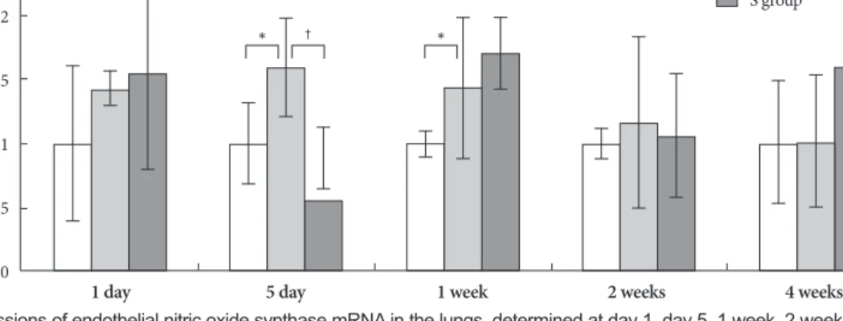
Gene expressions of endothelial nitric oxide synthase On days 5 and 7, expression of NOS3 significantly incre- ased in the M group compared to control group. On day 5, NOS3 was less expressed in the simvastatin group than in the M group (Fig. 6).
Gene expressions of matrix metalloproteinases
On days 1 and 28, expression of MMP2 significantly in-
creased in the M group compared to control group. On day 5, MMP2 was less expressed in the simvastatin group than in the M group (Fig. 7).
Gene expressions of tissue inhibitor of matrix metalloproteinases
On days 5, 14 and 28, expression of TIMP significantly in- creased in the M group compared to control group. On day 5, TIMP was less expressed in the simvastatin group than in the M group (Fig. 8).
Gene expressions of caspase 3
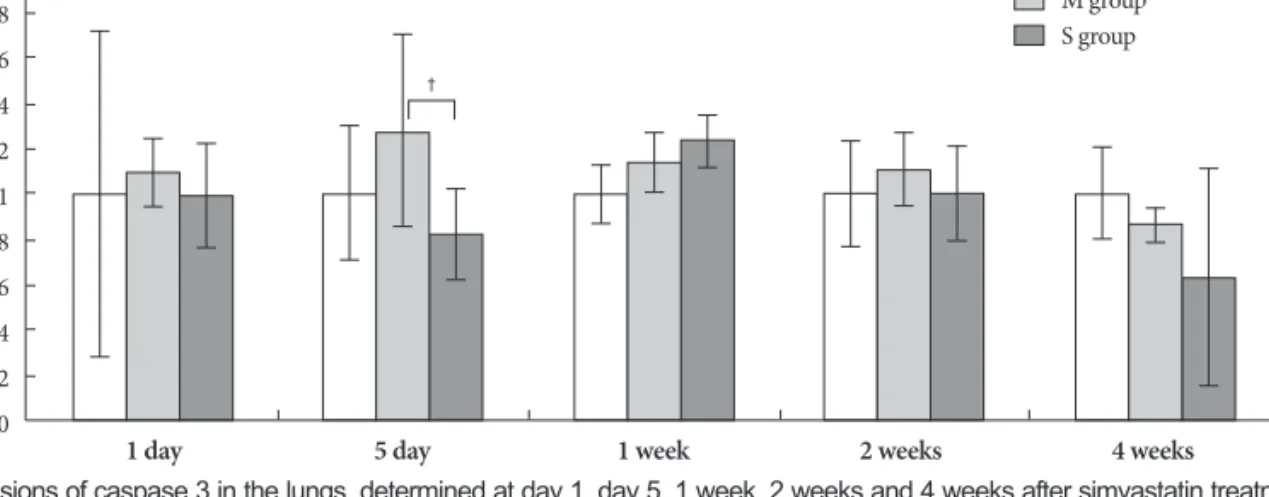
On day 5, expression of caspase 3 significantly decreased in the simvastatin group, compared to the M group (Fig. 9).
Liver enzymes and serum lipid profiles
On day 5, ALT significantly increased in the M group com- pared to control group. In contrast, on days 5 and 7, triglycer- ide significantly decreased in the M group compared to con- trol group.
On days 1 and 7, triglyceride also significantly decreased in the simvastatin group compared to the M group. On day 14, HDL-C significantly decreased in the M group compared to control group. LDL-C, AST and total cholesterol showed no significant changes after either M or simvastatin treatment (Table 6).
Discussion
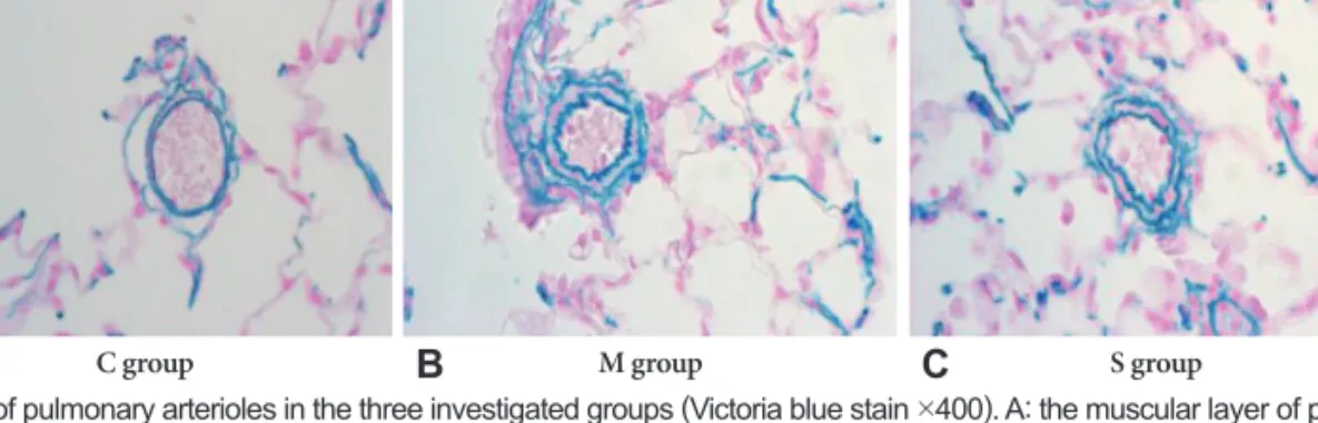
We confirmed that pulmonary hypertension was induced by M, as evidenced by pathologic finding and gene expres- sions. The evidence includes; first, a progressive increase in the ratio of RV to LV+S was significantly noted in both our previous study14-16) and in the present study. Second, the RVP progressively increased in the M group, compared with the control group. Third, in the M group, predominant changes in the pulmonary vasculature included the development of medial thickening in the pulmonary arterioles and increased number of intra-acinar muscular arteries. Fourth, the expres- Fig. 2. Photographs of pulmonary arterioles in the three investigated groups (Victoria blue stain ×400). A: the muscular layer of pulmonary arterioles was normal in C group. B: the medial layer of pulmonary arterioles was progressively thickened after M injection in M group. C: the medial wall thickness after M injection was significantly attenuated in the simvastatin group. C: control group, M: monocrotaline group, S: simvastatin group.
C group M group S group
A B C
Table 4. Ratio of the medial thickening of pulmonary artery in each group (%)
Day C group M group S group
01 9.2±0.7 08.2±0.8 08.6±0.6
05 7.8±0.9 09.0±0.9 10.6±1.3
07 9.4±1.1 13.8±1.7* 12.4±2.1
14 7.8±0.8 20.1±3.6* 19.8±1.9
28 7.6±0.8 23.8±2.5* 22.6±2.7
*p<0.05 vs. the corresponding value in the C group. C: control, M:
monocrotaline, S: simvastatin
Table 5. Number of muscular pulmonary arterioles in each group
Day C group M group S group
01 2.3±0.1 2.4±0.1 2.3±0.2
05 2.3±0.1 2.5±0.1 2.1±0.0
07 2.4±0.1 3.1±0.1* 2.2±0.2†
14 2.4±0.1 3.4±0.4* 2.8±0.1†
28 2.4±0.3 5.7±0.6* 4.9±0.3†
*p<0.05 vs. the corresponding value in the C group, †p<0.05 vs. the corresponding value in the M group. C: control, M: monocrotaline, S: simvastatin
sions of ET-1, ERA, NOS2, NOS3, MMP and TIMP genes were significantly increased in the M group. These pathologic find- ings were consistent with other results demonstrating a sig- nificant rise in pulmonary blood pressure and apparent RVH after M injection in the rat model.17)18)
During the development of M-induced pulmonary hyper- tension in rats, administration of simvastatin exerted inhibi- tory effects on RVH and on a number of muscular pulmo-
nary arterioles. On day 28, RVH significantly decreased in the simvastatin group compared to the M group. Starting with the first week, the number of muscular pulmonary arterioles was significantly reduced in the simvastatin group, com- pared to M group. However, the ratio of medial thickening of the pulmonary artery has not changed significantly in the simvastatin group. On day 5, expressions of ET-1, ERA, NOS2, NOS3, MMP and TIMP genes significantly decreased in the Fig. 3. Gene expressions of endothelin-1 in the lungs, determined at day 1, day 5, 1 week, 2 weeks and 4 weeks after simvastatin treatment.
*p<0.05 vs. the corresponding value in the C group, †p<0.05 vs. the corresponding value in the M group. C: control, M: monocrotaline, S:
simvastatin.
2.5
2
1.5
1
0.5
0
C group M group S group
* †
*
Expression change
1 day 5 day 1 week 2 weeks 4 weeks
Fig. 4. Gene expressions of endothelin receptor A in the lungs, determined at day 1, day 5, 1 week, 2 weeks and 4 weeks after simvastatin treat- ment. *p<0.05 vs. the corresponding value in the C group, †p<0.05 vs. the corresponding value in the M group. C: control, M: monocrotaline, S:
simvastatin.
3.5 3 2.5 2 1.5 1 0.5 0
C group M group S group
Expression change
1 day 5 day 1 week 2 weeks 4 weeks
*
†
Fig. 5. Gene expressions of inducible nitric oxide synthase in the lungs, determined at day 1, day 5, 1 week, 2 weeks and 4 weeks after simvas- tatin treatment. *p<0.05 vs. the corresponding value in the C group, †p<0.05 vs. the corresponding value in the M group. C: control, M: monocro- taline, S: simvastatin.
5 4 3 2 1 0 -1 -2
C group M group S group
Expression change
1 day 5 day 1 week 2 weeks 4 weeks
*
†
simvastatin group.
Statins, such as 3-hydroxyl-3-methylglutaryl (HMG)-CoA reductase inhibitors, have been developed as lipid-lowering drugs. In addition, recent experiments and clinical trials have demonstrated that statins also exert vasculoprotective effects, independent of their cholesterol-lowering effects.19) Thus, recent studies have suggested that statins may have potential
use in the treatment of pulmonary hypertension.8)9)
However, there is still controversy on the effect of simvas- tatin on pulmonary hypertension. Simvastatin attenuates ne- ointimal thickening of the smooth muscle.10) Simvastatin is effective in inducing apoptosis in hyperproliperative pulmo- nary vascular lesions and could be considered as a potential drug for treatment of human severe pulmonary hypertension.
Fig. 6. Gene expressions of endothelial nitric oxide synthase mRNA in the lungs, determined at day 1, day 5, 1 week, 2 weeks and 4 weeks after simvastatin treatment. *p<0.05 vs. the corresponding value in the C group, †p<0.05 vs. the corresponding value in the M group. C: control, M:
monocrotaline, S: simvastatin.
2.5
2
1.5
1
0.5
0
C group M group S group
Expression change
1 day 5 day 1 week 2 weeks 4 weeks
* † *
Fig. 7. Gene expressions of matrix metalloproteinase 2 in the lungs, determined at day 1, day 5, 1 week, 2 weeks and 4 weeks after simvastatin treat- ment. *p<0.05 vs. the corresponding value in the C group, †p<0.05 vs. the corresponding value in the M group. C: control, M: monocrotaline, S: simv- astatin.
2.5
2
1.5
1
0.5
0
C group M group S group
Expression change
1 day 5 day 1 week 2 weeks 4 weeks
*
*
†
Fig. 8. Gene expressions of tissue inhibitor of matrix metalloproteinases in the lungs, determined at day 1, day 5, 1 week, 2 weeks and 4 weeks after simvastatin treatment. *p<0.05 vs. the corresponding value in the C group, †p<0.05 vs. the corresponding value in the M group. C: control, M: monocro- taline, S: simvastatin.
4 3.5 3 2.5 2 1.5 1 0.5 0
C group M group S group
Expression change
1 day 5 day 1 week 2 weeks 4 weeks
*
*
* †
Nishimura et al.10) demonstrated impressive hemodynamic responses to simvastatin therapy, and also suggested that sim- vastatin promotes apoptosis of the smooth muscle cells in pul- monary arterioles. That data10) demonstrated the down-regul- ation of the inflammatory genes such as fos, jun, and TNF-a and the up-regulation of the cell cycle inhibitor p27Kip1, the NOS3 gene, and the bone morphogenetic protein receptor type 1. In contrast, our experiments demonstrated a significant down-regulation of NOS3 expression in simvastatin treated animals, suggesting that any increase in NO by simvastatin would relate to increased bioavailability, rather than enzyme expression.
However, McMurtry et al.11) reported no effect of simvasta- tin on pulmonary hypertension, i.e., simvastatin could not re- verse the M-induced pulmonary arterial hypertension. In that study, simvastatin improved echocardiographic parameters of pulmonary hypertension, including the pulmonary artery acceleration time and the RV thickness after 1-2 weeks, but
the effect was not sustained, and ultimately, rats treated with simvastatin developed severe pulmonary arterial hyperten- sion and expired. Our study revealed a weak inhibitory ef- fect of simvastatin on RVH and pulmonary hypertension. In our model, treatment with simvastatin initially appeared to im- prove lung pathologic findings, but did not reverse the M- induced pulmonary hypertension. In general the simvastatin effect of attenuation of RVH and pulmonary hypertension seems to be weaker than that of bosentan.14)
NO is best known for its marked vasodilator activity. Ex- ogenous NO is useful in the clinical management of acute pulmonary hypertensive crisis. However, NO is also a reac- tive radical capable of tissue injury, either directly or after re- acting with other radicals. For example, an extremely fast re- action of NO with superoxide yields the highly cytotoxic pe- roxynitrite.20) There are three subtypes of NOS {i.e., NOS2, NOS3 and neuronal NOS (nNOS)}. The NO can contribute to oxidative stress and, in association with tissue injury, NOS2 Fig. 9. Gene expressions of caspase 3 in the lungs, determined at day 1, day 5, 1 week, 2 weeks and 4 weeks after simvastatin treatment. †p<0.05 vs. the corresponding value in the M group. C: control, M: monocrotaline, S: simvastatin.
2 1.8 1.6 1.4 1.2 1 0.8 0.6 0.4 0.2 0
C group M group S group
Expression change
1 day 5 day 1 week 2 weeks 4 weeks
†
Table 6. Liver enzymes and lipid profiles in each group (mg/dL)
Group AST ALT TC TG HDL-C LDL-C
C01d 05d 07d 14d 28d
118.7±22.5 134.3±10.0 126.7±18.4 111.0±7.2 159.0±16.7
040.0±7.2 032.7±2.1 036.7±3.1 034.0±2.6 037.7±3.5
51.0±12.3 52.3±9.3 58.7±4.0 50.3±4.9 43.0±6.1
086.3±19.1 122.3±10.5 145.7±49.7 155.7±11.6 043.7±15.3
26.0±5.6 23.0±4.4 26.7±2.3 24.7±0.6 17.7±1.5
6.3±1.5 7.7±2.1 7.7±1.2 6.0±1.0 4.7±2.3 M01d
05d 07d 14d 28d
105.5±15.1 122.0±30.5 154.3±33.3 127.8±15.9 115.0±52.6
039.0±8.5 040.7±5.5*
044.7±6.1 035.8±5.1 058.7±38.9
56.3±9.6 52.7±9.9 53.5±8.8 50.8±8.0 48.0±10.6
122.7±37.7 059.8±13.7*
053.7±25.6*
109.2±53.8 039.0±21.7
26.3±5.2 21.5±3.6 23.0±3.9 22.0±1.7*
15.0±2.9
8.7±2.0 7.8±1.9 9.7±2.3 7.7±2.9 9.8±2.4 S01d
05d 07d 14d 28d
113.0±21.8 115.2±16.7 126.0±32.9 110.3±19.7 183.3±137.3
041.2±10.5 042.7±9.3 047.7±23.2 034.3±2.3 123.1±201.2
48.4±6.4 50.2±4.7 64.3±12.2 51.2±5.2 52.1±15.5
057.4±29.4† 055.7±19.4 054.7±16.4† 076.7±33.2 053.4±35.4
23.0±2.7 22.0±1.4 24.0±1.7 21.7±2.1 17.0±4.5
6.4±1.5 9.2±2.3 14.0±7.8 8.3±1.2 9.5±2.6
*p<0.05 vs. the corresponding value in the control group, †p<0.05 vs. the corresponding value in the M group. C: control, M: monocrotalin, S:
simvastatin, AST: aspartate aminotransterase, ALT: alanine aminotransferase, TC: total cholesterol, TG: triglyceride, HDL-C: high density li- poprotein-cholesterol, LDL-C: low density lipoprotein-cholesterol
is often up-regulated. Transient NOS2 induction in the pul- monary vascular wall at the beginning of chronic hypoxia participates in the pathogenesis of pulmonary hypertension.
In our study, on day 7, NOS2 significantly increased in the M group compared to control and significantly decreased in the simvastatin group, compared to the M group, on day 5. In chro- nic hypoxia, many authors have reported elevated NOS mRNA and protein in both the lungs and the pulmonary vessels.21) However, most of those studies either focused on NOS3 or did not discriminate between various NOS isoforms. In a study comparing NOS3, nNOS and NOS2 activity in mice, NOS3 showed the most important role in regulating pulmonary circulation.22) Similarly, the endothelial NO production has been shown to play a key role in vascular reactivity and in the modulation of smooth muscle cell proliferation, as well as inflammation.21) Therefore, at least, in part, the protective ef- fects of statins on the endothelium may contribute to recov- er pulmonary hypertension. In our study, NOS3 has not in- creased significantly in the simvastatin group.
Li et al.23) investigated the hypothesis that statins regulate vascular remodeling by modulation of MMP secretion in pri- mary cultured pulmonary artery smooth muscle cells. Thus, angiotension II induced a concentration-dependent MMP2 secretion. Pre-exposure of cells to simvastatin blocked the an- giotension II-stimulated MMP2 release. Simvastatin suppress- ed ET-1-induced MMP2 release through the Rho/ROCK pa- thway. In contrast, there was no effect of angiotensin II on the expression of TIMP genes. These results indicated a novel statins-regulation of Rho/ROCK signaling against pulmo- nary vascular remodeling, and suggested that statins could prove usefulness in targeting this pathway in pulmonary hy- pertension and other disease conditions.24)
In rats, during treatment of severe pulmonary hypertension with a HMG-CoA reductase inhibitor, simvastatin caused ap- optosis of the lumen-obliteration cells, which was associated with significant reduction of pulmonary artery pressure and of RVH.13) In normal rat lungs, blood vessels are negative for caspase 3 staining. Simvastatin treatment causes activation of caspase-3 and significantly decreases the number of pulmo- nary vascular lesions. Treatment with simvastatin induced ap- optosis of endothelial cells in obliterated vessels. Simvastatin reduced pulmonary hypertension through apoptosis-induc- ed reopening of the occluded vessels, as shown by caspase-3 immunohistochemistry.13) It is now becoming clear that st- atins can trigger apoptosis using both caspase-dependent and caspase-independent Rho kinase-dependent pathways.25) The ability of HMG-CoA reductase inhibitors to induce ap- optosis was demonstrated in both rat pulmonary vein endo- thelial cells26) and human hepatocytes.27) In our study, the si- mvastatin effect on caspase-3 mRNA was not apparent, suggest- ing that more study on high dose simvastatin treatment is needed.
Simvastatin has both anti-proliferative and pro-apoptotic effects on vascular smooth muscle cells and enhances the endothelial production of NO. Simvastatin stabilizes the en- dothelial barrier function and promotes anti-inflammatory effect, which leads to atherogenesis.
The dose of simvastatin, used both experimentally and cli- nically, was inconsistent among the various other investiga- tors. Girgis et al.24) used 20 mg/kg/day of simvastatin and fo- und that simvastatin attenuated and induced regression of chronic hypoxic pulmonary hypertension. Wilkins et al.28) gave the patients 80 mg/day of simvastatin for 6 months, and found that, when added to conventional therapy in patients with pulmonary arterial hypertension, simvastatin produced a small and transient early reduction in both the RV mass and the N terminal pro B type natriuretic peptide (NT-pro- BNP) level. Liu et al.29) used 2 mg/kg/d of simvastatin for 35 days in rats, and resulted in prevention of pulmonary hyper- tension. However, in McMurtry’s rat study,11) the same dose of 2 mg/kg/d of simvastatin did not reverse the M-induced pulmonary arterial hypertension.
In conclusion, treatment of simvastatin in M-induced hy- pertensive rats blunted the development of M-induced pul- monary hypertension and resulted in a weak attenuation of both RVH and vascular remodeling. Our results in M-in- duced pulmonary hypertension suggest that all seven genes increased, and all of those increases may play a role in RVH and remodeling. The sensitive markers of decreased expres- sion of ERA and NOS2 genes appeared earlier than other pathologic findings. Also, the decreased gene expression of MMP2 could be a marker of pulmonary hypertension treat- ment. Further studies are in progress to determine the effects of simvastatin on other gene expressions in the M-induced pulmonary hypertension rat model.
Larger sample sizes are needed to measure hemodynamics and follow-ups should extend for more than 4 weeks. The follow up duration is a critical factor to evaluate the effecti- veness of the simvastatin treatment on M-induced pulmona- ry hypertension. Finally, further studies are necessary to ex- plore the intricate mechanisms of simvastatin treatment in M-induced pulmonary hypertension.
Acknowledgments
This work was supported by the Korean Society of Cardiology (2009) and National Research Foundation of Korea (NRF) grant founded by the Korean government (MEST) (KRF-2009-0064784).
REFERENCES
1) Humbert M, Morrell NW, Archer SL, et al. Cellular and molecular pa- thobiology of pulmonary arterial hypertension. J Am Coll Cardiol 2004;
43(Suppl S):13S-24S.
2) Budhiraja R, Tuder RM, Hassoun PM. Endothelial dysfunction in pulmonary hypertension. Circulation 2004;109:159-65.
3) Meininger GA, Davis MJ. Cellular mechanisms involved in the vascu- lar myogenic response. Am J Physiol 1992;263:H647-59.
4) Keck EW. Pulmonary hypertension and pulmonary vascular disease in congenital heart defects. Z Kardiol 1989;78(Suppl 7):65-73.
5) Yamaki S, Endo M, Takahashi T. Different grades of medial hyper- trophy and intimal changes in small pulmonary arteries among vari- ous types of congenital heart disease with pulmonary hypertension.
Tohoku J Exp Med 1997;182:83-91.
6) Palmer RM, Ashton DS, Moncada S. Vascular endothelial cells syn- thesize nitric oxide from L-arginine. Nature 1988;333:664-6.
7) Todd L, Mullen M, Olley PM, Rabinovitch M. Pulmonary toxicity of monocrotaline differs at critical periods of lung development. Pediatr Res 1985;19:731-7.
8) Girgis RE, Li D, Zhan X, et al. Attenuation of chronic hypoxic pul- monary hypertension by simvastatin. Am J Physiol Heart Circ Physi- ol 2003;285:H938-45.
9) Nishimura T, Faul JL, Berry GJ, et al. Simvastatin attenuates smooth muscle neointimal proliferation and pulmonary hypertension in rats.
Am J Respir Crit Care Med 2002;166:1403-8.
10) Nishimura T, Vaszar LT, Faul JL, et al. Simvastatin rescues rats from fatal pulmonary hypertension by inducing apoptosis of neointimal smooth muscle cells. Circulation 2003;108:1640-5.
11) McMurtry MS, Bonnet S, Michelakis ED, Bonnet S, Haromy A, Ar- cher SL. Statin therapy, alone or with rapamycin, does not reverse mo- nocrotaline pulmonary arterial hypertension: the rapamycin-atorva- statin-simvastatin study. Am J Physiol Lung Cell Mol Physiol 2007;
293:L933-40.
12) Kim MY, Kim YK, Jung YW, Kim WT, Kwon TH, Lee DS. The pro- tective effect of simvastatin on monocrotaline-induced pulmonary hy- pertension in rats. Korean Circ J 2008;38:313-9.
13) Taraseviciene-Stewart L, Scerbavicius R, Choe KH, et al. Simvastatin causes endothelial cell apoptosis and attenuates severe pulmonary hy- pertension. Am J Physiol Lung Cell Mol Physiol 2006;291:L668-76.
14) Lim KA, Shim JY, Cho SH, Kim KW, Han JJ, Hong YM. Effect of endothelin receptor blokade on monocrotaline-induced pulmonary hypertension in rats. Korean J Pediatr 2009;52:689-95.
15) Lim KA, Kim KC, Cho MS, Lee BE, Kim HS, Hong YM. Gene ex- pression of endothelin-1 and endothelin receptor A on monocrotaline- induced pulmonary hypertension in rats after bosentan treatment. Ko- rean Circ J 2010;40:459-64.
16) Koo HS, Kim KC, Hong YM. Gene expression of nitric oxide syn- thase and matrix metalloproteinase-2 in monocrotaline-induced pulmo-
nary hypertension in rats after bosentan treatment. Korean Circ J 2010;41:83-90.
17) Miyauchi T, Yorikane R, Sakai S, et al. Contribution of endogenous endothelin-1 to the progression of cardiopulmonary alterations in rats with monocrotalin-induced pulmonary hypertension. Circ Res 1993;
73:887-97.
18) Itoh T, Nagaya N, Fujii T, et al. A combination of oral sildenafil and beraprost ameliorates pulmonary hypertension in rats. Am J Respir Crit Care Med 2004;169:34-8.
19) Maron DJ, Fazio S, Linton MF. Current perspectives on statins. Cir- culation 2000;101:207-13.
20) Beckman JS, Koppenol WH. Nitric oxide, superoxide and peroxyni- trite: the good, the bad, and the ugly. Am J Physiol 1996;271:C1424-37.
21) Hampl V, Herget J. Role of nitric oxide in the pathogenesis of chronic pulmonary hypertension. Physiol Rev 2000;80:1337-72.
22) Miller AA, Hislop AA, Vallance PJ, Haworth SG. Deletion of the eNOS gene has a greater impact on the pulmonary circulation of male than female mice. Am J Physiol Lung Cell Mol Physiol 2005;289:
L299-306.
23) Li M, Li Z, Sun X. Statins suppress MMP2 secretion via inactivation of RhoA/ROCK pathway in pulmonary vascular smooth muscle cells.
Eur J Pharmacol 2008;591:219-23.
24) Girgis RE, Mozammel S, Champion HC, et al. Regression of chronic hypoxic pulmonary hypertension by simvastatin. Am J Physiol Lung Cell Mol Physiol 2007;292:L1105-10.
25) Kim YC, Song SB, Lee MH, et al. Simvastatin induces caspase-in- dependent apoptosis in LPS-activated RAW264.7 macrophage cells.
Biochem Biophys Res Commun 2006;339:1007-14.
26) Kaneta S, Satoh K, Kano S, Kanda M, Ichihara K. All hydrophobic HMG-CoA reductase inhibitors induce apoptotic death in rat pulmo- nary vein endothelial cells. Atherosclerosis 2003;170:237-43.
27) Kubota T, Fujisaki K, Itoh Y, Yano T, Sendo T, Oishi R. Apoptotic injury in cultured human hepatocytes induced by HMG-CoA reduc- tase inhibitors. Biochem Pharmacol 2004;67:2175-86.
28) Wilkins MR, Ali O, Bradlow W, et al. Simvastatin as a treatment for pulmonary hypertension trial. Am J Respir Crit Care Med 2010;
181:1106-13.
29) Liu B, Wang XQ, Yu L, Zhou TF, Wang XM, Liu HM. Simvastatin restores down-regulated GATA-6 expression in pulmonary hyperten- sive rats. Exp Lung Res 2009;35:411-26.