ABSTRACT
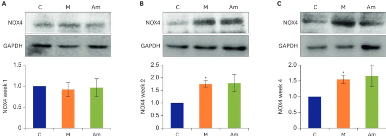
Background and Objectives: Elevated endothelin (ET)-1 level is strongly correlated with the pathogenesis of pulmonary arterial hypertension (PAH). Expression level of nicotinamide adenine dinucleotide phosphate oxidase (NOX) 4 is increased in the PAH patients.
Ambrisentan, a selective endothelin receptor A (ERA) antagonist, is widely used in PAH therapy. The current study was undertaken to evaluate the effects of ambrisentan treatment in the monocrotaline (MCT)-induced PAH rat model.
Methods: Rats were categorized into control group (C), monocrotaline group (M) and ambrisentan group (Am). The M and Am were subcutaneously injected 60 mg/kg MCT at day 0, and in Am, ambrisentan was orally administered the day after MCT injection for 4 weeks.
The right ventricle (RV) pressure was measured and pathological changes of the lung tissues were observed by Victoria blue staining. Protein expressions of ET-1, ERA, endothelial nitric oxide synthase (eNOS) and NOX4 were confirmed by western blot analysis.
Results: Ambrisentan treatment resulted in a recovery of the body weight and RV/left ventricle+septum at week 4. The RV pressure was lowered at weeks 2 and 4 after ambrisentan administration. Medial wall thickening of pulmonary arterioles and the number of intra- acinar arteries were also attenuated by ambrisentan at week 4. Protein expression levels of ET-1 and eNOS were recovered at weeks 2 and 4, and ERA levels recovered at week 4.
Conclusions: Ambrisentan administration resulted in the recovery of ET-1, ERA and eNOS protein expression levels in the PAH model. However, the expression level of NOX4 remained unaffected after ambrisentan treatment.
Keywords: Hypertension, pulmonary; Endothelin receptor antagonists; Monocrotaline;
Gene expression
Original Article
Received: Jan 6, 2019 Revised: Mar 4, 2019 Accepted: Apr 3, 2019 Correspondence to Young Mi Hong, MD, PhD
Department of Pediatrics, Ewha Womans University College of Medicine, 1071 Anyangcheon-ro, Yangcheon-gu, Seoul 07985, Korea.
E-mail: [email protected]
Copyright © 2019. The Korean Society of Cardiology
This is an Open Access article distributed under the terms of the Creative Commons Attribution Non-Commercial License (https://
creativecommons.org/licenses/by-nc/4.0) which permits unrestricted noncommercial use, distribution, and reproduction in any medium, provided the original work is properly cited.
ORCID iDs Hyeryon Lee
https://orcid.org/0000-0002-8566-7503 Arim Yeom
https://orcid.org/0000-0001-6343-4648 Kwan Chang Kim
https://orcid.org/0000-0001-8297-5415 Young Mi Hong
https://orcid.org/0000-0002-6600-7876
Hyeryon Lee , PhD
1, Arim Yeom , BA
1, Kwan Chang Kim , MD, PhD
2, and Young Mi Hong , MD, PhD
11
Department of Pediatrics, Ewha Womans University College of Medicine, Seoul, Korea
2
Department of Thoracic and Cardiovascular Surgery, Ewha Womans University College of Medicine, Seoul, Korea
Effect of Ambrisentan Therapy on the Expression of Endothelin Receptor, Endothelial Nitric Oxide Synthase and NADPH Oxidase 4 in Monocrotaline- induced Pulmonary Arterial
Hypertension Rat Model
Conflict of Interest
The authors have no financial conflicts of interest.
Author Contributions
Conceptualization: Hong YM, Kim KC;
Investigation: Lee H, Yeom A, Kim KC; Writing - original draft: Lee H; Writing - review & editing:
Hong YM.
INTRODUCTION
Pulmonary arterial hypertension (PAH) is a fatal disease, characterized by elevation of the pulmonary artery pressure.
1)PAH is a multifactorial disease involving endothelial dysfunction and pulmonary arteriole remodeling.
2)Many signaling molecules, including endothelin (ET)- 1, ET receptor, and the endothelial-derived vasoactive molecules such as nitric oxide, play a critical role in the pathophysiology of the disease.
3)ET-1 is a strong vasoconstrictor that induces proliferation of vascular smooth muscle cells,
4)and has been targeted for therapy of numerous hypertensive diseases. ET-1 exerts pathophysiological changes in PAH by stimulating the constriction of pulmonary vessels and uncontrolled
proliferation of vascular smooth muscle cells. The ET system is activated in plasma and lung tissues in both the PAH animal model as well as in PAH patients.
5)Cells exhibit 2 types of receptors to facilitate binding of ET: type A and B. Smooth muscle cells express both type A and B, whereas endothelial cells express only type A receptors. Stimulation of ET receptors type A and B on the surface of smooth muscle cell promotes vasoconstriction and proliferative effects of ET. However, stimulation of the ET receptor type B induces production of nitric oxide and prostacyclin, which in turn induce vasodilation and anti-proliferating effects.
6)Endothelial nitric oxide synthase (eNOS) and nicotinamide adenine dinucleotide phosphate oxidase (NOX) modulate reactive oxygen species, stimulation of vasoconstriction and vascular remodeling in PAH.
7)In the experimental PAH model, there is increased expression and activity of NOX4, which is identified to be responsible for modulating the production of the reactive oxygen species.
8)In our previous study, we noticed that NOX4 protein expression level was increased by ET-1 treatment in pulmonary arterial smooth muscle cells.
9)It has also been found that ET-1 increases vascular superoxide via NOX in deoxycorticosterone acetate–salt hypertension rat model.
10)Likewise, lung biopsy samples from PAH patients also reveal an imbalance in the levels of reactive oxygen species.
11)Ambrisentan, an orally active diphenyl propionic acid derivative, is a potent ET receptor type A selective antagonist, having a half-life of 9 to 15 hours, that enables a once-daily dosing.
12)13)However, to date, there are no reports on the effect of ambrisentan on NOX4. Therefore, we considered it worthwhile to investigate the effects of ambrisentan treatment on NOX4 production in animal models. This study therefore investigated the effects of ambrisentan by evaluating the pathophysiology in PAH.
METHODS
Animals
Six-week old male Sprague Dawley rats were purchased from Orient Bio (Orient Bio Inc., Seongnam, Korea). The rats were maintained under climate-controlled conditions with a 12:12-hour light:dark cycle, with free access to standard chow and water. All animals were treated in accordance with the guidelines of the Institutional Animal Care and Use Committee (IACUC) at the Ewha Womans University (IACUC approval No. ESM16-0356).
Experimental protocols
The rats were classified into 3 groups: control group (C), monocrotaline group (M) and
ambrisentan group (Am). Saline was injected subcutaneously to the C. To the both M and
Am, monocrotaline (MCT) 60 mg/kg were injected subcutaneously at day 0. Subsequently, from day 1 onwards, ambrisentan was administered (0.2 mg/kg, per os) to the Am group, every day for 4 weeks. The rats were sacrificed at weeks 2 and 4. Body and organ weight were measured, and the ratio was calculated as follows; organ / body weight (g) ×10000. The tissues were harvested and immediately frozen in liquid nitrogen for western blot, or fixed in 10% formalin for pathological examination.
Hemodynamics
To evaluate the effects of ambrisentan, right ventricle (RV) pressure was measured in anesthetized rats. Briefly, the rats were anesthetized and placed on a thermostatically controlled heating box for maintaining the body temperature. A catheter was inserted into the right external jugular vein, and the mean RV pressure was measured in this study.
Pathological changes in the pulmonary arteries
For histopathological evaluation, the lung tissues were embedded in paraffin and sectioned.
The sections were cut into 3–5 μm thickness and stained with hematoxylin-eosin and Victoria blue. More than 20 fields of pulmonary arterioles ranging 25–100 μm in diameter were observed. This method was based on our previous reports.
14)Western blot analysis
The harvested tissues were homogenized in lysis buffer (Proprep; IntRON Biotechnology, Gyeonggi, Korea) on ice. After 1 hour, the samples were centrifuged at 12,000 rpm in 4°C.
The protein content in supernatant was quantified with an enzyme-linked immunosorbent assay reader (Molecular Devices, Sunnyvale, CA, USA) at 562 nm, based on a bovine serum albumin (BSA) standard curve. The amount of supernatant containing 25–35 μg of protein was used for 8–12% sodium dodecyl sulfate polyacrylamide gel electrophoresis. Proteins resolved on the acrylamide gel by electrophoresis were transferred onto nitrocellulose membranes. The membranes were blocked with 5% BSA in tris-buffered saline containing 0.1% tween 20, for 1 hour at room temperature. Membranes were then washed, incubated overnight at 4°C with appropriate primary antibodies of ET-1 (Abcam, Cambridge, MA, USA), endothelin receptor A (ERA; Santa Cruz Biotechnology, Santa Cruz, CA, USA), eNOS (Santa Cruz Biotechnology) and NOX4 (Santa Cruz Biotechnology) separately, after which the probed membranes were incubated with the corresponding secondary antibodies.
Membranes were then washed and visualized by subjecting to a chemiluminescent reaction using an enhanced chemiluminescence detection kit (GE Healthcare, Piscataway, NJ, USA).
Statistical analysis
Statistical analysis was performed using PRISM 5 (GraphPad Software, San Diego, CA, USA).
A Kruskal-Wallis test was used for the comparison of differences in the 3 groups and a Mann- Whitney test was used for comparisons between groups with Bonferroni correction. Animals were randomized within each experimental group. All data are presented as mean±standard deviation. A p value of <0.05 is considered statistically significant.
RESULTS
RV pressure in 3 groups
MCT administration significantly increased the RV pressure in the M as compared to the
C at weeks 2 (C vs. M; 10.33±1.76 mmHg vs. 27.00±1.47 mmHg, p=0.001) and 4 (C vs. M;
12.33±0.33 mmHg vs. 32.40±0.76 mmHg, p<0.001). Early concurrent oral administration of ambrisentan significantly decreased the right ventricular systolic pressure at weeks 2 (M vs.
Am; 27.00±1.47 mmHg vs. 16.60±0.40 mmHg, p<0.001) and 4 (M vs. Am; 32.40±0.76 mmHg vs. 14.67±0.80 mmHg, p<0.001) (Figure 1).
Changes of the body weight in 3 groups
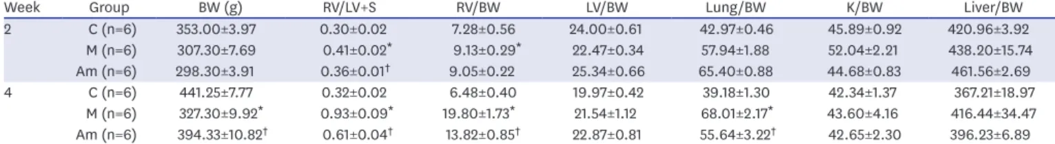
At week 4, the body weight (BW) decreased significantly in the M group as compared to the C group (C vs. M; 441.25±7.77 g vs. 327.30±9.92 g, p<0.001). Recovery in the BW was observed in the Am group at week 4 (M vs. Am; 327.30±9.92 g vs. 394.33±10.82 g, p<0.001) (Table 1). With the onset of RV hypertrophy, the RV/[left ventricle (LV)+septum (S)] weight ratio increased in the M at week 2 (C vs. M; 0.30±0.02 vs. 0.41±0.02, p=0.005) and 4 (C vs. M; 0.32±0.02 vs. 0.93±0.09, p<0.001), which decreased significantly in the Am at weeks 2 (M vs. Am; 0.41±0.02 vs. 0.36±0.01, p=0.035) and 4 (M vs. Am; 0.93±0.09 vs. 0.61±0.04, p=0.007). Furthermore, the RV weight/BW ratio decreased in the Am at week 4 (M vs. Am; 19.80±1.73 vs. 13.82±0.85, p=0.010).
The lung weight/BW ratio increased in the M at week 4 (C vs. M; 39.18±1.30 vs. 68.01±2.17, p<0.001), and decreased in the Am (M vs. Am; 68.01±2.17 vs. 55.64±3.22, p=0.014) (Table 1).
Lung pathology in 3 groups
MCT-induced PAH resulted in pulmonary arteriole remodeling, characterized by medial wall thickening and angiogenesis. The lung pathology data are presented in Figure 2. Compared to the C, increased medial wall thickness was observed in the M at weeks 2 (C vs. M; 21.99±1.95 vs. 38.43±0.94, p<0.001) and 4 (C vs. M; 23.60±2.17 vs. 38.25±0.66, p<0.001). The number of intra-acinar arteries also increased in the M at weeks 2 (C vs. M; 0.70±0.14 vs. 1.60±0.12, p=0.002) and 4 (C vs. M; 0.77±0.14 vs. 2.05±0.08, p<0.001). Conversely, medial wall thickening
Week 2 Week 4
0 15 35 30 25 20
10
RV pr essur e (mmHg) 5 C M
Am
*
*
† †
Figure 1. RV pressure in MCT-induced PAH model after ambrisentan treatment. RV pressure increased in the M compared to the C at weeks 2 and 4. The RV pressure decreased in the Am compared with the M at weeks 2 and 4.
RV = right ventricle; MCT = monocrotaline; PAH = pulmonary arterial hypertension; C = control group;
M = monocrotaline group; Am = ambrisentan group.
* p<0.05 as compared with the C;
†p<0.05 as compared with the M.
Table 1. Changes of BW and organ weight/BW ratio after low-dose ambrisentan treatment
Week Group BW (g) RV/LV+S RV/BW LV/BW Lung/BW K/BW Liver/BW
2 C (n=6) 353.00±3.97 0.30±0.02 7.28±0.56 24.00±0.61 42.97±0.46 45.89±0.92 420.96±3.92
M (n=6) 307.30±7.69 0.41±0.02 * 9.13±0.29 * 22.47±0.34 57.94±1.88 52.04±2.21 438.20±15.74
Am (n=6) 298.30±3.91 0.36±0.01
†9.05±0.22 25.34±0.66 65.40±0.88 44.68±0.83 461.56±2.69
4 C (n=6) 441.25±7.77 0.32±0.02 6.48±0.40 19.97±0.42 39.18±1.30 42.34±1.37 367.21±18.97
M (n=6) 327.30±9.92 * 0.93±0.09 * 19.80±1.73 * 21.54±1.12 68.01±2.17 * 43.60±4.16 416.44±34.47 Am (n=6) 394.33±10.82
†0.61±0.04
†13.82±0.85
†22.87±0.81 55.64±3.22
†42.65±2.30 396.23±6.89 BW = body weight; RV = right ventricle; LV = left ventricle; S = septum; K = kidney; C = control group; M = monocrotaline group; Am = ambrisentan group.
* p<0.05 as compared with the C;
†p<0.05 as compared with the M.
and angiogenesis decreased in the Am. Medial wall thickness decreased in the Am at week 4 (M vs. Am; 38.25±0.66 vs. 33.58±1.78, p=0.008), and the number of intra-acinar arteries also decreased in the Am at week 4 (M vs. Am; 2.05±0.08 vs.1.50±0.13, p=0.008).
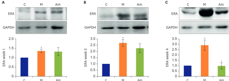
ET-1 and ERA protein expression levels in lung tissues in 3 groups
Administration of MCT injection significantly increased the ET-1 and ERA protein expression levels from week 1 (Figure 3 and 4). However, ambrisentan treatment resulted in decreased
Week 2 Week 4
A
C
0 10 50 30 40
20
Medical w all thickness
C M Am
Week 2 Week 4
B
0 1 2
No . of intr a- acinar art eries
C M Am C
M Am
*
*
* *
††
Figure 2. Pulmonary pathology in MCT-induced PAH model after ambisentan treatment (400× images). (A, C) Pulmonary arteriole medial wall thickness was decreased in the Am at week 4. (B) The number of intra-acinar arteries was reduced in the Am at week 4.
MCT = monocrotaline; PAH = pulmonary arterial hypertension; C = control group; M = monocrotaline group; Am = ambrisentan group.
* p<0.05 as compared with the C;
†p<0.05 as compared with the M.
ET-1 GAPDH
C M Am
B
C M Am
0 2.5 2.0 1.5
0.5
ET -1 week 2 1.0
†*
ET-1 GAPDH
C M Am
C
C M Am
0 2.0 1.5
0.5 1.0
ET -1 week 4
†
*
ET-1 GAPDH
C M Am
C M Am
A
0 2.0 1.5
0.5 1.0
ET -1 week 1 *
Figure 3. ET-1 protein expression level in lung tissues of MCT-induced PAH model after ambrisentan treatment. ET-1 protein expression decreased significantly in lung tissues of PAH rat model 2 weeks after ambrisentan treatment.
ET = endothelin; MCT = monocrotaline; PAH = pulmonary arterial hypertension; C = control; M = monocrotaline; Am = ambrisentan.
* p<0.05 as compared with the C;
†p<0.05 as compared with the M.
ET-1 expression as compared to the M at week 2 (M vs. Am; 2.03±0.43 vs. 1.28±0.19, p=0.025) and 4 (M vs. Am; 1.45±0.21 vs. 0.96±0.06, p=0.043). The protein expression levels of ERA also decreased at week 4 (M vs. Am; 2.93±0.48 vs. 0.98±0.30, p=0.006).
eNOS protein expression levels in the lung tissues in 3 groups
Decrease in the eNOS protein level was observed in the M, as compared with the C, from week 1 (Figure 5). The decreased eNOS levels were dramatically recovered in the Am at weeks 2 (M vs. Am; 0.46±0.02 vs. 0.87±0.07, p<0.001) and 4 (M vs. Am; 0.10±0.02 vs.
0.69±0.21, p=0.004).
ERA GAPDH
C M Am
B
C M Am
0 3.0 2.5 2.0 1.5
0.5 ERA week 2 1.0
*
ERA GAPDH
C M Am
C
C M Am
0 3.5
2.5
0.5 1.5 3.0
2.0
1.0
ERA week 4
†
*
ERA GAPDH
C M Am
C M Am
A
0 2.0 1.5
0.5 1.0
ERA week 1 *
Figure 4. ERA protein expression level in lung tissues of MCT-induced PAH model after ambrisentan treatment. ERA protein expression decreased in lung tissues of PAH rat model 4 weeks after ambrisentan treatment.
ERA = endothelin receptor A; MCT = monocrotaline; PAH = pulmonary arterial hypertension; C = control; M = monocrotaline; Am = ambrisentan.
* p<0.05 as compared with the C;
†p<0.05 as compared with the M.
eNOS GAPDH
C M Am
B
C M Am
0 0.5 1.0
eNOS week 2
*
eNOS GAPDH
C M Am
C
C M Am
0 0.5 1.0
eNOS week 4
† †