113
Open Access
Correspondence: Gi-Byoung Nam, MD, Department of Internal Medi- cine, Asan Medical Center, University of Ulsan College of Medicine, 86 Asan- byeongwon-gil, Songpa-gu, Seoul 138-736, Korea
Tel: 82-2-3010-3159, Fax: 82-2-486-5918 E-mail: [email protected]
• The author has no financial conflicts of interest.
cc This is an Open Access article distributed under the terms of the Cre- ative Commons Attribution Non-Commercial License (http://creativecom- mons.org/licenses/by-nc/3.0) which permits unrestricted non-commer- cial use, distribution, and reproduction in any medium, provided the origi- nal work is properly cited.
Introduction
The importance of exercise for maintenance of a healthy life is well appreciated. Regular physical activity offers a variety of health benefits not only in the general, healthy population, but also in subjects with cardiac diseases. This idea is support- ed by solid scientific evidence compiled over the past 50 years.
The health benefits encompass all the aspects of life, from mere improvement in the physiologic functions, such as the cardio- vascular/respiratory capacity and metabolic status, to achieve- ment of hard end points, including reduction of the stroke and cancer risks, or even decrease in total mortality.
1)In spite of this general positive effect of exercise, exercise-related cardiac events are not infrequently reported during highly competitive sports activity or vigorous exercises.
2)In addition, certain groups of
patients who have structural or genetic ion channel diseases are especially prone to develop life-threatening cardiac arrhythmias precipitated by vigorous physical exercise.
3)4)This review de- scribes the general health benefit of regular physical exercise, as well as the potential adverse effects of vigorous exercise; it also identifies the subgroup of patients who are at higher risk of exercise-related sudden death, and finally screening meth- ods to avoid the potential hazard of exercise.
Benefits of Exercise
Health benefits of physical exercise Prevention of coronary artery disease
Numerous epidemiologic studies have demonstrated a pro- tective effect of exercise on coronary artery disease (CAD).
5)A recent meta-analysis showed that a moderate-to-high level of leisure time physical activity was associated with a reduced risk of CAD. Specifically, vigorous and moderate exercise decreased the risk of CAD by 27% and 12%, respectively, compared with individuals with low or nil exercise activity.
5)Possible contribu- tion of exercise-induced blood pressure lowering, improved body composition, glucose tolerance, insulin sensitivity, and platelet function has been suggested.
6)Exercise, Heart and Health
Gi-Byoung Nam, MD
Department of Internal Medicine, Asan Medical Center, University of Ulsan College of Medicine, Seoul, Korea
ABSTRACT
Regular physical activity provides a variety of health benefits, including improvement in cardiopulmonary or metabolic sta- tus, reduction of the risk of coronary artery disease or stroke, prevention of cancer, and decrease in total mortality. Exercise- related cardiac events are occasionally reported during highly competitive sports activity or vigorous exercises. However, the risk of sudden death is extremely low during vigorous exercise, and habitual vigorous exercise actually decreases the risk of sudden death during exercise. The cause of sudden death is ischemic in older subjects (≥35 years old), while cardiomyopathies or genetic ion channel diseases are important underlying pathology in younger (<35 years old) victims. The subgroup of pa- tients who are particularly at higher risk of exercise-related sudden death may be identified in different ways, such as pre-par- ticipation history taking, physical examination and/or supplementary cardiac evaluation. Limitations exist because current di- agnostic tools are not sufficient to predict a coronary artery plaque with potential risk of disruption and/or an acute thrombotic occlusion. Proper and cost-effective methods for identification of younger subjects with cardiac structural problems or genet- ic ion channel diseases are still controversial.
(Korean Circ J 2011;41:113-121)KEY WORDS:
Exercise; Sudden cardiac death; Health; Coronary arteries.
Prevention of stroke
Studies on stroke prevention and meta-analysis of the stud- ies
7-10)indicated that moderate- to high-intensity exercise was associated with a reduced stroke risk. This protective effect is not only confined to ischemic stroke, but also extends to re- duction of hemorrhagic and, therefore, the total stroke risk.
7)Overall, moderately active individuals had a 20% lower risk and highly active individuals had a 27% lower risk of stroke in- cidence or mortality than the low-active individuals.
8)Consid- ering the pooled relative risk from studies on CAD and stroke prevention, the protective effect on stroke seemed comparable or even greater than the effect on CAD.
7)Hypertension is a risk factor for both ischemic and hemor- rhagic strokes, and there is a direct dose-response relationship between blood pressure and stroke risk. Physical activity low- ers blood pressure, improves lipid profiles, and also improves endothelial function, which enhances vasodilation and vaso- motor function in the vessels. In addition, physical activity can play an antithrombotic role by reducing blood viscosity, fi- brinogen levels, and platelet aggregability, all of which might reduce cardiac and cerebral events.
8)Prevention of cancers
There have been numerous epidemiologic studies on physi- cal activity and cancer prevention.
11)The available data indi- cate that physical activity has a different association with dif- ferent types of cancers.
11)Most studies focused on commonly occurring cancers (e.g., prostate, lung, colorectal for men, br- east, lung, colorectal for women). The effect on the risk of co- lon cancer varied from 80% reduction to 60% increase. Over- all, exercise was associated with a lower risk of colon cancer among both men and women. In addition, a dose-response relationship in cancer prevention was observed across levels of physical activity.
11)In contrast, the available data show no clear association between physical activity and rectal cancer rates in men and women.
11)It also appears that physically ac- tive women have a 20-30% reduced risk of breast cancer.
12)Al-
though exercise appears to be related with a lower risk of lung cancer, the confounding effect of smoking (passive smoking, depth of inhalation, use of filter tips etc) could not be complete- ly controlled. There is no clear data supporting that physical activity decreases the risk of prostate cancer, with the median relative risk around 0.9. The data for other cancers, such as ovarian, testicular, pancreatic, kidney or bladder cancer, are limited. Although the beneficial effect of physical activity on the colon and breast cancers is obvious, the amount of exer- cise, the duration or frequency of exercise, as well as the dose- response relationship, is less clear.
Reduction of mortality
A significant relationship between physical activity and re- duction in mortality has been reported, with a mortality reduc- tion reaching up to 20-40%.
13-16)A clear dose-response rela- tionship was established and a larger volume of physical activity was related with a lower all-cause mortality.
14)15)This inverse dose-response relationship has been shown both in men and women, in younger and older subjects.
16)However, fewer data are available about the relationship between com- ponents of exercise dose (duration, intensity, frequency) and the improvement in longevity.
15)Other benefits
Other beneficial effects of exercise include modification of cardiovascular risk profiles, such as control of hypertension, improved lipid profile, prevention of type 2 diabetes, benefit on the bone-mineral metabolism and body composition.
17)Dose-response relationship
Earlier studies evaluating the role of exercise focused primar- ily on the beneficial effect of vigorous, sustained, aerobic exer- cise. Later, it became clear from epidemiology and controlled experiments that moderate intensity physical activity can also show substantial health benefits.
17)Participation in physical ac- tivities above minimum recommended amounts provides ad-
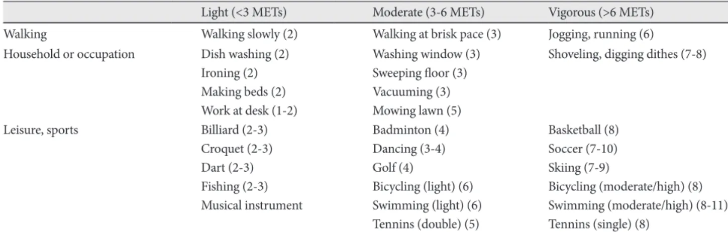
Table 1. Classification of physical activity according to the intensity of METs
Light (<3 METs) Moderate (3-6 METs) Vigorous (>6 METs)
Walking Walking slowly (2) Walking at brisk pace (3) Jogging, running (6)
Household or occupation Dish washing (2) Ironing (2) Making beds (2) Work at desk (1-2)
Washing window (3) Sweeping floor (3) Vacuuming (3) Mowing lawn (5)
Shoveling, digging dithes (7-8)
Leisure, sports Billiard (2-3)
Croquet (2-3) Dart (2-3) Fishing (2-3) Musical instrument
Badminton (4) Dancing (3-4) Golf (4)
Bicycling (light) (6) Swimming (light) (6) Tennins (double) (5)
Basketball (8) Soccer (7-10) Skiing (7-9)
Bicycling (moderate/high) (8) Swimming (moderate/high) (8-11) Tennins (single) (8)
Modified from table 2, reference 25. METs: metabolic equivalents
ditional health benefits in a dose-dependent manner. Howev- er, the point of maximum benefit for most health benefits has not been established, and may vary with many factors, such as age, sex, genetic or body composition.
18)Even more, recent re- ports from the Women’s Health Initiative or Women’s Health Study confirmed that a physical activity of only one hour of walking per week may predict a significantly lower risk of CAD.
19-21)An important quotation regarding the amount of ex- ercise beneficial for health is “Even a little is good, more is better.”, although this amount of activity might fall below the minimum requested in current guidelines.
22)23)Exercise Recommendation
Exercise recommendations from the 1970s prescribed con- tinuous, vigorous exercise for 20 minutes, 3 days per week.
24)Later consensus guideline recommended at least 30 minutes of accumulated, moderate-intensity activity per day, most days of the week.
18)The updated recommendation specified that “All healthy adults aged 18-65 need moderate-intensity aerobic physical activity for a minimum of 30 minutes on five days each week or vigorous-intensity aerobic activity for a minimum of 20 minutes on three days each week”.
25)A convenient way to estimate the energy expenditure dur- ing exercise is to calculate metabolic equivalents (METs) multi- plied by exercise duration in minutes, where 1 MET corresponds to energy expenditure during sitted rest. The total, accumulat- ed energy expenditure is the sum of the multiplication of METs of a specific physical activity and its duration. To meet the current recommendation, the minimum expenditure should be in the range of 450-750 MET×minutes per week.
25)One may calculate the approximate requirement of physical activity by this multiplication of intensity and duration, and thus be able to combine different levels of exercise to meet the recommend- ed dose of exercise. For example, if one walked briskly for 30 minutes three days a week (3 MET×30 minutes×3=270 MET×minutes), and played soccer for 30 minutes twice a week (8 MET×30 minutes×2=480 MET×minutes), the total energy
expenditure would be 750 MET×minutes (Table 1).
25)Accumulation of shorter bouts of exercise (e.g., 10 minutes) towards the 30-minutes minimum can be as effective as sin- gle, longer bouts.
25)26)Moreover, exercise may be compressed into fewer days of the week (e.g., exercise once or twice a week or on weekends only), with each activity session prolonged suf- ficiently enough to fall within current guidelines.
21)25)The 2007 physical activity recommendations for healthy adults devel- oped by the American Heart Association and the American College of Sports Medicine is summarized in Table 2.
25)Hazards of Exercise
Exercise and sudden cardiac death (Cardiovascular risk of vigorous exercise)
Regular physical activity substantially reduces the incidence of CAD and may improve survival. However, it has also been known that vigorous physical activity can precipitate acute myocardial infarction (AMI) or sudden cardiac death.
2)27-29)A prospective study from the Veneto Region of Italy states that adolescents and young adults (<35 y.o.) engaged in competi- tive sports have an increased risk of SCD, compared with their non-athletic counterparts (the annual incidence of SCD was 2.3 in 100,000 in competitive athletes compared with 0.9 in non- athletes, with an estimated relative risk of 2.5).
29)About 90% of all SCD were exercise-related in athletes, while only 9% of all SCD were related with exercise in the non-athletes group. It is generally accepted that sports, per se, is not a cause of increased mortality; rather, it acts as a trigger for cardiac arrest in the pres- ence of underlying cardiovascular diseases predisposing to life- threatening ventricular arrhythmias.
29)For healthy adults, Thompson et al.
30)reported 1 death per year for every 7,620 joggers, and Siscovick et al.
2)estimated an annual rate of exercise-related cardiac arrest of 1 in 18,000 in previously healthy men.
The relative risk of SCD during vigorous exertion (mara- thon running) was significantly elevated at 16.9, compared with other situations.
31)However, even with such intense
Table 2. Physical activity recommendations for healthy adults aged 18-65 year1.. To promote and maintain good health, adults aged 18-65 year should perform either moderate-intensity aerobic (endurance)
physical activity for a minimum of 30 minutes on five days each week or vigorous-intensity aerobic activity for a minimum of 20 minutes on three days each week.
2.. Combinations of moderate- and vigorous-intensity activity can be performed to meet this recommendation. For example, a person can meet the recommendation by walking briskly for 30 minutes twice during the week and then jogging for 20 minutes on two other days.
3.. Moderate-intensity aerobic activity, which is generally equivalent to a brisk walk and noticeably accelerates the heart rate, can be accumulated toward the 30-minutes minimum by performing several bouts, each lasting for 10 or more minutes.
4.. In addition, at least twice each week adults will benefit by performing activities using the major muscles of the body that maintain or increase muscular strength and endurance.
5.. Because of the dose-response relation between physical activity and health, persons who wish to further improve their personal fitness, reduce their risk for chronic diseases and disabilities, or prevent unhealthy weight gain will likely benefit by exceeding the minimum recommended amount of physical activity.
Modified from table 4, reference 25
physical efforts, the absolute risk for SCD was exceedingly small (1 per 1.51 million episodes of exertion, or 1 in 215,000 hours of running).
31)32)In fact, the U.S. Physicians’ Health Study, by comparing the relative risk of SCD according to the frequency of habitual vigorous exercise, demonstrated that habitual vigorous exercise diminishes the risk of sudden death during vigorous exertion.
31)Precipitation of AMI follows a sim- ilar pattern. Although there is an increased risk of develop- ment of AMI with vigorous exercise, the risk is highest in in- dividuals with low levels of habitual activity.
27)33)Overall, the relative risk is lowest in the most physically active, while the relative risk is highest in sedentary individuals.
Cardiac pathology of exercise-related sudden cardiac death
It has been shown that acute cardiac events during exercise occur predominantly in individuals with structural heart dis- eases, and the distribution of cardiac diseases is dependent on ages. In individuals aged <35 years, cardiomyopathies or con- genital heart diseases were responsible for the majority of the SCD victims, whereas CAD was the most frequent cause of death among older individuals (≥35 years).
27)28)34-37)The differ- ent distribution of cardiovascular diseases between the two countries may be attributable to systematic pre-participation screening, leading to identification of athletes with hypertro- phic cardiomyopathy (HCM) in Italian athletes (Table 3).
Mechanism of sudden cardiac death or ventricular fibrillation during sympathetic stimulation
Acute ischemia and sympathetic activation
CAD is the most common cause of SCD in older subjects.
Even in previously asymptomatic adults, coronary artery pla- que disruption (rupture or erosion) with acute thrombotic oc- clusion can be found. However, the mechanism by which vig- orous exercise precipitates cardiac events is not clearly defined.
Exercise-related physiologic changes, such as elevated blood pressure, increased heart rate, coronary artery spasm in the af- fected artery segment, may contribute to the plaque disrup- tion. Other contributing factors include deepening of the coro- nary fissures, as well as catecholamine-induced platelet acti- vation and aggregation. Once myocardial blood flow is severe- ly impaired in acute ischemia, ATP-sensitive potassium channels open and, together with hyperkalemia or cellular acidosis, lead to a markedly increased dispersion of repolarization and cell- to-cell conduction, which ultimately leads to ventricular fibril- lation (VF).
38)Congestive heart failure
The most prominent cellular electrophysiological alteration seen in failing heart is prolongation of the action potential du- ration. This is caused by down-regulation of the K
+currents
and alteration in the inward Na
+and Ca
++currents. This action potential prolongation occurs inhomogeneously throughout the transmural myocardium, and serves as a substrate for re- entrant ventricular tachyarrhythmias.
39)40)In addition, altered channel gating property of the RyR receptor, reduced sarco- plasmic reticulum (SR) Ca
++ATPase function and increased Na-Ca exchange contribute to spontaneous Ca
++leak, decreas- ed SR Ca
++contents, and reduced Ca
++transient. These chang- es, in association with up-regulated Na-Ca exchange and re- duced inward rectifier K
+current, lead to contractile dysfunc- tion and development of delayed afterdepolarization.
41)42)Upon sympathetic stimulation, spontaneous Ca
++release and enhanced Na-Ca exchange, together with I
K1reduction, re- sults in greater propensity for developing delayed after-depo- larization.
43)Hypertrophic cardiomyopathy
In a large multicenter ICD registry, ventricular tachyar- rhythmias appeared to play a major role in the pathogenesis of SCD in HCM, while bradyarrhythmia-mediated events could not be excluded as a cause of SCD.
44)As sinus tachycar- dia was often the initiating rhythm before development of ven- tricular tachyarrhythmias, high sympathetic drive seemed to be proarrhythmic in patients with HCM.
45)It has been shown that the histological features of HCM are a markedly disorganized myocardial architecture, medial
Table 3. Causes of sudden death in young adultsU.S. athletes (%) Italy, athletes &
non-athletes (%)
HCM 215 (36.2) 17 (6.3)
CA anomaly 119 (17.2) 07 (2.6)
Possible HCM 57 (8.2)
Myocarditis 41 (5.9) 22 (8.2)
ARVC 30 (4.3) 029 (10.8)
Ion channel disease 25 (3.6)
MVP 24 (3.4) 26 (9.7)
LAD bridge 23 (3.3) 07 (2.6)
CAD 23 (3.3) 045 (16.7)
Aortic rupture 19 (2.8) 12 (4.5)
AS 17 (2.5)
Dilated CM 14 (2.0) 10 (3.7)
WPW 11 (1.6)
Conduction system disease 24 (8.9)
Other 36 (5.2) 070 (29.0)
Total 690 (100). 269 (100).
Modified from references 28, 35. ARVC: arrhythmogenic right ven- tricular cardiomyopathy, AS: aortic stenosis, CA: coronary artery, CAD: coronary artery disease, CM: cardiomyopathy, CV: cardio- vascular, HCM: hypertrophic cardiomyopathy, LAD: left anterior descending coronary artery, MVP: mitral valve prolapse, WPW:
Wolff-Parkinson-White
hypertrophy with luminal narrowing of the intramural coro- nary arteries, and replacement fibrosis after bursts of myocar- dial ischemia or myocyte necrosis.
46)This highly heteroge- neous substrate may be prone to develop ventricular tachyar- rhythmias during exercise, due to outflow tract obstruction, myocardial ischemia or sympathetic surge, as previously de- monstrated.
47-49)Arrhythmogenic right ventricular dysplasia
Arrhythmogenic right ventricular dysplasia (ARVD) is an inherited cardiomyopathy in which the right ventricular myo- cardium is progressively replaced by fat and fibrosis. SCD is fre- quently the first manifestation of the disease, and it occurs rel- atively frequently during exercise or stress. RV dilation, precordial repolarization abnormalities, and LV involvement have been associated with higher risk of sudden death.
50)Coronary artery anomaly
Congenital coronary artery anomaly indicates either the left main coronary artery arising from the right aortic sinus/
pulmonary artery, or the right coronary artery arising from the left aortic sinus. Overall, it is an important cause of sud- den death in young athletes. Cardiac events occur mainly dur- ing intense exertion probably provoked by myocardial isch- emia from angulation of the coronary ostium or compression of the coronary artery. Premonitory symptoms (syncope, chest pain) may occur shortly before the onset of sudden death.
Echocardiography or stress electrocardiogram (ECG) with ma- ximal exercise may not disclose this anomaly, and a careful history taking revealing the presence of syncope or chest pain during exercise is crucial in the pre-participation screening of competitive athletes.
51)Myocardial bridge
Myocardial bridging is defined as a segment of a major epi- cardial coronary artery running intramurally through the myo- cardium, and is a common angiographic or autopsy finding. It is generally a benign congenital coronary artery anomaly, but it has been associated with angina, arrhythmia, depressed left ventricular (LV) function, myocardial stunning, and sudden death.
52)Medical treatment with beta-blockers seems to be the first choice. However, intracoronary stents and surgery have been attempted in selected patients with refractory symptoms.
Additional study is necessary to define the role of the myocar- dial bridge in the occurrence of sudden death, and to select the patients requiring invasive or surgical management.
52)53)Myocarditis
Myocarditis accounts for about 10% of the SCD in the young population. Infection or exposure to toxic chemicals/radiation can cause myocardial inflammation, and SCD results from conduction abnormalities or malignant ventricular tachyar-
rhythmias.
50)Mitral valve prolapse
The mitral valve prolapase (MVP) is a common echocardio- graphic abnormality, observed in about 3-4% of the general population. It is not a single disease entity, but a spectrum of different conditions, ranging from simple bowing of the mi- tral valve leaflet to redundant myxomatous leaflet and flail MV.
54)55)Although SCD frequently occurs during exercise, the MVP per se. may not be related with an increased risk of SCD. In- stead, the risk of SCD is strongly associated with accompany- ing mitral regurgitation.
54)55)The mechanism of SCD in pa- tients with MVP is not clear. Ventricular tachyarrhythmias have been reported to occur more frequently in patients with MVP than in control subjects. However, this may be due to the presence of mitral regurgitation, not due to MVP itself. There- fore, the cause of SCD seems multifactorial, including ven- tricular tachyarrhythmias or atrioventricular block, hemody- namic dysfunction with severe hypotension, coronary artery spasm, hidden myocarditis or myocardial dysplasia.
56)The Wolff-Parkinson-White syndrome
The risk of SCD associated with ventricular preexcitation is less than 0.15% per year.
57)The main mechanism of SCD seems to be atrial fibrillation conducting rapidly over an accessory pathway and degenerating into VF.
58)Exercise is associated with an increased risk of VF. The approach to asymptomatic patients with ventricular preexcitation varies from simple observation to routine electrophysiological study and catheter ablation of the accessory pathway in patients with inducible atrial tachyar- rhythmia.
58)Considering the low risk of SCD in this aymptom- atic population, the clinical decision should be individualized.
Athletes with ventricular preexcitation should be evaluated for inducibility of supraventricular tachycardia and refractori- ness of the accessory pathway before participation in compet- itive sports.
69)Long QT syndrome
It has been well demonstrated that subgroups of patients with long QT syndrome (LQTS) are sensitive to sympathetic stimulation. Arrhythmic events are often associated with physi- cal exercise, emotional stress or auditory stimulation in long QT type 1 or 2.
59)Swimming is a relatively genotype-specific ar- rhythmogenic trigger for both type 1 of the long QT syndrome and catecholaminergic polymorphic ventricular tachycardia (CPVT). In contrast, type 2 LQTS patients are susceptible to auditory stimulation or alarm.
59)60)Catecholaminergic polymorphic ventricular tachycardia
CPVT is an inherited disease characterized by a normal rest-
ing ECG, adrenergically-mediated bigemini premature ven-
tricular contraction (PVC) and ventricular tachyarrhythmias.
Mutation of the cardiac Ryanodine receptor 2 with impaired calcium-handling and delayed afterdepolarization was demon- strated to be the electrophysiological mechanism of fatal ven- tricular tachyarrhythmias.
61)62)Other genetic arrhythmia syndromes
Cardiac events in patients with short QT syndrome may oc- cur during effort or loud noise, but this association appears to be weaker than in both LQTS and CPVT.
63-65)The arrhythmo- genic mechanism in patients with Brugada syndrome or early repolarization syndrome is strongly dependent on the vagal nerve activity, and exercise is less strictly prohibited compared with patients with LQTS, CPVT or HCM.
66)Exercise recommendations in patients with genetic cardiovascular diseases
Patients with Exercise recommendations in patients with ge- netic cardiovascular diseases (GCVDs) are at higher risk for SCD during exercise compared with normal subjects. The con- flict between the known benefits and potential adverse out- comes of exercise requires careful weighing of the perceived risk to benefit ratio for each patient. It is generally recommend- ed that patients with GCVDs can safely participate in most forms of recreational exercise of moderate or low intensity (i.e., equivalent to or lower than 6 METs). Activities requiring sudden acceleration or deceleration (burst exertion), exercise under extreme adverse environmental conditions, intense static exertion (weight lifting), exercises with potential risk of trau- matic injury associated with loss of consciousness (free-weight lifting, rock climbing or ice hockey) or with possibilities of im- paired consciousness during water-related activities are gen- erally either not advised or strongly discouraged in all forms of genetic cardiovascular diseases.
34)66)In addition, individual clinical judgment is required for patients with high risk clini- cal features, such as history of important cardiac symptoms, including syncope, prior cardiac operation, heart transplanta-
tion, presence of an implanted cardioverter-defibrillator or pace- maker, potentially life-threatening arrhythmias or other evi- dence of high-risk status.
Preparticipation screening
Due to the risk of exercise-related sudden death or other car- diac events, pre-participation screening for the susceptible sub- jects has emerged as an important issue in exercise recommen- dation. Brief guidelines for preparticipation are as follows.
Athletes
In Italy, a national pre-participation screening mandates annual evaluations that routinely include 12-lead ECG, in ad- dition to medical history and physical examination. Wide im- plementation of the Italian screening model has been promot- ed by the endorsement of the European Society of Cardiology in 2005. Recent scientific evidence demonstrated this screen- ing strategy to be useful for identification of HCM and other cardiomyopathies in asymptomatic athletes,
67)and implemen- tation of this program in Italy has been associated with sub- stantial reduction in mortality due to cardiomyopathies.
68)On the other hand, customary screening strategies for U.S. high school and college athletes is confined to medical history and physical examination at 2- to 4-year intervals.
34)This is due to current lack of medical and financial resources, and more im- portantly, due to potentially deleterious effects to many ath- letes by virtue of false-positive test results that would lead to unnecessary further evaluation or disqualification.
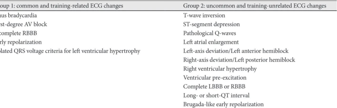
To overcome this controversy, a new classification of the ath- lete ECG was proposed. ECG may be abnormal in up to 50% of athletes, which hinders the widespread use of ECG as part of the pre-participation screening. This new guideline classifies the changes seen on the athlete ECG into physiological (com- mon and training-related) and pathological (uncommon and training-unrelated), to lower the traditionally high number of false-positive results and thus reducing unnecessary inves- tigations
69)(Table 4).
Table 4. Classification of abnormalities seen on the electrocardiogram recorded in subjects with athlete’s heart
Group 1: common and training-related ECG changes Group 2: uncommon and training-unrelated ECG changes Sinus bradycardia
First-degree AV block Incomplete RBBB Early repolarization
Isolated QRS voltage criteria for left ventricular hypertrophy
T-wave inversion ST-segment depression Pathological Q-waves Left atrial enlargement
Left-axis deviation/Left anterior hemiblock Right-axis deviation/Left posterior hemiblock Right ventricular hypertrophy
Ventricular pre-excitation Complete LBBB or RBBB Long- or short-QT interval Brugada-like early repolarization Adapted from references 69. ECG: electrocardiogram, RBBB: right bundle branch block, LBBB: left bundle branch block
Non-athletes
Guidelines from the AHA indicated that exercise testing is not necessary for all people beginning a moderate intensity physical activity program. The rationale is based on the ex- tremely low rate of cardiovascular complications in asymp- tomatic persons while performing moderate-intensity physi- cal activity, on the poor predictive value of exercise testing for acute cardiac events and on the high costs of mass exercise test- ing. Symptomatic subjects or those suffering from any cardio- vascular disease, diabetes, other active chronic disease, or with any medical concern in general, should consult a physician prior to any substantive increase in physical activity, particu- larly vigorous-intensity activity.
25)Other issues or adverse effects of exercise;
the athletes’ heart and commotio cordis The athlete’s heart
Exercise-related cardiac events occur predominantly in sub- jects with underlying structural abnormalities. However, exer- cise may cause a deleterious effect in individuals without un- derlying cardiac diseases. Athlete’s heart denotes exercise- related electrical and structural remodeling. Intense resistance training increases LV wall thickness and mass with little or no change in the LV diameter, while aerobic exercise is associated with asymmetric LV hypertrophy.
70)71)Clinical implications of the athlete’s heart include differen- tial diagnosis from hypertrophic/dilated/RV cardiomyopathy, or possible long-term consequences of the extreme LV remod- eling. As exercise itself causes alteration in the cardiac chamber size and morphology resembling those of cardiomyopa- thies, differential diagnosis of the athlete’s heart (i.e., a physio- logical adaptation) from cardiomyopathies has long been a di- agnostic dilemma. Athletes with benign right ventricular out- flow tract VT may show wall motion abnormalities mimicking ARVD. In addition, concern has been raised that uninterrupt- ed training per se, might ultimately cause irreversible cardiac dilatation,
71)systolic dysfunction,
72)or even ventricular tachyar- rhythmias.
73)A recent report, however, demonstrated that even young Olympic athletes exposed to extreme endurance train- ing over long periods of time (up to 17 years) did not show de- terioration in LV function and morphology or any occurrence of cardiovascular events. These findings confirmed the concept that the athlete’s heart is a physiological adaptation to exercise, not causing irreversible cardiac remodeling.
74)Commotio cordis
Commotio cordis refers to VF and SCD triggered by a blunt, non-penetrating, and often innocent-appearing blow to the chest wall, without visible damage to the ribs, sternum, or heart, in individuals without previous structural heart diseases. It is an important cause of exercise-related SCD in young athletes,
accounting for 3% of all sudden deaths in young, competitive athletes.
75)A direct blow to the chest walls falling on a critical time of the cardiac cycle (i.e., 10-20 msec on the upstroke of the T wave) can precipitate VF. The cellular mechanism of VF ini- tiation appears to be multifactorial. Instantaneous increase in the left ventricular intra-cavitary pressure and opening of st- retch-activated ion channels, including ATP-sensitive potassi- um channels, may result in inhomogeneous repolarization of the myocardium, thus creating the vulnerable substrate for VF.
76)Conclusion
Regular physical activity provides a variety of health bene- fits, including improvement in the cardiopulmonary or meta- bolic status, reduction of the risk of CAD or stroke, prevention of cancer, and decrease in total mortality. These benefits offset the small but significant increase in the risk of SCD during vig- orous exercise. There is a subgroup of patients, however, par- ticularly at higher risk of sudden death during exercise. Proper identification of patients with hidden CADs, as well as screen- ing of young subjects with structural or genetic ion channel diseases, may prove important for the prevention of exercise- related sudden death.
Acknowledgments
This study was supported by a grant of the Korea Healthcare technolo- gy R& D Project, Ministry of Health & Welfare, Republic of Korea (A100607).
REFERENCES
1) Blair SN, Morris JN. Healthy hearts and the universal benefits of being physically active: physical activity and health. Ann Epidemiol 2009;
19:253-6.
2) Siscovick DS, Weiss NS, Fletcher RH, Lasky T. The incidence of pri- mary cardiac arrest during vigorous exercise. N Engl J Med 1984;311:
874-7.
3) Maron BJ, Doerer JJ, Haas TS, Tierney DM, Mueller FO. Sudden deaths in young competitive athletes: analysis of 1866 deaths in the United States, 1980-2006. Circulation 2009;119:1085-92.
4) Wever EF, Robles de Medina EO. Sudden death in patients without structural heart disease. J Am Coll Cardiol 2004;43:1137-44.
5) Sofi F, Capalbo A, Cesari F, Abbate R, Gensini GF. Physical activity during leisure time and primary prevention of coronary heart dis- ease: an updated meta-analysis of cohort studies. Eur J Cardiovasc Prev Rehabil 2008;15:247-57.
6) Sesso HD, Paffenbarger RS Jr, Lee IM. Physical activity and coronary heart disease in men: the Harvard Alumni Health Study. Circulation 2000;102:975-80.
7) Wendel-Vos GC, Schuit AJ, Feskens EJ, et al. Physical activity and stroke: a meta-analysis of observational data. Int J Epidemiol 2004;33:
787-98.
8) Lee CD, Folsom AR, Blair SN. Physical activity and stroke risk: a meta-analysis. Stroke 2003;34:2475-81.
9) Lee IM, Paffenbarger RS Jr. Physical activity and stroke incidence: the Harvard Alumni Health Study. Stroke 1998;29:2049-54.
10) Lee IM, Hennekens CH, Berger K, Buring JE, Manson JE. Exercise and risk of stroke in male physicians. Stroke 1999;30:1-6.
11) Lee IM. Physical activity and cancer prevention: data from epidemio- logic studies. Med Sci Sports Exerc 2003;35:1823-7.
12) Lee IM, Cook NR, Rexrode KM, Buring JE. Lifetime physical activ- ity and risk of breast cancer. Br J Cancer 2001;85:962-5.
13) Oguma Y, Sesso HD, Paffenbarger RS Jr, Lee IM. Physical activity and all cause mortality in women: a review of the evidence. Br J Sports Med 2002;36:162-72.
14) Lee IM, Hsieh CC, Paffenbarger RS Jr. Exercise intensity and longevi- ty in men. The Harvard Alumni Health Study. JAMA 1995;273:1179- 15) Lee IM, Skerrett PJ. Physical activity and all-cause mortality: what is 84.
the dose-response relation? Med Sci Sports Exerc 2001;33(6 Suppl):
S459-71.
16) Vogel T, Brechat PH, Leprӗtre PM, Kaltenbach G, Berthel M, Lons- dorfer J. Health benefits of physical activity in older patients: a review.
Int J Clin Pract 2009;63:303-20.
17) Blair SN, Morris JN. Healthy hearts and the universal benefits of be- ing physically active: physical activity and health. Ann Epidemiol 2009;
19:253-6.
18) Pate RR, Pratt M, Blair SN, et al. Physical activity and public health:
a recommendation from the Centers for Disease Control and Preven- tion and the American College of Sports Medicine. JAMA 1995;273:
402-7.
19) Lee IM. Dose-response relation between physical activity and fitness:
even a little is good; more is better. JAMA 2007;297:2137-9.
20) Manson JE, Greenland P, LaCroix AZ, et al. Walking compared with vigorous exercise for the prevention of cardiovascular events in wom- en. N Engl J Med 2002;347:716-25.
21) Lee IM, Sesso HD, Oguma Y, et al. The “weekend warrior” and risk of mortality. Am J Epidemiol 2004;160:636-41.
22) Lee IM. Physical activity in women: how much is good enough? JAMA 2003;290:1377-9.
23) Church TS, Earnest CP, Skinner JS, Blair SN. Effects of different dos- es of physical activity on cardiorespiratory fitness among sedentary, overweight or obese postmenopausal women with elevated blood pres- sure: a randomized controlled trial. JAMA 2007;297:2081-91.
24) American College of Sports Medicine position statement on the rec- ommended quantity and quality of exercise for developing and main- taining fitness in healthy adults. Med Sci Sports 1978;10:vii-x.
25) Haskell WL, Lee IM, Pate RR, et al. Physical activity and public health:
updated recommendation for adults from the American College of Sports Medicine and the American Heart Association. Circulation 2007;116:
1081-93.
26) Lee IM, Sesso HD, Paffenbarger RS Jr. Physical activity and coro- nary heart disease risk in men: does the duration of exercise episodes predict risk? Circulation 2000;102:981-6.
27) Thompson PD, Franklin BA, Balady GJ, et al. Exercise and acute car- diovascular events placing the risks into perspective: a scientific state- ment from the American Heart Association Council on Nutrition, Physi- cal Activity, and Metabolism and the Council on Clinical Cardiology.
Circulation 2007;115:2358-68.
28) Maron BJ, Doerer JJ, Haas TS, Tierney DM, Mueller FO. Sudden deaths in young competitive athletes: analysis of 1866 deaths in the United States, 1980-2006. Circulation 2009;119:1085-92.
29) Corrado D, Basso C, Rizzoli G, Schiavon M, Thiene G. Does sports ac- tivity enhance the risk of sudden death in adolescents and young adults?
J Am Coll Cardiol 2003;42:1959-63.
30) Thompson PD, Funk EJ, Carleton RA, Sturner WQ. Incidence of death during jogging in Rhode Island from 1975 through 1980. JAMA 1982;
247:2535-8.
31) Albert CM, Mittleman MA, Chae CU, Lee IM, Hennekens CH, Manson JE. Triggering of sudden death from cardiac causes by vigorous exer- tion. N Engl J Med 2000;343:1355-61.
32) Maron BJ, Poliac LC, Roberts WO. Risk for sudden cardiac death as- sociated with marathon running. J Am Coll Cardiol 1996;28:428-31.
33) Siscovick DS, Ekelund LG, Johnson JL, Truong Y, Adler A. Sensitiv- ity of exercise electrocardiography for acute cardiac events during mod- erate and strenuous physical activity: the lipid research clinics coro- nary primary prevention trial. Arch Intern Med 1991;151:325-30.
34) Maron BJ, Thompson PD, Ackerman MJ, et al. Recommendations and considerations related to pre-participation screening for cardiovascu- lar abnormalities in competitive athletes: 2007 update: a scientific state- ment from the American Heart Association Council on Nutrition, Physi- cal Activity, and Metabolism: endorsed by the American College of Cardiology Foundation. Circulation 2007;115:1643-455.
35) Corrado D, Basso C, Schiavon M, Thiene G. Screening for hypertro- phic cardiomyopathy in young athletes. N Engl J Med 1998;339:364-9.
36) Corrado D, Basso C, Thiene G. Sudden cardiac death in young people with apparently normal heart. Cardiovasc Res 2001;50:399-408.
37) Corrado D, Basso C, Pavei A, Michieli P, Schiavon M, Thiene G. Trends in sudden cardiovascular death in young competitive athletes after im- plementation of a pre-participation screening program. JAMA 2006;
296:1593-601.
38) Luqman N, Sung RJ, Wang CL, Kuo CT. Myocardial ischemia and ventricular fibrillation: pathophysiology and clinical implications. Int J Cardiol 2007;119:283-90.
39) Aiba T, Tomaselli GF. Electrical remodeling in the failing heart. Curr Opin Cardiol 2010;25:29-36.
40) Akar FG, Yan GX, Antzelevitch C, Rosenbaum DS. Unique topographi- cal distribution of M cells underlies reentrant mechanism of torsade de pointes in the long-QT syndrome. Circulation 2002;105:1247-53.
41) Yano M, Ono K, Ohkusa T, et al. Altered stoichiometry of FKBP12.6 versus ryanodine receptor as a cause of abnormal Ca(2+) leak through ryanodine receptor in heart failure. Circulation 2000;102:2131-6.
42) Yano M, Yamamoto T, Kobayashi S, Ikeda Y, Matsuzaki M. Defective Ca2+ cycling as a key pathogenic mechanism of heart failure. Circ J 2008;72(Suppl A):A22-30.
43) Pogwizd SM, Schlotthauer K, Li L, Yuan W, Bers DM. Arrhythmogen- esis and contractile dysfunction in heart failure: roles of sodium-calci- um exchange, inward rectifier potassium current, and residual beta-ad- renergic responsiveness. Circ Res 2001;88:1159-67.
44) Maron BJ, Spirito P, Shen WK, et al. Implantable cardioverter-defibril- lators and prevention of sudden cardiac death in hypertrophic cardio- myopathy. JAMA 2007;298:405-12.
45) Cha YM, Gersh BJ, Maron BJ, et al. Electrophysiologic manifestations of ventricular tachyarrhythmias provoking appropriate defibrillator in- terventions in high-risk patients with hypertrophic cardiomyopathy. J Cardiovasc Electrophysiol 2007;18:483-7.
46) Maron BJ. Hypertrophic cardiomyopathy. Lancet 1997;350:127-33.
47) Nicod P, Polikar R, Peterson KL. Hypertrophic cardiomyopathy and sudden death. N Engl J Med 1988;318:1255-7.
48) McKenna WJ, Firoozi S, Sharma S. Arrhythmias and sudden death in hypertrophic cardiomyopathy. Card Electrophysiol Rev 2002;6:26-31.
49) Ly HQ, Greiss I, Talakic M, et al. Sudden death and hypertrophic car- diomyopathy: a review. Can J Cardiol 2005;21:441-8.
50) Zipes DP, Camm AJ, Borggrefe M, et al. ACC/AHA/ESC 2006 guide- lines for management of patients with ventricular arrhythmias and the prevention of sudden cardiac death: a report of the American College of Cardiology/American Heart Association Task Force and the Euro- pean Society of Cardiology Committee for Practice Guidelines (writing committee to develop Guidelines for Management of Patients With Ven- tricular Arrhythmias and the Prevention of Sudden Cardiac Death):
developed in collaboration with the European Heart Rhythm Associ- ation and the Heart Rhythm Society. Circulation 2006;114:e385-484.
51) Basso C, Maron BJ, Corrado D, Thiene G. Clinical profile of congenital coronary artery anomalies with origin from the wrong aortic sinus leading to sudden death in young competitive athletes. J Am Coll Cardiol 2000;35:1493-501.
52) Barcin C, Kursaklioglu H, Kose S, Amasyali B. Coronary myocardial bridge constitutes a risk: but how to manage it? Int J Cardiol 2010;138:
215-6.
53) Alegria JR, Herrmann J, Holmes DR Jr, Lerman A, Rihal CS. Myo- cardial bridging. Eur Heart J 2005;26:1159-68.
54) Plicht B, Rechenberg W, Kahlert P, Buck T, Erbel R. Mitral valve pro- lapse: identification of high-risk patients and therapeutic management.
Herz 2006;31:14-21.
55) Hayek E, Gring CN, Griffin BP. Mitral valve prolapse. Lancet 2005;
365:507-18.
56) Alliot E, Clementy J, Prystowsky E. Fighting sudden cardiac death: a worldwide challenge. 1st ed. New York: Futura Publishing Co, 2000.
57) Munger TM, Packer DL, Hammill SC, Feldman BJ, Bailey KR, Bal- lard DJ, Holmes DR Jr, Gersh BJ. A population study of the natural history of Wolff-Parkinson-White syndrome in Olmsted County, Min- nesota, 1953-1989. Circulation 1993;87:866-73.
58) Pappone C, Manguso F, Santinelli R, et al. Radiofrequency ablation in children with asymptomatic Wolff-Parkinson-White syndrome. N Engl J Med 2004;351:1197-205.
59) Wilde AA, Roden DM. Predicting the long-QT genotype from clini- cal data: from sense to science. Circulation 2000;102:2796-8.
60) Shimizu W, Antzelevitch C. Differential effects of beta-adrenergic ag- onists and antagonists in LQT1, LQT2 and LQT3 models of the long QT syndrome. J Am Coll Cardiol 2000;35:778-86.
61) Priori SG, Napolitano C, Memmi M, et al. Clinical and molecular char- acterization of patients with catecholaminergic polymorphic ventricu- lar tachycardia. Circulation 2002;106:69-74.
62) Liu N, Colombi B, Memmi M, et al. Arrhythmogenesis in catechol- aminergic polymorphic ventricular tachycardia: insights from a RyR2 R4496C knock-in mouse model. Circ Res 2006;99:292-8.
63) Gaita F, Giustetto C, Bianchi F, et al. Short QT Syndrome: a familial cause of sudden death. Circulation 2003;108:965-70.
64) Maury P, Extramiana F, Sbragia P, et al. Short QT syndrome: update on a recent entity. Arch Cardiovasc Dis 2008;101:779-86.
65) Giustetto C, Di Monte F, Wolpert C, et al. Short QT syndrome: clinical findings and diagnostic-therapeutic implications. Eur Heart J 2006;27:
2440-7.
66) Maron BJ, Chaitman BR, Ackerman MJ, et al. Recommendations for physical activity and recreational sports participation for young pa- tients with genetic cardiovascular diseases. Circulation 2004;109:
2807-16.
67) Pelliccia A, Di Paolo FM, Corrado D, et al. Evidence for efficacy of the Italian national pre-participation screening programme for identifi- cation of hypertrophic cardiomyopathy in competitive athletes. Eur Heart J 2006;27:2196-200.
68) Corrado D, Basso C, Pavei A, Michieli P, Schiavon M, Thiene G. Trends in sudden cardiovascular death in young competitive athletes after implementation of a preparticipation screening program. JAMA 2006;
296:1593-601.
69) Corrado D, Pelliccia A, Heidbuchel H, et al. Recommendations for in- terpretation of 12-lead electrocardiogram in the athlete. Eur Heart J 2010;31:243-59.
70) Maron BJ, Pelliccia A. The heart of trained athletes: cardiac remodel- ing and the risks of sports, including sudden death. Circulation 2006;
114:1633-44.
71) Pelliccia A, Maron BJ, De Luca R, Di Paolo FM, Spataro A, Culasso F.
Remodeling of left ventricular hypertrophy in elite athletes after long- term deconditioning. Circulation 2002;105:944-9.
72) Nishimura T, Yamada Y, Kawai C. Echocardiographic evaluation of long-term effects of exercise on left ventricular hypertrophy and func- tion in professional bicyclists. Circulation 1980;61:832-40.
73) Ector J, Ganame J, van der Merwe N, et al. Reduced right ventricular ejection fraction in endurance athletes presenting with ventricular ar- rhythmias: a quantitative angiographic assessment. Eur Heart J 2007;
28:345-53.
74) Pelliccia A, Kinoshita N, Pisicchio C, et al. Long-term clinical conse- quences of intense, uninterrupted endurance training in Olympic ath- letes. J Am Coll Cardiol 2010;55:1619-25.
75) Maron BJ, Doerer JJ, Haas TS, Tierney DM, Mueller FO. Sudden deaths in young competitive athletes: analysis of 1866 deaths in the United States, 1980-2006. Circulation 2009;119:1085-92.
76) Maron BJ, Estes NA 3rd. Commotio cordis. N Engl J Med 2010;362:
917-27.