β-glucan Stimulates Release of TNF-α in Human Monocytic THP-1 Cells
Bo Ram Keum1, Jin Yi Hyeon1, So Hui Choe1, Ji Young Jin1, Ji Woo Jeong2, Jong Min Lim3, Dong Chan Park3, Kwang Keun Cho4, Eun Young Choi2* and In Soon Choi1,2*
1Department of Bioscience, Silla University, Busan 46958, Korea
2Department of Life Science, Silla University, Busan 46958, Korea
3Glucan Corporation, Gijang-gun, Busan 46048, Korea
4Department of Animal Resources Technology, Gyeongnam National University of Science and Technology, Jinju 52725, Korea Received September 18, 2017 /Revised November 21, 2017 /Accepted November 27, 2017
β-glucan is a constituent of the cell wall of fungi, yeast and plants. It plays an important role in the immune system such as activation of immunocyte, release of pro-inflammatory and anti-cancer effect.
The immune system maintains a healthy immune homeostasis. However, when pathogenic substances enter the body, immune homeostasis can break down and disease can be triggered. Therefore, we studied a substance that regulates immune homeostasis. The purpose of the study we demonstrated whether the β-glucan can be applied to the immune-modulation effects in human monocytic THP-1 cells. β-glucan was incubated in THP-1 cells at various concentrations. The TNF-α mRNA expression and protein levels were analyzed by ELISA and Real-time PCR. Additionally, the expression of MAPKs (p38 and JNK), IκB-α and NF-κB p50 were analyzed by western blot. β-glucan enhanced the production of TNF-α mRNA expression and protein levels in human monocytic THP-1 cells. In addi- tion, activation of MAPKs (p38 and JNK) and NF-κB p50 induced by β-glucan were increased. The study suggests that β-glucan contributes to immune-stimulation effect by production TNF-α in human monocytic THP-1 cells, and that MAPKs and NF-κB p50 are involved in the process. Synthetically, we have suggested β-glucan may be improved to immune system effect in human monocytic THP-1 cells.
Key words : β-glucan, immunomodulation, NF-κB p50, THP-1, TNF-α
*Corresponding authors
*Tel : +82-51-999-5906, Fax : +82-51-999-5644
*E-mail : [email protected] (Eun Young Choi)
*Tel: +82-51-999-5348, Fax: +82-51-999-5644
*E-mail: [email protected] (In Soon Choi)
This is an Open-Access article distributed under the terms of the Creative Commons Attribution Non-Commercial License (http://creativecommons.org/licenses/by-nc/3.0) which permits unrestricted non-commercial use, distribution, and reproduction in any medium, provided the original work is properly cited.
Journal of Life Science 2017 Vol. 27. No. 11. 1256~1261 DOI : https://doi.org/10.5352/JLS.2017.27.11.1256
서 론
β-glucan은 β-glycosidic 결합에 의해 연결된 D-glucose 단 량체로 이루어진 다당류이다. 자연적으로 발생된 다당류로 세 포벽의 구성물질 및 박테리아, 효모, 균류, 식물 등에서 다양하 게 생성된다[1, 23]. 효능으로는 항암, 면역활성, 항산화, 피부 보습, 장내 환경 개선 등이 알려져 있다[5, 9, 13, 14, 17, 18].
포유동물은 β-glucan을 분해할 수 있는 효소를 가지고 있지 않기 때문에 체내에서 분해되지 않고, 소장까지 이동하여 면 역세포인 대식세포와 수지상 세포를 활성화시켜 다양한 사이 토카인, 케모카인 등을 분비하도록 한다. β-glucan은 신체면역 반응을 변화시켜 면역증강효과를 나타내는 생물학적 반응조 절물질(BRM, biologic response modifier)으로 알려져 있으며
[16], 또한, 신체 장내에 유익한 효과를 주는 미생물인 pro- biotics 생육을 향상시키는 비소화성 식품 prebiotics으로 이용 되고 있다[3].
면역계(immune system)는 내부 및 외부 병원성 물질로부 터 신체를 보호하기 위해 다양한 세포들이 self, non-self를 인 식하고 식별하여 병원성 물질 제거를 통해 신체의 항상성을 유지하는 체계이다. 면역계는 면역을 억제·조절하는 면역 관 용(tolerance)과 면역을 증진하는 면역 반응(immunity)으로 이루어져 있으며[24], 이러한 면역 관용과 면역 반응의 균형을 통해 신체의 항상성을 유지한다. 하지만, 면역 반응이 높아질 경우에는 바이러스, 세균, 병원성 물질이 신체 내부로 침입함 으로써 발병되는 질환, 감염을 완화시키고, 면역 관용이 높아 질 경우에는 면역 세포들이 신체의 장기, 조직을 손상시키는 자가 면역 질환과 같은 질병을 발생시킨다[15]. 그러므로, 면역 계는 면역 관용과 면역 반응의 적절한 균형이 이루어져야 신 체의 항상성 즉, 건강함을 유지할 수 있다.
면역계는 내부 병원성 물질 생성 또는 외부 병원성 물질이 신체 내로 침입하게 되면, 혈액에 존재하는 림프구(lympho- cyte), 단핵구(monocyte) 등과 같은 면역 세포들이 이를 인식 하고, 다른 면역 세포에게 외부 물질을 인식·제시시킨다. 이러 한 면역 세포들은 세포 표면에 있는 수용체를 통해 외부 물질
을 인식하고, 세포 내 MAPKs, PI3/Akt, NF-κB 신호전달 등과 같은 신호 경로와 전사 인자를 통해 활성화된다. 활성화된 면 역 세포들은 신체의 항상성을 유지하기 위해서 Nitric Oxide (NO), Tumor necrosis factor-α (TNF-α), Interleukin-12p40 (IL-12p40) 등과 같은 사이토카인을 분비하여 감염과 질병에 대한 신체 면역 방어 역할 또는 면역 증강 역할을 한다고 알려 져 있다[19, 20].
단핵구는 골수에서 유래되어 혈액을 순환하는 백혈구 세포 중 하나로, 사이토카인 또는 미생물 분자 등 자극에 따라 대식 세포와 수지상 세포로 분화된다. 감염 및 질환이 일어나지 않 은 상태 즉, 정상적인 신체 상태에서 단핵구는 항미생물 효능 을 나타내며, 감염 발생 경우, 단핵구 및 단핵구 유래 세포는 림프절로 이동 및 사이토카인과 케모카인 분비 등으로 면역 반응을 일으킨다[22].
본 연구는 β-glucan의 면역 조절 효능을 평가하기 위하여, β-glucan을 인간 단핵구 세포주인 THP-1 cells에 β-glucan을 처리하여 TNF-α 생성 및 발현에 미치는 영향을 보았다. 또한 β-glucan의 활성 기전을 알아보기 위해 Mitogen-activated protein kinases (MAPKs), Nuclear factor-kappa B (NF-κB)와 Inhibitor of kappa B (IκB) 활성을 확인하였다. 이를 통해 면역 조절 효능을 확인하고 기능성 소재로서 가능성을 확인하고자 하였다.
재료 및 방법
실험재료
본 실험에 사용한 polycan은 Aureobasidium pullans SM- 2001으로 발효된 β-glucan으로, 이전 연구에 따르면 주요 구성 물질은 15% β-1,3/1,6-glucan이 첨가된 다당류로 알려져 있다 [8]. 실험에 사용한 β-glucan은 ㈜글루칸에서 제공받아 실험에 사용하였다.
세포 배양
인간 단핵구 세포주인 THP-1 cell (American Type Culture Collection, Rockville, MD, USA)은 10% Heat-inactivation fe- tal bovine serum (FBS) (Gibco, BRL,. USA), 100 unit/ml Penicillin, 100 μg/ml Streptomycin (Lonza, Walkersville, MA, USA), 1 mM Sodium Pyruvate (Sigma-Aldrich Co., St.
Louis, USA), 0.05 mM 2-Mercaptoethanol (Sigma-Aldrich Co., St. Louis, USA)이 첨가된 Roswell Park Memorial Institute (RPMI) 1640 (Lonza, walkersville, MD, USA)을 사용 하였다. THP-1 cell을 5×10⁵ cells/ml로 seeding한 다음 50 μ g/ml PMA(Sigma-Aldrich Co., St. Louis, USA)를 처리하여 72시간 동안 37℃, 5% CO2 배양기를 이용하여 배양하였다 [17]. RPMI 1640 serum-free medium로 배지 교환하여 37℃, 5% CO2 incubator에 24시간 배양한 후, β-glucan을 농도 별로
처리하여 배양하였다.
세포 생존율 평가
THP-1 cells에 대한 β-glucan의 세포 생존에 미치는 영향을 알아보기 위해 측정하였다. THP-1 cells에 β-glucan이 농도 별로 처리된 상층액을 제거하였다. 각 well에 5 mg/ml MTT (3-(4,5-Dimethylthiazol-2-yl)-2,5-Diphenyltetrazolium Bromide)을 처리한 다음 37℃, 5% CO2 배양기에 3시간 동안 배양하였다. 그 이후, 상층액을 제거하고 DMSO (Dimethyl Sulfoxide)을 이용하여 비 수용성 MTT formazan을 용해시켜 Spectra Max 250 ELISA Reader (Molecular Devices, Sunnyvale, CA, USA)을 이용하여 570 nm에서 흡광도를 측정 하였다[6].
Enzyme-linked immunosorbent assay (ELISA) THP-1 cells에 β-glucan이 처리된 상층액으로 commercial ELISA kit (OptEIA; BD Pharmingen, San Diego, CA, USA)를 이용하여 TNF-α를 측정하였다. 제조사의 지시에 따라, mono- clonal antibody로 microtiter plate에 코팅하고 quantitative solid-phase sandwich enzyme immunoassay를 진행하였다.
발색 반응 결과는 Spectra max 250 ELISA Reader (Molecular Devices, Sunnyvale, CA, USA)를 이용하여 450 nm에서 흡광 도를 측정하였다[6].
Western blot
THP-1 cells을 60 mm dish에 5×10⁵ cells/ml로 seeding한 후, β-glucan을 농도별로 처리하였다. PBS로 3회 세척한 후 cell sample buffer (62.5 mM Tris-HCl, pH 6.8, 2% w/v SDS, 10% glycerol, 50 mM DTT, 0.01% w/v bromophenol blue)를 이용하여 세포를 용해시키고, sonicate하여 점성을 떨어트린 후 100℃에서 5분간 끓여주었다. 30 μg Cell lysates을 10%
SDS-PAGE (Sodium dodecyl sulfate-polyacrylamide gel electrophoresis)에 전기 영동하여 분리하였다. nitrocellulose paper로 transfer하고, 5% skim milk로 상온에서 1시간 동안 blocking 후 PBS-Tween 20으로 세척하였다. primary anti- body를 처리하여 4℃에서 24시간 반응시켰다. PBS-Tween 20 으로 세척한 다음 secondary antibody 상온에서 24시간 처리 하여 주었다. 그 후, Enhanced chemiluminescence (ECL) 반응 을 이용해 분석하였다[6].
RNA extraction and real-time polymerase chain reaction (Real-time PCR)
Total RNAs는 RNeasy Mini kit (Qiagen, Valencid, CA, USA)를 이용하여, 제조사의 지시에 따라 분리하였다. iScript cDNA Synthesis Kit (Bio-Rad, Hercules, CA, USA)를 이용하 여 complementary DNA를 역전사하였다. quantitative Real-
Fig. 1. Effect of β-glucan on cell viability in THP-1 cells. The cells were treated with various concentration (0.0, 0.3, 0.75, 1.5 mg/ml) of β-glucan for 24 hr. Cell viability was determined using the MTT (3-(4,5-Dimethylthiazol-2- yl)-2,5-Diphenyltetrazolium Bromide) assay. The results are means ± standard error of the mean.
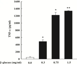
Fig. 2. Effect of β-glucan on the TNF-α production by THP-1 cells. Cells were treated with various concentration of β-glucan for 24 hr (for TNF-α). The concentration of re- leased into the culture supernatant were analyzed by enzyme-linked immunosorbent assay. The results are means ± SD. *p<0.05 versus control; **p<0.01 versus control.
Fig. 3. Effect of β-glucan on TNF-α mRNA expression induced in THP-1 cells. Cells were incubated with various con- centration (0.0, 0.3, 0.75, 1.5 mg/ml) of β-glucan for 24 hr, after which Real-time PCR was performed with EvaGreen Supermix. *p<0.05 versus control; **p<0.01 versus control.
time PCR은 CFX96 quantitative Real-time PCR detection system (Bio-Rad, Hercules, CA, USA)을 사용하였으며, Inter- nal control로서는 β-actin을 이용하여 측정하였다. Real-time PCR amplification condition은 98℃로 30초간 진행한 후, de- naturation 95℃로 1초간, annealing/extending 60℃로 5초간 수행하여 총 45 cycle을 수행하였다[6]. TNF-α, β-actin primer 는 다음과 같다. TNF-α, 5’-TGT GAG GAG GAC GAA CAT C-3’(sense), 5’-TTG AGC CAG AAG AGG TTG AG-3’(anti- sense); β-actin, 5’-GTT GCG TTA ACC TTT TTG-3’(sense), 5’-CAC TTC CCG TCC GTT-3’(anti-sense).
DNA-binding activities of NF-κB subunits
THP-1 cells을 60 mm dish에 4×10⁶ cells/dish로 seeding한 후, 배양된 cells에 β-glucan을 농도 별로 처리하여 배양하였 다. ELISA-based NF-κB transcription factor assay kit (Active Motif, Carlsbad, CA, USA) 제조사 설명에 따라, Nuclear ex- tract의 NF-κB DNA 결합 활성을 분석하였다[6].
통계학적 분석
본 실험의 값은 평균 ± 표준 오차를 표시하였으며, 실험 결과 는 통계적인 유의성을 측정하기 위해서 Student’s t-test를 실 시하였다. 통계적으로 p<0.05 수준에서 유의성을 확인하였다.
결과 및 고찰
β-glucan의 THP-1 cells에 대한 세포 생존율
β-glucan이 THP-1 cells에 미치는 세포생존율 평가하기 위
해서 MTT assay를 이용하였다. 결과는 Fig. 1에 나타낸 바와 같이 β-glucan 0.0, 0.3, 0.75, 1.5 mg/ml 농도에서 모두 90%
이상의 세포 생존율을 나타냈다. 높은 세포 생존율을 나타낸 바와 같이 THP-1 cells에 대한 세포 독성이 나타나지 않음을 확인하였다. 따라서, 본 실험은 0.0, 0.3, 0.75, 1.5 mg/ml 농도 로 분석하였다.
A
B
Fig. 4. Effects of β-glucan on the phosphorylation of MAPKs ((A)p38, (B) JNK1/2) induced in THP-1 cells. Cells were incubated with various concentration (0.0, 0.3, 0.75, 1.5 mg/ml) of β-glucan for 30 min, after which cells lysates were subjected to western blot assay using specific anti- bodies.
β-glucan의 TNF-α 발현에 미치는 영향
TNF-α는 다양한 생물학적 효과를 나타내는 사이토카인으 로, 대식세포 활성, T 림프구, 수지상 세포, 단핵구 등에서 생성 된다고 알려져 있다. 또한, TNF-α는 IFN-γ을 증가시킨다고 알려져 있으며, IFN-γ는 Th1 subtype으로 세포성 면역반응을 활성화시키며, 기도 염증을 완화시킨다고 알려져 있다[4, 7].
또한, TNF-α는 미성숙 수지상 세포의 표면 단백질 발현을 촉 진시켜, 활성화된 수지상 세포로 만들어 T 림프구와 상호 작용 으로 항암, 면역조절, 염증, 세포독성, 혈관생성, 성장 촉진 또 는 억제와 같은 다양한 효능을 나타낸다고 알려져 있다[2, 25].
본 연구는 THP-1 cells에 β-glucan의 농도별 처리에 따른 TNF-α 분비량을 알아보고자 하였다. THP-1 cells에 β-glucan 을 처리하여 48시간 동안 배양한 상층액으로 ELISA을 이용하 여 분석하였다. Fig. 2A에 나타난 바와 같이 TNF-α 분비량이 농도 의존적으로 유의하게 증가하였다. 또한, mRNA 수준에 서 TNF-α 발현을 Real-time PCR 이용하여 확인한 결과, TNF-α mRNA 발현량이 농도 의존적으로 증가하는 경향을 나타냈으 며, 1.5 mg/ml 농도에서 유의성이 높게 증가하였다(Fig. 3).
β-glucan의 MAP kinases 발현에 미치는 영향
세포 표면 수용체에 리간드가 결합하게 되면, 세포 내 신호 전달에 관여하는 MPAKs를 통해 세포의 성장, 이동, 사멸, 분 화, cytokine 생성 등에 관여한다. MAPKs는 p38 kinase, c-Jun N-terminal kinase (JNK/SAPK), extracellular signal-regu- lated kinase (ERK)로 구성되어 있으며, 이러한 조절인자가 인 산화됨으로써 사이토카인, 유전자 발현을 조절한다[11].
신호 전달 물질인 MAPKs에 β-glucan이 미치는 영향을 알아 보기 위해서 β-glucan을 처리하여 p38, JNK 단백질 인산화 정도를 western blot analysis을 이용하여 알아보고자 하였다.
A
B
C
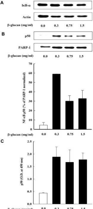
Fig. 5. Effects of β-glucan on activation NF-κB p50 induced in THP-1 cells. (A, B) Cells were incubated with various concentration (0.0, 0.3, 0.75, 1.5 mg/ml) of β-glucan in THP-1 cells. (A) After 30 min of incubation, IκB-α degra- dation was determined by western bolt analysis of cell lysates using antibody against IκB-α. (B) After 8 hr of in- cubation, the nuclear fraction was isolated form THP-1 cells. Nuclear translocation of NF-κB subunit was assessed by Western blot analysis using antibodies against NF-κB p50. (C) Cells were incubated with various concentration of β-glucan. After 8 hr of incubation, the nuclear fraction was isolation from cells. DNA-binding activity of NF-κB in nuclear extracts was assessed by using the enzyme- linked immunosorbent assay-based NF-κB p50 tran- scription factor assay kits. The results are means ± S.D.
THP-1 cells에 β-glucasn을 농도별(0.0, 0.3, 0.75, 1.5 mg/ml) 로 처리하여 30분간 배양하였다. 그 결과, Fig. 4에 나타난 바와 같이 phospho-p38, phospho-JNK는 β-glucan 처리에 따른 농 도별에 따라 의존적으로 증가되는 것을 확인하였다.
β-glucan의 처리에 따른 IκB-α 및 NF-κB p50에 미치는 영향
NF-κB는 TNF 수용체, T cells receptor (TCR), B cells re- ceptor (BCR) 등의 면역 반응에 대한 다양한 수용체의 자극으 로 인하여 세포 활성 물질, 성장 인자의 발현을 조절한다[10].
NF-κB는 세포 내에서 비활성상태로 존재하다가 NF-κB가 사 이토카인과 같은 면역 활성 인자들에 의해 자극을 받으면 IκB-α 가 인산화되고 NF-κB는 핵 안으로 이동하여 산화질소, 사이토 카인, 케모카인 등과 같은 다양한 면역 활성 물질의 유전자 발현을 유도한다[20].
β-glucan이 THP-1 cell을 통해 생성되는 TNF-α가 NF-κB 전사인자에 미치는 영향을 알아보기 위해서 β-glucan을 처리 하여 IκB-α 단백질을 Western blot을 통해 측정하였다. Fig.
5A에 나타난 바와 같이 β-glucan에 의해 활성화된 THP-1 cells 에서 유도되는 IκB-α가 처리구에 따른 변화가 나타나지 않았 다. 그러나, β-glucan 처리에 따라 NF-κB subunit인 NF-κB p50의 핵 내 전위 활성이 감소되는 것을 확인하였다(Fig. 5B).
그 다음, NF-κB가 DNA에 결합하는 능력을 확인하기 위해서 NF-κB transcription factor assay kit을 이용하여 단백질 발현 을 확인하였다. Fig. 5C에서 나타낸 바와 같이 β-glucan 처리 로 THP-1 cells에서 DNA binding 활성은 감소되는 것을 확인 하였으나, 유의한 효과는 나타나지 않았다.
β-glucan은 대식세포, 호중구, 단핵구 및 수지상 세포의 세 포 표면에 있는 receptor인 Toll-like receptor 2 (TLR 2)와 dec- tin-1, complement receptor 3 (CR3)을 통해 활성화되면, 핵 내로 신호가 전달되어 면역을 촉진하는 등의 효능과 TNF-α, IL-12, IL-10 등의 사이토카인을 분비한다고 나타나있다[5, 12].
본 연구 결과 종합적으로, β-glucan은 THP-1 cells을 자극하 여 면역을 활성시키는 인자로 작용되는 것으로 사료된다. 이 러한 면역 활성은 TNF-α 증가로 일어나며, NF-κB p50와 MAPKs 신호전달경로를 통해 면역 활성을 일으킴을 확인하 였다. 이에 따라, 면역 증강에 대한 기능성 소재로 될 수 있다 고 사료된다.
감사의 글
본 연구는 산업기술혁신사업(NO.10063302,10049026)의 지 원에 의해 이루어진 것입니다.
References
1. Akramiene. D., Kondrotas, A., Didziapetriene, J. and Keve-
laitis, E. 2007. Effects of beta-glucans on the immune system.
Medicina (Kaunas). 43, 597-606.
2. Alvarez, B., Quinn, L. S., Busquets, S., Lopez-Soriano, F. J.
and Argiles, J. M. 2001, Direct effects of tumor necrosis fac- tor alpha (TNF-alpha) on murine skeletal muscle cell lines.
Bimodal effects on protein metabolism. Eur. Cytokine Netw.
12, 399-410.
3. Arena, M. P., Spano, G. and Fiocco, D. 2017. β-Glucans and Probiotics. Am. J. Immunol. 13, 34-44.
4. Cavalcanti, Y. V., Brelaz, M. C., Neves, J. K., Ferraz, J. C.
and Pereira, V. R. 2012. Role of TNF-alpha, IFN-gamma, and IL-10 in the development of pulmonary tuberculosis. Pulm.
Med. 2012. 745483-745493.
5. Chan, G. C., Chan, W. K. and Sze, D. M. 2009. The effect of β-glucan on human immune and cancer cells. J. Hematol Oncol. 2, 1-11.
6. Choi, E. Y., Lee, S. S., Hyeon, J. Y., Choe, S. H., Keum, B.
R., Lim, J. M., Park, D. C., Choi, I. S. and Cho, K. K. 2016.
Effects of β-glucan on the release of nitric oxide by macro- phages stimulated with lipopolysaccharide. Asian-Australas.
J. Anim. Sci. 29, 1664-1674.
7. Choi, M. W., Park, I. D., Park, K. Y. and Kim, K. H. 2011.
Effects of β-lapachone on the production of inflammatory cytokines in mice. Cancer Prev. Res. 16, 155-160.
8. Dapat, I. C., Pascapurnama, D. N., lwasaki, H., Labayo, H.
K., Haorile, C. Y., Egawa, S. and Hattori, T. 2017. Secretion of galectin-9 as a DAMP during dengue virus infection in THP-1 cells. Int. J. Mol. Sci. 18, 1644-1653.
9. Hong, K. H., Jang, K. H. and Kang, S. A. 2016. Effects of dietary β-glucan on short chain fatty acids composition and intestinal environment in rats. Kor. J. Food Nutr. 29, 162-170.
10. Joo, J. D. 2009. The use of intra-cellular signaling pathways in anesthesiology and pain medicine field. Kor. J. Anesthesiol.
57, 277-283.
11. Kang, S. W. 2013. Role of reactive oxygen species in cell death pathways. Hanyang Med. Rev. 33, 77-82.
12. Kim, H. W. 2014. Immune mechanism of mushroom beta glucan. J. Mushrooms. 18, 31-38.
13. Kim, W. J., Yoon, T. J., Kim, D. W., Moon, W. K. and Lee, K. H. 2010. Immunostimulating activity of beta-glucan iso- lated from the cell wall of mutant Saccharomyces cerevisiae, and its anti-tumor application in combination with cisplatin.
Kor. J. Food Nutr. 23, 141-146.
14. Kofuji, K., Aoki, A., Tsubaki, K., Konishi, M., Isobe, T. and Murata, Y. 2012. Antioxidant activity of β-glucan. ISRN Pharm. 2012, 10.5402/2012/125864.
15. Kwon, H. K., Hwang, J. S., So, J. S. and Im, S. H. 2008.
Immunological homeostasis and inflammatory immune disorders. Kor. Soc. Mol. Cells. 3. 48-69.
16. Lee, J. S., Lee, S. H., Jang, Y. M., Lee, J. D., Lee, B. H. and Jung, J. Y. 2011. Macrophage and anticancer activities of feed additives on β-glucan from Schizophyllum commune in breast cancer cells. J. Kor. Soc. Food Sci. Nutr. 40, 949-955.
17. Lei, N., Wang, M., Zhang, L., Xiao, S., Fei, C., Wang, X., Zhang, K., Zheng, W., Wang, C., Yang, R. and Xue, F. 2015.
Effects of low molecular weight yeast β-glucan on anti-
초록:인간 단핵구 THP-1 세포에서 β-glucan으로 인한 TNF-α 분비 증가 효과
금보람1․현진이1․최소희1․진지영1․정지우2․임종민3․박동찬3․조광근4․최은영2*․최인순1,2*
(1신라대학교 일반대학원 바이오과학과, 2신라대학교 생명과학과, 3(주)글루칸, 4경남과학기술대학교 동물소재과학과)
β-glucan은 균류의 세포벽, 귀리, 효모, 식물의 구성물질로, 면역 세포의 활성, 전염증성 사이토카인 분비, 항암 효능과 같은 면역 체계에 중요한 역할을 한다. 면역계는 건강한 몸 상태의 항상성을 유지한다. 하지만, 병원성 물질이 신체 내로 들어오게 되면 면역 항상성이 무너지게 되고, 질병이 유발될 수 있다. 따라서, 본 연구는 β-glu- can이 인간 단핵구 THP-1 세포에서 면역 조절 효과에 이용될 수 있는지를 확인하였다. β-glucan은 THP-1 세포에 다양한 농도를 처리하여 배양하였으며, TNF-α mRNA 발현과 단백질 수준을 Real-time PCR와 ELISA을 이용하여 분석하였다. 또한 전사 인자 NF-κB p50와 MAPKs 신호 기작 활성을 western blot을 이용하여 분석하였다. β-glu- can으로 유도된 MAPKs와 NF-κB p50 활성이 증가하였다. β-glucan이 인간 단핵구 THP-1 세포에서 TNF-α 생성 에 의해 면역 증강 효과를 나타내며, 이는 MAPKs와 NF-κB p50 신호 전달을 통해 나타내는 것을 제시한다. 종합 적으로, 본 연구는 β-glucan이 인간 단핵구 THP-1 세포를 통해 면역 체계를 향상시킬 것이라고 사료된다.
oxidant and immunological activities in mice. Int. J. Mol.
Sci. 16, 21575-21590.
18. Park, E. K., Kang, S. M. and Leem, M. H. 2003. A study on the variation of skin moisture, oil (sebum), melanin and erythema index after application of β-glucan. Asian J. Beauty Cosmetol. 1, 83-94.
19. Park, K. S. and Kim, K. J. 2010. Effects of atopic dermatitis induced materials on the expression of cytokine genes in human monocytes and mast cells. J. Kor. Med. Ophthalmol.
Otolaryngol. Dermatol. 23, 41-56.
20. Park, W. Y., Sung, N. Y., Byun, E. H., Oh, K. H., Byun, M. W. and Yoo, Y. C. 2015. Immuno-modulatory activities of polysaccharides separated from Jubak in macrophage cells. J. Kor. Soc. Food Sci. Nutr. 44, 1079-1083.
21. Seo, H. P., Kim, J. M., Shin, H. D., Kim, T. K., Chang, H.
J., Park, B. R. and Lee, J. W. 2002. Production of be
ta-1,3/1,6-glucan by Aureobasidium pullulans SM-2001.
Kor. J. Biotechnol. Bioeng. 17, 376-380.
22. Shi, C. and Parmer, E. G. 2011. Monocyte recruitment dur- ing infection and inflammation. Nat. Rev. Immunol. 11, 762-774.
23. Stier, H., Ebbeskotte, V. and Gruenwald, J. 2014. Immune- modulatory effects of dietary Yeast Beta-1,3/1,6-D-glucan.
Nutr. J. 13, 38-47.
24. Yoo, S. A., Kim, O. K., Nam, D. E., Kim, Y. J., Bae, H. Y., Jum, W. J. and Lee, J. M. 2014. Immunomodulatory effects of fermented Curcuma longa L. extracts on RAW 264.7 cells.
J. Kor. Soc. Food Sci. Nutr. 43, 216-223.
25. Yu, A. R., Park, H. Y., Kim, Y. S., Ha, S. K., Hong, H. D.
and Choi, H. D. 2012. Immuno-enhancing effect of seed ex- tracts on a RAW 264.7 macrophage cell line. J. Kor. Soc. Food Sci. Nutr. 41, 1671-1676.