Phosphodiesterase III Inhibitor Cilostazol Protects Amyloid β -Induced Neuronal Cell Injury via Peroxisome Proliferator-Activated Receptor-γ Activation
Sun Haeng Park
1, Ji Hyun Kim
1, Sun Sik Bae
2, Ki Whan Hong
2, Byung Tae Choi
1and Hwa Kyoung Shin
1*
1
Division of Meridian and Structural Medicine, School of Korean Medicine, Pusan National University, Yangsan 626-870, Korea
2
Department of Pharmacology, School of Medicine, Pusan National University, Yangsan 626-870, Korea
Received February 21, 2011 /Accepted March 9, 2011The neurotoxicity of aggregated amyloid β (Aβ) has been implicated as a critical cause in the patho- genesis of Alzheimer’s disease (AD). It can cause neurotoxicity in AD by evoking a cascade of apopto- sis to neuron. Here, we investigated the neuroprotective effects of cilostazol, which acts as a phospho- diesterase III inhibitor, on Aβ
25-35-induced cytotoxicity in mouse neuronal cells and cognitive decline in the C57BL/6J AD mouse model via peroxisome proliferator-activated receptor (PPAR)-γ activation.
Aβ
25-35significantly reduced cell viability and increased the number of apoptotic-like cells. Cilostazol treatment recovered cells from Aβ-induced cell death as well as rosiglitazone, a PPAR-γ activator.
These effects were suppressed by GW9662, an antagonist of PPAR-γ activity, indicative of a PPAR-γ -mediated signaling. In addition, cilostazol and rosiglitazone also restored PPAR-γ activity levels that had been altered as a result of Aβ
25-35treatment, which were antagonized by GW9662. Furthermore, cilostazol also markedly decreased the number of apoptotic-like cells and decreased the Bax/Bcl-2 ratio. Intracerebroventricular injection of Aβ
25-35in C57BL/6J mice resulted in impaired cognitive function. Oral administration of cilostazol (20 mg/kg) for 2 weeks before Aβ
25-35injection and once a day for 4 weeks post-surgery almost completely prevented the Aβ
25-35-induced cognitive deficits, as did rosiglitazone. Taken together, our findings suggest that cilostazol could attenuate Aβ
25-35-induced neuronal cell injury and apoptosis as well as promote the survival of neuronal cells, subsequently im- proving cognitive decline in AD, partly because of PPAR-γ activation. The phosphodiesterase III in- hibitor cilostazol may be the basis of a novel strategy for the therapy of AD.
Key words : Alzheimer’s disease, amyloid β, apoptosis, cell injury
*Corresponding author
*Tel:+82-51-510-8476, Fax:+82-51-510-8437
*E-mail : [email protected]
Introduction
Alzheimer’s disease (AD) is neuropathological charac- terized by deposition of amyloid β (Aβ)-containing plaques and intracellular neurofibrillary tangles and loss of neurons in the brain, along with progressive cognitive impairment [7,8]. Excessive accumulation of Aβ in the brain is a possible cause of neurodegeneration [6,28], although the precise mechanism by which Aβ induces neuronal death remains unknown [11,31]. Therefore, therapeutic intervention with attenuating Aβ-mediated neurotoxicity may help in prevent- ing neurodegeneration and clinical decline in AD patients.
Cilostazol [OPC-13013, 6-[4-(1-cyclohexyl-1H-tetrazol-5-yl) butoxy]-3,4-dihydro-2-(1H)-quinolinone] increases intra- cellular cyclic AMP (cAMP) levels by inhibiting type III phosphodiesterase. Previous studies have shown that cil-
ostazol possesses many pharmacological activities, including anti-inflammatory, anti-oxidative, and anti-apoptotic effects in the brain [12]. Cilostazol was shown to prevent cerebral hypoperfusion-induced cognitive impairment and white matter damage in a rat model in which the occlusion of the common carotid artery via bilateral ligation [18]. A more re- cent preliminary study conducted in human patients with moderate AD reported that a combination therapy of done- pezil and cilostazol maintained improvement or preserved the current status until the end of the follow-up period [2].
Therefore, cilostazol may prevent the neuronal death and cognitive impairment caused by AD.
Peroxisome proliferator-activated receptor (PPAR)-γ is a nuclear transcription factor belonging to the PPAR family.
Although considerable research has shown a role for PPAR-
γ in adipose differentiation [27] and in the regulation of
inflammation [15], little is known about its possible func-
tions in neurons. Recently, PPAR-γ agonists have raised the
hope that PPAR-γ could become a drug target for the treat-
ment of neurological disease such as AD. This beneficial ef- fect of PPAR-γ agonists in AD was mediated by controlling neuroinflammation and reducing neuronal death [19,34]. We recently reported that cilostazol significantly elevated en- dogenous PPAR-γ transcriptional activity in COS-7 cells and human umbilical vein endothelial cells [22] and sup- pressed proinflammatory markers via activation of PPAR-γ transcription in db/db mice [23]. Therefore, cilostazol could decrease neuronal death and improve cognitive deficit via PPAR-γ activation in AD. However, there is no direct evi- dence to support the neuroprotective properties of cilostazol in case of Aβ insult. The aim of this study was to examine whether cilostazol has neuroprotective effects against Aβ -induced cell death and cognitive deficits.
We hypothesized that the phosphodiesterase III inhibitor cilostazol decreased neuronal cell death via PPAR-γ activa- tion, subsequently improving the cognitive decline in AD.
To test this hypothesis, we investigated the neuroprotective effects of the cilostazol on Aβ-induced cell death and Aβ -induced expression of apoptotic proteins in mouse neuronal cells and on spatial learning and memory in Aβ
25-35-injected mice compared with rosiglitazone, a PPAR-γ activator.
Materials and Methods Neuronal primary cell culture
Primary neural cell cultures from E14 ICR mouse brains were prepared as described previously [29]. Briefly, the brain tissues were dissected and were incubated in HBSS (Sigma-Aldrich, St. Louis, MO) containing 0.5% trypsin at 37°C for 30 min. Dissociated cells were plated on poly–D–
lysine–coated dishes. After 1 day, the plates were washed extensively with HBSS to remove residual non-adherent cells. Adherent cells were harvested and cultured in Dulbecco’s modified Eagle’s medium (DMEM, Gibco, Carlsbad, CA) with 10% fetal bovine serum, 100 U/ml pen- icillin, 100 μg/ml streptomycin (Hyclone, Logan, UT), 10 ng/ml basic fibroblast growth factor (bFGF), and 10 ng/ml epidermal growth factor (EGF) in a humidified atmosphere containing 5% CO
2in air at 37°C. To induce cell injury, cells were incubated with 30 μM Aβ
25-35for 24 hr.
MTT assay
The MTT assay was a standard method used to assess cell viability. Neuronal cells (1×10
5cells/well) were seeded in 96-well microtiter plates. The cells were treated with 30
μ M Aβ
25-35and various concentration cilostazol (1, 3, 10 or 30 μM) for 24 hr. Subsequently, 20 μl MTT solution (5 mg/ml in PBS) was added to each well, and the plates were in- cubated at 37°C for 4 hr, and 150 μl dimethyl sulfoxide (DMSO) was added to dissolve the formazan crystals. The absorbance was measured at 540 nm using a spectropho- tometer (Molecular devices, Sunnyvale, CA). Cilostazol was donated by Otsuka Pharmaceutical (Tokushima, Japan).
Promotor assay
The PPARγ (Peroxisome proliferator-activated receptor) activity was measured by luciferase reporter assays.
Neuronal cells in 12-well plates were co-transfected with a firefly luciferase gene tagged with pGL3-PPRE and renilla luciferase using Lipofectamine 2000 (Invitrogen, Carlsbad, CA). Medium was replaced with fresh medium after 6 hr.
Twenty-four hours post-transfection, cells were stimulated with 30 μM Aβ
25-35and various concentration cilostazol (1, 3, 10 or 30 μM). Luciferase activity was assayed 24 hr later using a dual-luciferase reporter assay system (Promega, Madison, WI).
Hoechst 33258 staining
The characteristic features of apoptotic nuclei were as- sessed by Hoechst 33258 fluorescent dye. Neuronal cells were fixed with 4% paraformaldehyde and then stained with 10 μg/ml of Hoechst 33258 (Sigma-Aldrich) for 10 min.
Nuclear morphology was visualized using Fluorescence mi- croscope (Axio Imager, Carl Zeiss, Jena, Germany). The number of cells with apoptotic morphology appearing con- densed or fragmented nuclei was counted.
Western blotting
Proteins from mouse neuronal cells were isolated accord- ing to standard techniques, separated by 10% sodium do- decyl sulfate-polyacrylamide gel electrophoresis (SDS- PAGE), and transferred onto a nitrocellulose membrane (Amersham Biosciences, Piscataway, NJ). Blots were probed for Bcl-2 (Calbiochem, Gibbstown, NJ) and Bax (Santa Cruz Biotechnology, Santa Cruz, CA) followed by incubation with secondary antibody conjugated with horseradish peroxidase.
The intensity of chemiluminescence was measured by an
ImageQuant LAS 4000 apparatus (GE Healthcare Life
Sciences, Buckinghamshire, UK). The membrane was re-
probed with an anti-β-actin antibody (Sigma-Aldrich) as an
internal control.
Aβ
25-35injection model
To evaluate the effects of cilostazol on Aβ-induced cogni- tive impairment, a mouse model produced with mod- ification of previously reported methods [21,30] was used.
Male C57BL/6J mice (20-25 g) were housed under diurnal lighting conditions and allowed food and tap water ad libitum. All animal procedures were in accordance with Pusan National University guidelines for animal research, and were approved by the university’s Animal Care and Use Committee (PNU-2009-0043). Mice were anesthetized with chloral hydrate (45 mg/kg, intraperitoneally) and were al- lowed spontaneous respiration throughout the surgical procedures. The depth of anesthesia was checked by the ab- sence of cardiovascular changes in response to tail pinch.
Rectal temperature was kept at 36.5-37.5°C using a thermo- statically controlled heating mat (Panlab, Harvard Apparatus, Holliston, MA). Aβ
25-35(Sigma-Aldrich; 10 nmol in 5 μl of saline) was injected intracerebroventricularly (icv) into the mice, aimed at 1 mm lateral to the midline, 0.5 mm posterior to the bregma and 3 mm deep using a 25 μl Hamilton syringe with a 26 gauge needle (Hamilton, Reno, NV) at a rate of 0.5 μl/min using a stereotaxic injector (KD Scientific, Holliston, MA). The sham group of mice received icv injections of an equal volume of saline. Either cilostazol (20 mg/kg), rosiglitazone (10 mg/kg) or an equal volume of DMSO (the vehicle) was given orally 2 weeks before Aβ
25-35
injection and daily for 4 weeks post-surgery.
Morris water maze task
Spatial learning and memory deficits were assessed using the Morris water maze task as described previously [3,30]
with minor modification. The experiment was performed on mice after 4 weeks Aβ
25-35injection period. The maze con- sisted of a 1.15 m-diameter pool which was painted flat white. A 10 cm-diameter platform was placed halfway be- tween the center of the pool and the edge, and was posi- tioned 1 cm below the surface of the water. The water in the pool was made opaque by the addition of powdered milk. The water temperature was 19-21°C. The water tank was located in a test room, in which there were many cues external to the maze. The position of the cues remained un- changed throughout the water-maze task. Each mouse was subjected to a series of five trials per day. For each trial, mice were randomized to one of four directional starting locations (north, south, east and west) and were placed in
the pool facing the wall. During the four subsequent training days, the mice were given three or five trial sessions per day with the platform in place before an Aβ
25-35injection.
When a mouse located the platform, it was permitted to re- main on it for 10 sec. If the mouse did not locate the platform within 180 sec, it was placed on the platform for 10 sec.
Mice were given a maximum of 180 sec to find the sub- merged platform. Swimming was video tracked, and latency time from the platform was analyzed by Smart software (Panlab, Barcelona, Spain).
Data analysis
The data were expressed as mean±SEM. Statistical com- parisons were performed using paired or unpaired Student’s t test and one-way analysis of variance (ANOVA) or two-way ANOVA for repeated measures followed by Fisher’s protected least significant difference test. p<0.05 was considered statistically significant.
Results
Cilostazol protected neuronal cells against Aβ
25-35
-induced cytotoxicity
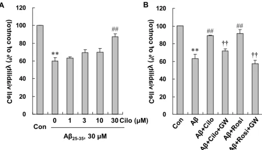
MTT assay was used to test the effect of cilostazol on the toxicity of Aβ
25-35. At concentrations between 1 and 30 μM, cilostazol alone did not cause any apparent cytotoxicity (data not shown). 30 μM Aβ
25-35significantly decreased the cell viability (60.09±3.52%, p<0.01 vs. control group), which were significantly attenuated by 30 μM cilostazol to 89.25±0.62%
(p<0.01 vs. Aβ
25-35-treated alone group) and 10 μM rosiglita- zone, a PPAR-γ agonist to 91.44±4.25% (p<0.01 vs. Aβ
25-35
-treated alone group) (Fig. 1). Co-treatment with 5 μM GW9662, a PPAR-γ antagonist significantly reversed the in- creased cell viability induced by cilostazol (71.59±2.50%, p<0.01 vs. cilostazol-treated group) and rosiglitazone (57.49±3.48%, p<0.01 vs. rosiglitazone-treated group). Thus, the results of the MTT assay showed that cilostazol could block cytotoxic effects of Aβ
25-35via PPAR-γ activation in mouse neuronal cells.
Cilostazol restored PPAR-γ activation in neuronal cells
Aβ
25-35significantly decreased PPARγ transcription activ-
ity (0.57±0.02 fold of the control, p<0.01 vs. control group),
which was markedly reversed by cilostazol in dose-depend-
0 20 40 60 80 100 120
0 20 40 60 80 100 120
Aβ25-35, 30 μM 1
0 3 10
Con Cilo (μM)
A B
Cell viability (% of control) Cell viability (% of control)
30
**
##
**
##
††
##
††
A B
Fig. 1. Cilostazol protects Aβ25-35-induced neurotoxicity. (A) Mouse neuronal cells treated with or without cilostazol (Cilo; 1–30 μM), followed by incubation with 30 μM Aβ25-35 for 24 hr. After this incubation, cell viability was determined using the MTT assay. (B) The effect of 30 μM cilostazol against Aβ-induced neurotoxicity was assessed in the absence and presence of 5 μM GW9662 (GW) compared with 10 μM rosiglitazone (Rosi). Results are shown as the mean±SEM and represent four independent experiments. **,
p
<0.01 vs. control group (Con); ##,p
<0.01 vs. Aβ25-35-treated alone group; ††,p
<0.01 vs. Aβ+cilostazol group or Aβ+rosiglitazone group.ent manners (Fig. 2A). The activity induced by 30 μM cil- ostazol and 10 μM rosiglitazone increased to 2.42±0.16 fold and 1.35±0.107 fold (p<0.01 vs. Aβ
25-35-treated alone group).
Increased PPAR-γ activities stimulated by cilostazol and ro- siglitazone were significantly antagonized by GW9662 (5 μM) (Fig. 2B). Cilostazol alone (30 μM) or rosiglitazone alone (10 μ M) without treatment of Aβ
25-35also significantly elevated PPARγ transcription activity to 2.28±0.28 fold and 1.60±0.08 fold (p<0.01 vs. control group).
Cilostazol suppressed Aβ
25-35-induced apoptosis in neuronal cells
Antiapoptotic properties of cilostazol were verified by quantitation of Hoechst 33258 stained apoptotic nuclei. In the control group, the nuclei of neuronal cells were round and homogeneously stained. After 24 hr exposure of 30 μM Aβ
25-35, the cells exhibited highly condensed and fragmented nuclei morphology, which are the typical characteristics of apoptosis (7.50±0.79% and 44.67±1.46% in the control group and Aβ
25-35-treated alone group, respectively, p<0.01, Fig. 3).
Treatment with 30 μM cilostazol decreased the number of apoptotic cells compared to the cells treated with Aβ
25-35alone (7.00±1.30%, p<0.01, Fig. 3). Thus, the results showed that cilostazol suppressed Aβ
25-35-induced DNA damage in neuronal cells.
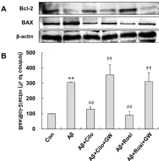
Effect of cilostazol on the expression of proa- poptotic proteins in Aβ
25-35-induced neuronal cells
To explore the molecular mechanisms underlying Aβ
25-35
-induced cell apoptosis, we examined the expression of Bcl-2 and Bax to determine whether the regulation of these cell death-associated proteins might be responsible for the protective effect of cilostazol. Exposure cells to Aβ
25-35in- duced a robust increase in the protein level of Bax, and a strong change in the protein level of Bcl-2, and the ratio of Bax/Bcl-2 expression increased approximate 3-fold in Aβ
25-35treatment alone compared with the control using western blot analysis (Fig. 4). While, cilostazol (30 μM) significantly reduced the Aβ
25-35-induced increase of the Bax/Bcl-2 ratio to 129.21±16.01% (p<0.01 vs. control) in a similar degree as did rosiglitazone (10 μM), both of which were significantly antagonized by GW9662 (5 μM). The results suggested that cilostazol could prevent Aβ
25-35-induced apoptosis, at least in part, via PPAR-γ-mediated regulation of Bcl-2 and Bax expression.
Cilostazol improved cognitive impairment in Aβ
25-35-injected mice
Learning and memory deficits are the early and critical
symptoms of AD [16]. We investigated the preventive effect
of cilostazol, by administering cilostazol beginning 2 weeks
0.0 0.5 1.0 1.5 2.0 2.5 3.0
0.0 0.5 1.0 1.5 2.0 2.5 3.0
Aβ25-35, 30 μM 1
0 3 10
Con 30Cilo (μM)
PPRE activity (% of control) PPRE activity (% of control)
**
## ##
##
**
##
††
##
††
A
AB
BFig. 2. Cilostazol restores PPARγ activation. (A) Mouse neuronal cells were transiently transfected with PPRE-pGL3, a renilla lucifer- ase control reporter vector, and then treated with or without cilostazol (Cilo; 1–30 μM), followed by incubation with 30
μM Aβ25-35for 24 hr. After this incubation, PPARγ transcription activity was determined using Luciferase reporter assay.
(B) The effect of 30 μM cilostazol against Aβ-induced decrease of PPARγ transcription activity was assessed in the absence and presence of 5 μM GW9662 (GW) compared with 10 μM rosiglitazone (Rosi). Results are shown as the mean±SEM and represent five independent experiments. **,
p
<0.01 vs. control group (Con); ##,p
<0.01 vs. Aβ25-35-treated alone group; ††,p
<0.01 vs. Aβ+cilostazol group or Aβ+rosiglitazone group.B A
Fig. 3. Cilostazol prevents Aβ25-35-induced apoptosis. (A) Mouse neuronal cells, with or without treated 30 μM cilostazol (Cilo) which were exposed to Aβ25-35for 24 hr, and then were subjected to Hoechst 33258 staining and viewed un- der a fluorescence microscope. Apoptotic cells were iden- tified by morphological changes, such as nuclei con- densation and fragmentation (arrows). (B) Quantification of abnormal nuclei after exposure of Aβ25-35in the pres- ence or absence of 30 μM cilostazol. Results are shown as the mean±SEM and represent six independent experiments. **,
p
<0.01 vs. control group (Con); ##,p
<0.01 vs. Aβ25-35-treated alone group. Scale bar is 10 μm.0 100 200 300 400 500
A
B
Bcl-2
Bax/Bcl-2/actin (% ofcontrol)
BAX β-actin
**
##
††
##
††
A
B
Fig. 4. Effect of cilostazol on the expression of Bcl-2 family pro- teins in Aβ25-35-induced neuronal cells. (A) Effect of 30 μM cilostazol (Cilo) in comparison with 10 μM rosiglita- zone (Rosi) on Aβ25-35-induced Bcl-2 and Bax protein ex- pressions in mouse neuronal cells by western blotting.
(B) Effect of cilostazol on the ratio of values of Bax/Bcl-2/actin. Densitometric analysis is mean±SEM of four independent experiments. **,
p
<0.01 vs. control group (Con); ##,p
<0.01 vs. Aβ25-35-treated alone group;††,
p
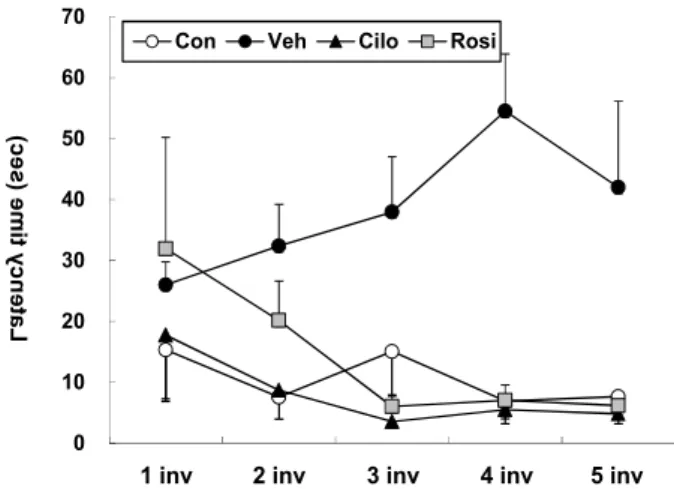
<0.01 vs. Aβ+cilostazol group or Aβ+rosiglitazone group.0 10 20 30 40 50 60 70
Con Veh Cilo Rosi
1 inv 2 inv 3 inv 4 inv 5 inv
Latency time (sec)
Fig. 5. Effect of cilostazol on spatial learning and memory in Aβ25-35-injected mice by the Morris water maze test. The changes in escape latency time to reach the platform in mice treated with DMSO (Veh), cilostazol (Cilo; 20 mg/kg) or rosiglitazone (Rosi; 10 mg/kg) at 4 weeks after Aβ25-35injection. Aβ25-35significantly increased es- cape latency time at 4 weeks after Aβ25-35 injection (
p
<0.05 vs control, Two-way ANOVA), which were re- versed by cilostazol (p
<0.05 vs vehicle, Two-way ANOVA). Results are shown as the mean±SEM and rep- resent five independent experiments.before Aβ
25-35injection and once a day for 4 weeks post-surgery. Unilateral icv injection of Aβ
25-35resulted in a significantly increased escape latency time in the target quadrant compared to control group at 4 weeks after Aβ injection (Fig. 5, p<0.05 vs. control). Treatment with cil- ostazol (20 mg/kg) or rosiglitazone (10 mg/kg) significantly reduced the escape latency time in Aβ
25-35-injected mice (p<0.05 vs. vehicle).
Discussion
Here, we report that a phosphodiesterase III inhibitor cil- ostazol strongly protected Aβ
25-35-induced cytotoxicity in mouse neuronal cells and significantly improved spatial learning and memory in Aβ
25-35-injected mice as did rosigli- tazone, a PPAR-γ activator. Cilostazol also directly in- creased PPAR-γ activity levels and restored the levels of proapoptotic Bax and antiapoptotic Bcl-2 that had been al- tered as a result of Aβ
25-35treatment as did rosiglitazone, which were antagonized by GW9662, a PPAR-γ antagonist.
Therefore, the beneficial effects of cilostazol may contribute to the PPAR-γ activity.
Increased Aβ deposition is believed to have a central role in the pathogenesis of AD [8]. Many researchers have dem-
onstrated that Aβ triggered apoptotic degeneration [9,14].
To induce AD-like in vitro and in vivo model, we used Aβ
25-35in this study. Aβ
25-35is known as a toxic core fragment of full-length Aβ
1-40[36,25] and a number of studies have dem- onstrated that Aβ
25-35can mimic the neurotoxicity of Aβ
1-42and dramatically decrease neuronal viability in multiple cel- lular systems [1,37]. Consistent with these reports, we ob- served Aβ
25-35significantly reduced cell viability and in- creased the number of apoptotic-like cells in mouse neuronal cells and icv injection of Aβ
25-35in C57BL/6J mice resulted in impaired cognitive function. Therefore, we believe that these in vitro and in vivo models are suitable for determining whether cilostazol affords protection against Aβ-induced cy- totoxicity and cognitive dysfunction.
Activation of PPAR-γ prevents Aβ-induced neuro- degeneration [13]. Recently, PPAR-γ activation has been re- ported to induce clearance of the Aβ peptide [4] and re- pression of γ-secretase (γ-site amyloid precursor protein cleaving enzyme) [10]; therefore, PPAR-γ agonists have been expected to be effective for the prevention of AD.
Several studies using animal models of AD have shown that treatment with PPAR-γ agonist attenuated learning and memory deficits [5,20,24]. In fact, it has been reported that treatment with rosiglitazone, a PPAR-γ agonist, prevented cognitive impairment in patients with early AD in a prelimi- nary study [26,33]. This beneficial effect of PPAR-γ agonists in AD was mediated by controlling neuroinflammation and reducing neuronal death [19,34].
Cilostazol increases intracellular cAMP levels by inhibit-
ing type III phosphodiesterase. 8-Bromo-cAMP and forskolin
were reported to increase PPAR-γ transcriptional activity
[32] and protein kinase A pathway is an important modu-
lator of PPAR-γ transcriptional activity [17]. Recently, we
reported that cilostazol significantly elevated endogenous
PPAR-γ transcriptional activity in COS-7 cells and human
umbilical vein endothelial cells [22] and suppressed proin-
flammatory markers via activation of PPAR-γ transcription
in db/db mice [23]. Therefore, cilostazol could decrease neu-
ronal cell death and improve cognitive deficit via PPAR-γ
activation in AD. As expect, in the present study, we ob-
served that cilostazol increased PPAR-γ transcriptional ac-
tivity, as did rosiglitazone, which were antagonized by
GW9662 in mouse neuronal cells. These results suggest that
cilostazol exerts a pharmacological action similar to rosiglita-
zone for the activation of PPAR-γ transcription. In addition,
cilostazol treatment recovered cells from Aβ-induced cell
death as did rosiglitazone and these effects were suppressed by GW9662. Furthermore, oral administration of cilostazol prevented the Aβ
25-35-induced spatial learning and memory deficits, as did rosiglitazone. These findings suggest that cil- ostazol could attenuate Aβ
25-35-induced neuronal cell injury, subsequently improving the cognitive decline in AD, partly because of PPAR-γ activation.
In the present study, we also found that cilostazol treat- ment attenuated the biochemical alterations associated with
Aβ
25-35-induced apoptotic cell death. The MTT assay in-
dicated that 30 μM cilostazol significantly protected neuronal cells from Aβ toxicity. The neuroprotective effects were also confirmed by analysis of morphological nuclear changes (Fig. 3). The Bcl-2 family includes antiapoptotic members such as Bcl-2, and proapoptotic members such as Bax. Bax is potent regulators of cytochrome c release from mitochon- dria under a variety of stress conditions and Bcl-2 prevents release of cytochrome c by heterodimerizing with Bax [35].
The ratio of Bax to Bcl-2 has been reported to be correlated with apoptosis. Our results showed that cilostazol markedly decreased Bax/Bcl-2 ratio in a similar degree as did rosiglita- zone, both of which were significantly inhibited by GW9662.
The results suggest that cilostazol could prevent Aβ
25-35-in- duced apoptosis, at least in part, via PPAR-γ-mediated reg- ulation of Bcl-2 and Bax expression.
In conclusion, a phosphodiesterase III inhibitor cilostazol rescues neuronal cells from Aβ
25-35-induced cell death and improves cognitive decline in AD mouse model through ac- tivation of PPAR-γ transcription. Further study of the an- ti-apoptotic and anti-inflammatory properties of cilostazol may provide opportunities for novel pharmacological inter- ventions aimed at preventing or palliating the consequences of AD.
Acknowledgement
This work was supported for two years by Pusan National University Research Grant.
References
1. Agostinho, P., J. P. Lopes, Z. Velez, and C. R. Oliveira. 2008.
Overactivation of calcineurin induced by amyloid-beta and prion proteins.
Neurochem. Int.
52, 1226-1233.2. Arai, H. and T. Takahashi. 2009. A combination therapy of donepezil and cilostazol for patients with moderate Alzheimer disease: pilot follow-up study.
Am. J. Geriatr.
Psychiatry
17, 353-354.3. Bermpohl, D., Z. You, E. H. Lo, H. H. Kim, and M. J. Whalen.
2007. TNF alpha and Fas mediate tissue damage and func- tional outcome after traumatic brain injury in mice.
J. Cereb.
Blood Flow Metab.
27, 1806-1818.4. Camacho, I. E., L. Serneels, K. Spittaels, P. Merchiers, D.
Dominguez, and B. De Strooper. 2004. Peroxisome-pro- liferator-activated receptor gamma induces a clearance mechanism for the amyloid-beta peptide.
J. Neurosci.
24, 10908-10917.5. Escribano, L., A. M. Simon, A. Perez-Mediavilla, P.
Salazar-Colocho, J. Del Rio, and D. Frechilla. 2009.
Rosiglitazone reverses memory decline and hippocampal glucocorticoid receptor down-regulation in an Alzheimer's disease mouse model.
Biochem. Biophys. Res. Commun.
379, 406-410.6. Hardy, J. 1997. Amyloid, the presenilins and Alzheimer's disease.
Trends Neurosci.
20, 154-159.7. Hardy, J. 2006. A hundred years of Alzheimer's disease research.
Neuron
52, 3-13.8. Hardy, J. and D. J. Selkoe. 2002. The amyloid hypothesis of Alzheimer's disease: progress and problems on the road to therapeutics.
Science
297, 353-356.9. Harkany, T., T. Hortobagyi, M. Sasvari, C. Konya, B. Penke, P. G. Luiten, and C. Nyakas. 1999. Neuroprotective ap- proaches in experimental models of beta-amyloid neuro- toxicity: relevance to Alzheimer's disease.
Prog.
Neuropsychopharmacol. Biol. Psychiatry
23, 963-1008.10. Heneka, M. T., M. Sastre, L. Dumitrescu-Ozimek, A. Hanke, I. Dewachter, C. Kuiperi, K. O'Banion, T. Klockgether, F.
Van Leuven, and G. E. Landreth. 2005. Acute treatment with the PPARgamma agonist pioglitazone and ibuprofen re- duces glial inflammation and Abeta1-42 levels in APPV717I transgenic mice.
Brain
128, 1442-1453.11. Hensley, K., J. M. Carney, M. P. Mattson, M. Aksenova, M.
Harris, J. F. Wu, R. A. Floyd, and D. A. Butterfield. 1994.
A model for beta-amyloid aggregation and neurotoxicity based on free radical generation by the peptide: relevance to Alzheimer disease.
Proc. Natl. Acad. Sci. USA
91, 3270-3274.12. Hong, K. W., J. H. Lee, K. Y. Kima, S. Y. Park, and W. S.
Lee. 2006. Cilostazol: therapeutic potential against focal cere- bral ischemic damage.
Curr. Pharm. Des.
12, 565-573.13. Inestrosa, N. C., J. A. Godoy, R. A. Quintanilla, C. S. Koenig, and M. Bronfman. 2005. Peroxisome proliferator-activated receptor gamma is expressed in hippocampal neurons and its activation prevents beta-amyloid neurodegeneration: role of Wnt signaling.
Exp. Cell Res.
304, 91-104.14. Jang, J. H. and Y. J. Surh. 2002. beta-Amyloid induces oxida- tive DNA damage and cell death through activation of c-Jun N terminal kinase.
Ann. N Y Acad. Sci.
973, 228-236.15. Jiang, C., A. T. Ting, and B. Seed. 1998. PPAR-gamma ago- nists inhibit production of monocyte inflammatory cytokines.
Nature
391, 82-86.16. Kaskie, B. and M. Storandt. 1995. Visuospatial deficit in de- mentia of the Alzheimer type.
Arch. Neurol.
52, 422-425.17. Lazennec, G., L. Canaple, D. Saugy, and W. Wahli. 2000.
Activation of peroxisome proliferator-activated receptors (PPARs) by their ligands and protein kinase A activators.
Mol. Endocrinol.
14, 1962-1975.18. Lee, J. H., S. Y. Park, Y. W. Shin, K. W. Hong, C. D. Kim, S. M. Sung, K. Y. Kim, and W. S. Lee. 2006. Neuroprotection by cilostazol, a phosphodiesterase type 3 inhibitor, against apoptotic white matter changes in rat after chronic cerebral hypoperfusion.
Brain Res.
1082, 182-191.19. Luna-Medina, R., M. Cortes-Canteli, M. Alonso, A. Santos, A. Martinez, and A. Perez-Castillo. 2005. Regulation of in- flammatory response in neural cells
in vitro
by thiadiazolidi- nones derivatives through peroxisome proliferator-activated receptor gamma activation.J. Biol. Chem.
280, 21453-21462.20. Mogi, M., J. M. Li, K. Tsukuda, J. Iwanami, L. J. Min, A.
Sakata, T. Fujita, M. Iwai, and M. Horiuchi. 2008.
Telmisartan prevented cognitive decline partly due to PPAR-gamma activation.
Biochem. Biophys. Res. Commun.
375, 446-449.
21. Nitta, A., A. Itoh, T. Hasegawa, and T. Nabeshima. 1994.
beta-Amyloid protein-induced Alzheimer's disease animal model.
Neurosci. Lett.
170, 63-66.22. Park, S. Y., J. H. Lee, K. Y. Kim, E. K. Kim, S. J. Yun, C.
D. Kim, W. S. Lee, and K. W. Hong. 2008. Cilostazol in- creases 3T3-L1 preadipocyte differentiation with improved glucose uptake associated with activation of peroxisome proliferator-activated receptor-gamma transcription.
Atherosclerosis
201, 258-265.23. Park, S. Y., H. K. Shin, J. H. Lee, C. D. Kim, W. S. Lee, B. Y. Rhim, and K. W. Hong. 2009. Cilostazol ameliorates metabolic abnormalities with suppression of proin- flammatory markers in a db/db mouse model of type 2 dia- betes via activation of peroxisome proliferator-activated re- ceptor gamma transcription.
J. Pharmacol. Exp. Ther.
329, 571-579.24. Pedersen, W. A., P. J. McMillan, J. J. Kulstad, J. B. Leverenz, S. Craft, and G. R. Haynatzki. 2006. Rosiglitazone attenuates learning and memory deficits in Tg2576 Alzheimer mice.
Exp. Neurol.
199, 265-273.25. Pike, C. J., A. J. Walencewicz-Wasserman, J. Kosmoski, D.
H. Cribbs, C. G. Glabe, and C. W. Cotman. 1995.
Structure-activity analyses of beta-amyloid peptides: con- tributions of the beta 25-35 region to aggregation and neurotoxicity.
J. Neurochem.
64, 253-265.26. Risner, M. E., A. M. Saunders, J. F. Altman, G. C. Ormandy, S. Craft, I. M. Foley, M. E. Zvartau-Hind, D. A. Hosford, and A. D. Roses. 2006. Efficacy of rosiglitazone in a genet- ically defined population with mild-to-moderate Alzheimer's disease.
Pharmacogenomics J.
6, 246-254.27. Rosen, E. D., P. Sarraf, A. E. Troy, G. Bradwin, K. Moore, D. S. Milstone, B. M. Spiegelman, and R. M. Mortensen. 1999.
PPAR gamma is required for the differentiation of adipose tissue
in vivo
andin vitro. Mol. Cell
4, 611-617.28. Selkoe, D. J. 2000. Toward a comprehensive theory for Alzheimer's disease. Hypothesis: Alzheimer's disease is caused by the cerebral accumulation and cytotoxicity of amyloid beta-protein.
Ann. N Y Acad. Sci.
924, 17-25.29. Sung, S. M., D. S. Jung, C. H. Kwon, J. Y. Park, S. K. Kang, Y. K. Kim. 2007. Hypoxia/reoxygenation stimulates pro- liferation through PKC-dependent activation of ERK and Akt in mouse neural progenitor cells.
Neurochem. Res.
32, 1932-1939.30. Takeda, S., N. Sato, K. Niisato, D. Takeuchi, H. Kurinami, M. Shinohara, H. Rakugi, M. Kano, and R. Morishita. 2009.
Validation of Abeta1-40 administration into mouse cere- broventricles as an animal model for Alzheimer disease.
Brain Res.
1280, 137-147.31. Troy, C. M., S. A. Rabacchi, Z. Xu, A. C. Maroney, T. J.
Connors, M. L. Shelanski, and L. A. Greene. 2001. be- ta-Amyloid-induced neuronal apoptosis requires c-Jun N-terminal kinase activation.
J. Neurochem.
77, 157-164.32. Watanabe, M., K. Inukai, H. Katagiri, T. Awata, Y. Oka, and S. Katayama. 2003. Regulation of PPAR gamma transcrip- tional activity in 3T3-L1 adipocytes.
Biochem. Biophys. Res.
Commun.
300, 429-436.33. Watson, G. S., B. A. Cholerton, M. A. Reger, L. D. Baker, S. R. Plymate, S. Asthana, M. A. Fishel, J. J. Kulstad, P. S.
Green, D. G. Cook, S. E. Kahn, M. L. Keeling, and S. Craft.
2005. Preserved cognition in patients with early Alzheimer disease and amnestic mild cognitive impairment during treatment with rosiglitazone: a preliminary study.
Am. J.
Geriatr. Psychiatry
13, 950-958.34. Yan, Q., J. Zhang, H. Liu, S. Babu-Khan, R. Vassar, A. L.
Biere, M. Citron, and G. Landreth. 2003. Anti-inflammatory drug therapy alters beta-amyloid processing and deposition in an animal model of Alzheimer's disease.
J. Neurosci.
23, 7504-7509.35. Yang, J., X. Liu, K. Bhalla, C. N. Kim, A. M. Ibrado, J. Cai, T. I. Peng, D. P. Jones, and X. Wang. 1999. Prevention of apoptosis by Bcl-2: release of cytochrome c from mitochon- dria blocked.
Science
275, 1129-1132.36. Yankner, B. A., L. K. Duffy, and D. A. Kirschner. 1990.
Neurotrophic and neurotoxic effects of amyloid beta pro- tein: reversal by tachykinin neuropeptides.
Science
250, 279-282.37. Zhang, H. Y., Y. H. Liu, H. Q. Wang, J. H. Xu, and H. T.
Hu. 2008. Puerarin protects PC12 cells against beta-amy- loid-induced cell injury.