Article Info
Received October 7, 2020 Revised October 31, 2020 Accepted November 2, 2020 Corresponding Author Tae-lim Yoon
E-mail: [email protected] https://orcid.org/0000-0002-1718-2205
Key Words Electromyography MyoVIDEO Orthotic devices
Background: Because a forward-leaning posture can cause increased back muscle activity and pain. Therefore, an innovative method to reduce back muscle activity and pain is required.
Objects: This study aimed to investigate the effects of a head support on muscle activity and pain in a forward-leaning posture.
Methods: A total of 14 male and 16 female students (average age, 21.65 ± 2.37 years;
height, 166.15 ± 7.90 cm; and weight, 60.65 ± 9.00 kg) were recruited for the experiment.
Two of them were excluded due to musculoskeletal disorders. The muscle activity and pain in the forward-leaning posture were assessed while participants washed dishes for 7 minutes with and without a head support. The condition of using a head support was randomly per- formed with a 5-minutes break. To confirm a lumbar flexion angle of 30° during the experi- ment, myoVIDEO was used, and surface electromyography was used to measure muscle activ- ity. Pain was assessed using a 10-point visual analog scale (VAS). The Wilcoxon signed-rank test was used to analyze the data, with p < 0.05 indicating statistical significance.
Results: The cervical, thoracic, and lumbar erector spinae muscle activities significantly de- creased with the use of the head support, but there was no significant change in the gluteus maximus. There was a significant decrease in the VAS score for the lumbar erector spinae (p <
0.05), but there was no significant change in the VAS score for the cervical region.
Conclusion: The use of a head support in a forward-leaning posture reduced cervical, tho- racic, and lumbar erector muscle activity and pain. Therefore, it could be recommended dur- ing working in a forward-leaning posture, such as during dishwashing, cooking, and working as a factory employee.
Copyright ⓒ Korean Research Society of Physical Therapy
This is an Open Access article distributed under the terms of the Creative Commons Attribution Non-Commercial License (http://creativecommons.org/licenses/by-nc/4.0) which permits unrestricted non-commercial use, distribution, and reproduction in any medium, provided the original work is properly cited.
INTRODUCTION
When standing and working for an extended duration, a forward-leaning posture can cause musculoskeletal disorders.
A forward-leaning posture causes the center of the body mass to move forward and causes excessive contractions of the back muscles to maintain the posture [1]. Prolonged standing at work and a forward-leaning posture have been associated with serious health consequences, such as leg or back pain, cardiovascular problems, fatigue, and discomfort, and ap- proximately 50% of healthy participants report low back pain (LBP) after two hours of standing long-term [2,3]. People who use large, deep sinks, such as chefs, bend forward for long durations when preparing food or washing dishes, which pro- vides a heavy workload on the low back, which is a risk fac-
tor for back pain [4]. Repetitive bending or inconsistent body postures, which increase the amount of work of the waist and legs, are also major risk factors for the development and exac- erbation of LBP [5].
To prevent LBP that can occur while working in a forward- leaning posture, a short break to relieve muscle fatigue or height adjustment of the work surface or kitchen counter have been used to reduce back muscle activity and discomfort [6,7].
One study prevented LBP by adjusting the height of the legs of the work surface, but this strategy was disadvantageous when several people worked and had a limitation in the adjustment of the worktable or care taken for long-term use [6]. In an- other study, a kitchen counter was used to relieve the activity of the lumbar erector spinae in the forward-leaning posture [7].
This strategy reduced discomfort in the low back and low back
Physical Therapy Korea
PTK https://doi.org/10.12674/ptk.2020.27.4.264 pISSN: 1225-8962 eISSN: 2287-982X Phys Ther Korea. 2020;27(4):264-271
Original Article
The Effects of Head Support on Muscle Activity and Pain in a Forward-leaning Posture
Kang-hee Kim, BS, Yoon-hee Ko, BS, Tae-lim Yoon, PT, PhD
Department of Physical Therapy, College of Health and Medical Science, Cheongju University, Cheongju, Korea
muscles [8]. When using the kitchen counter, the muscle load on the low back and leg muscles decreases, but neck and back flexion are increased, which can cause back pain and subjec- tive discomfort [9].
Using a head support to reduce the mass of the body in a forward-leaning posture reduces muscle activity and pain in the cervical and lumbar regions. A previous study reported that supporting the body using a chest support, which was designed for surgeons who often lean forward over the surgi- cal field, was effective in reducing muscle activity (40%) in the low back muscles in a forward-leaning posture. The reduced low back muscle activity also minimized LBP [10,11]. However, the chest support disrupted participants’ breathing because of the pressure on the chest and only affected the lumbar region.
Using a head support with a different design could solve these problems.
The purpose of this study was to evaluate the effects of a head support on cervical, thoracic, and lumbar erector muscle activity and pain in the cervical and lumbar regions in a forward-leaning posture. The hypothesis of this study was that there are significant changes in the activity of muscles and pain in the cervical, thoracic, and lumbar erector spinae with the use of a head support.
MATERIALS AND METHODS
1. Participants
The participants were male and female students who were recruited with a recruitment notice from September 2019 to November 2019 at a University. Before the experiment, all participants received a full explanation of how they would be measured and provided written informed consent. The selec- tion criteria were as follows: 1) Those who had not had back pain in the last 6 months; 2) those who did not have a history of lumbar or other musculoskeletal disorders [12,13]; 3) those who did not have psychiatric problems; and 4) those who were not pregnant. A total of 32 students were recruited at the beginning of the study, two of whom were excluded for a di- agnosis of lumbar intervertebral disc escape. The participants’
characteristics are shown in Table 1.
2. Equipment 1) Head support
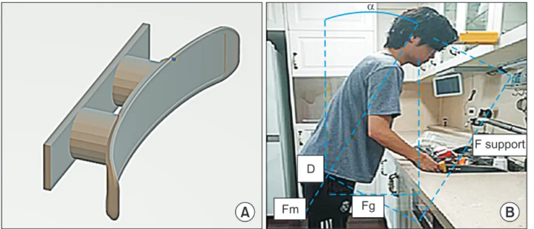
A head support was used to distribute the weight of the body along the support during dishwashing (Figure 1A). Figure 1B shows the details of the model for bending forward while leaning against a chest support. The upper body weight (Fg), the back muscle force (Fm) at the level of L5 (lumbar), and the supporting force (F support) are included in this biomechani-
Table 1.
Table 1. The descriptive characteristic of the participant
Variable Male (n = 14) Female (n = 16) Total (N = 30)
Age (y) 22.00 ± 3.37 21.30 ± 0.87 21.65 ± 2.37
Height (cm) 172.70 ± 5.67 159.60 ± 2.75 166.15 ± 7.90
Weight (kg) 66.10 ± 8.40 55.20 ± 6.04 60.65 ± 9.00
Body mass index (kg/m
2) 22.07 ± 2.05 21.70 ± 2.46 21.83 ± 2.25
Values are presented as mean ± standard deviation.
Figure 1.
Figure 1. Applying of head support (A,
model of head support; B, model of for- ward leaning posture and reaction forces).
Fg, upper body weight; Fm, back muscle force; F support, supporting force; D, the equilibrium of moment of forces in the sagittal plane at position D.
A B
D
Fm Fg
F support
cal model. Note that the model is limited to the sagittal plane and describes a static equilibrium. The mass center of gravity of the upper body is located near the axillae. Considering the equilibrium of the moment of forces in the sagittal plane at position D, the lower Fm can be calculated as follows:
Without Support (F support = 0) Fm = Fg Equation (1) With Support (F support = 0) Fm = Fg – F support Equation (2)
According to Equation (2), a head support might be most ef- fective in reducing the Fm in the low back, since distance c is at a maximum (and thus F support is maximized) [14]. Based on this information, the head restraint can achieve more than 65% efficiency. The head support is installed perpendicular to the wall and is designed to adjust the height according to the height of the participant. The participants were ordered to bend 30° from an upright posture and stand with their fore- heads against the head support. If the head support fell or the participants’ posture collapsed during dishwashing, the mea- surements were stopped and remeasured.
2) MyoVIDEO
The Noraxon MyoVIDEO system was used with a NiNox 125/250 camera (Noraxon USA Inc., Scottsdale, AZ, USA) to record and monitor the lumbar flexion angle during the dish- washing motion [15,16]. MyoVIDEO was used to continuously collect displacement data from all four markers at the upper part of the sacrum, greater trochanter of the femur, spinous process of C7, and lower part of the ear [17]. The sensors were attached to the skin using tape. The angular direction was normalized to a standing upright position, which was recorded before each test. Therefore, the participants were instructed to maintain lumbar flexion to 30° during the task in a forward- leaning posture. If there was an angle change of more than ± 10° during the task in the forward-leaning posture, the mea- surement was stopped and remeasured the following day.
3) Surface EMG
In this study, surface electromyography (sEMG) was used to measure the muscle activity of the erector spinae (cervical, lumbar, and thoracic) and the gluteus maximus. A Noraxon DTS (Noraxon Inc., Scottsdale, AZ, USA) was used to collect sEMG data. To measure muscle activity, the electrodes were at- tached while maintaining the distance between the electrodes
2 cm perpendicular to the muscle fiber. An sEMG attachment site was attached to 2 cm of C4, T12, L1, L5, the great tro- chanter, and S2 to one-third of the crypt line (Figure 2). To minimize skin resistance, all areas were cleaned with alcohol and completely dried before electrode attachment. The follow- ing parameters were set: sampling rate, 1,500 Hz; notch filter, 60 Hz; and band-pass filter, 20 –500 Hz. All sEMG data were calculated using Noraxon MR 3.8 software [18]. In addition, maximum voluntary isometric contraction (MVIC) was used to determine the normalized value of muscle activity. When mea- suring the MVIC of each muscle, the sEMG of the maximum effort state was repeatedly measured three times for 5 seconds each. The value for 3 seconds in the middle except for the first and the last of the root mean square values measured for 5 seconds was selected, and the average value of the repeated EMG over 3 seconds was obtained and used as a normalized value (%MVIC).
4) Visual analogue scale
Subjective ratings of cervical and low back pain were col- lected at the completion of the task in each condition. Low back and cervical pain were assessed using a fixed 10-point visual analogue scale (VAS) from no pain to extreme pain [19].
3. Procedures
Initially, the test participants practiced through a famil- iarization process for 5 minutes. Participants performed the dishwashing motion for 7 minutes with the head support and
Figure 2.
Figure 2. Placement of surface electromyography electrode.
7 minutes without head support. The sequence for trials was randomized (Figure 3) [20]. During the test, the participants’
lumbar angles were maintained at 30°. Verbal cues were pro- vided to maintain the angle while the researcher continuously observed the body posture. If a change of more than ±10° oc- curred for more than 5 seconds, the experiment was stopped and resumed the following day. The participants did not lean on the sink with their waists, and their feet were placed 10 cm away from the bottom of the sink. They were instructed to stand with their feet apart at pelvic width. The participants
were instructed to do the dishes without washing any special sections. The dishes were washed at a speed of 20 bowls per minute. Tape markings on the floor were used to maintain a consistent foot position and posterior distance from the sink throughout testing (Figure 4A). The head was supported by the head support (Figure 4B and C). The remaining tasks were performed with the same procedure as above. Tasks with and without the head support were conducted in random order, and 5 min of rest was provided between the tasks [21]. In ad- dition, EMG was simultaneously measured. After the two tests,
A B C
Figure 4.
Figure 4. Posture of experiment (A, side
view with no head support; B, side view with head support; C, back view with head support).
Recruited subject (n = 32)
Head support progress task in forward learning posture with
head support (sEMG, VAS)
Head support progress task in forward learning posture without
head support (sEMG, VAS)
Refusal of participation (n = 2)
Participant (n = 30)
Maximum voluntary isometric contraction, sEMG (cervical, thoracic, lumbar,
gluteus maximus)
Proceed in random order
Data analysis
Figure 3.
Figure 3. Fowchart: procedure with andwithout head support. sEMG, surface elec-
tromyography; VAS, visual analog scale.
pain in the low back and cervical spine were measured using a 10-point VAS [19].
4. Statistics
Statistical analysis of all measurements was performed us- ing SPSS ver. 25.0 for Windows (IBM Co., Armonk, NY, USA).
The data did not follow a normal distribution according to the Kolmogorov–Smirnov test. The Wilcoxon signed-rank test was used to compare muscle activity and VAS scores with and with- out the head support. Statistical significance was set at p < 0.05.
RESULTS
There were significant differences in the activity of the cer- vical, thoracic, and lumbar erector spinae muscles with and without use of the head support (p < 0.05). However, the mus- cle activity in the gluteus maximus did not differ for the two conditions significantly (Table 2).
There was a significant difference in the VAS scores in the lumbar region with and without the use of the head support (p
< 0.05). However, the VAS scores in the cervical region did not significantly differ for the two conditions (Table 3).
DISCUSSION
In this study, the effects of a head support on the activity of the cervical, thoracic, and lumbar erector spinae were assessed during the task performed in a forward-leaning position. In the forward-leaning posture, the participants demonstrated changes in muscle activity and pain in the cervical and low
back when washing dishes using a head support according to sEMG and survey findings. When the head support was used, there were significant differences in the activity of the cervi- cal, thoracic, and lumbar erector spinae but not in the gluteus maximus. When the head support was used, the VAS scores in the low back significantly decreased. There was a trending decrease in the VAS scores in the neck, although these results were not statistically significant.
There were significant decreases in the activity of cervical, thoracic, and lumbar erector spinae with the head support.
A previous study that examined the effects of muscle activ- ity with a chest support on a dental stool demonstrated a significant decrease in the sEMG activity of both the thoracic (50%) and lumbar (33%) regions during a simulated operating position when compared with a standard dental stool. These findings suggest that the use of a chest support may reduce erector spinae fatigue in dental practitioners [22]. In another study that examined changes in muscle activity with a chest support while operating in a forward-leaning posture, the use of a chest support suggested systematic reduction in muscle activity in the low back and leg muscles [11]. In another previ- ous study, which examined the effects of a passive exoskeleton brace on muscle activity and pain in the lumbar region, the back and leg muscle activity tended to decrease [23]. Specifi- cally, reductions in the thoracic erector spinae activity ranging from 21% to 31% and reductions in the lumbar erector spinae activity ranging from 10.3% to 13.7% were observed with the use of these devices [23]. However, Parsell et al. [24] showed that a chest support with rigid and mobile arm supports was not effective for reducing low back muscle activity. This find-
Table 2.
Table 2. Comparison of muscle activities of erector spinae and gluteus maximus with and without head support while performing dishwashing
Muscle %MVIC
Z-value p-value
Head support No head support
Cervical ES 5.30 ± 2.42 10.47 ± 4.27 –4.78 < 0.001
Thoracic ES 15.57 ± 13.26 22.20 ± 14.52 –3.75 < 0.001
Lumbar ES 12.63 ± 10.53 16.83 ± 10.65 –3.44 < 0.001
Gmax 4.78 ± 3.48 3.48 ± 5.79 –1.14 0.254
Values are presented as mean ± standard deviation. Gmax, gluteus maximus; ES, erector spinae.
Table 3.
Table 3. Comparison of visual analog scale on the cervical and lumbar region with and without head support after dishwashing