Surveillance of viable Acanthamoeba spp. and Naegleria fowleri in major water sources for tap water in Korea
§Min-jeong Kim1,2, Gyu-Cheol Lee1, Kunwoo Kim1, Hyunji Lee1, Min Young Kim1, Dae Keun Seo2, Jeong Yeob Lee1, and Young-Cheol Cho3*
1Water Quality Research Center, 2Drinking Water Quality and Management Team, K-water, Daejeon 34350, Republic of Korea
3Department of Environmental Engineering, Chungbuk National University, Cheongju 28644, Republic of Korea
한국 주요 상수원수에서의 가시아메바와 파울러자유아메바 조사
김민정1,2, 이규철1, 김건우1, 이현지1, 김민영1, 서대근2, 이정엽1, 조영철3*
1한국수자원공사 수질연구센터, 2한국수자원공사 수돗물품질부, 3충북대학교 환경공학과
(Received June 27, 2018; Revised July 6, 2018; Accepted July 6, 2018)
§Supplemental material for this article may be found at
http://www.kjom.org/main.html.
*For correspondence. E-mail: choy@chungbuk.ac.kr;
Tel.: +82-43-261-3577; Fax: +82-43-264-2465
The pathogenic free-living amoebas (FLAs), Acanthamoeba spp. and Naegleria fowleri, can cause fatal infections, including amoebic encephalitis. They are ubiquitously distributed in nature, including in diverse bodies of water. In order to survey Acanthamoeba spp. and N. fowleri in source water in Korea, we used culture-based real-time PCR to detect viable FLAs in 52 source water samples collected between July 2017 and December 2017. Acanthamoeba spp. and N. fowleri were detected in 42 samples (80.8%) and 6 samples (11.5%), respectively. Acanthamoeba spp. were detected at approximately the same frequency in all seasons, but N. fowleri was mainly detected in summer and autumn, with no N. fowleri detected in winter. These results demonstrate that these pathogenic FLAs, especially N. fowleri, which has caused deaths in the United States and China, are widely distributed in the Korean aquatic environment.
Keywords: free-living amoebas, pathogenic amoebas, protozoan parasites, real-time PCR, source water
Free-living amoebas (FLAs) are widely distributed in natural environments throughout the world (da Rocha-Azevedo et al., 2009). Among the many genera of free-living amoebas, four genera, Acanthamoeba, Naegleria, Balamuthia, and Sappinia, have been reported to cause diseases in humans (Schuster and Visvesvara, 2004; da Rocha-Azevedo et al., 2009). Several species of Acanthamoeba are pathogenic, and can cause serious amoebic keratitis in healthy people (Marciano-Cabral and Cabral, 2003). In addition, in immunodeficient people, Acanthamoeba spp. may cause the opportunistic infection granulomatous amoebic encephalitis (Siddiqui and Khan, 2012). Naegleria fowleri, widely known as the ‘brain-eating amoeba’, is commonly distributed in soil and warm water, including rivers, lakes, and hot springs (De Jonckheere, 2012). Among the more than 30 species of Naegleria, N. fowleri is the only recognized species that causes infections in humans (De Jonckheere, 2004; Visvesvara et al., 2007). Acute infection with N. fowleri, known as primary amoebic meningoencephalitis, can be fatal. This infection is appeared through exposure during aquatic activities (Martinez and Visvesvara, 1997).
These pathogenic FLAs are classified as pathogenic protozoa in the contaminants candidate list (CCL) of the U.S. Environmental
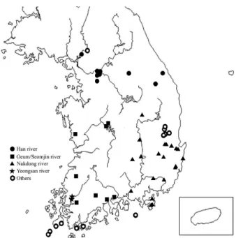
Fig. 1. Sampling locations of water sources in Korea.
water for water supplies (Jeong and Yu, 2005; Jung et al., 2008).
In addition to being ubiquitous and pathogenic, these FLAs are highly resistant to sterilization. This makes them a risk factor in treated drinking water. De Jonckheere and van de Voorde (1976) and Sarkar and Gerba (2012) reported that Acanthamoeba spp. are highly resistant to both chlorine and UV light, with a level of UV resistance even higher than that of Cryptosporidium oocysts. De Jonckheere and van de Voorde (1976) found that N. fowleri also showed high sterilization resistance, and Cursons et al. (1980) found that inactivation of cysts required exposure to a residual chorine concentration of 0.74 mg/L for 30 min. Thus, for the safety of drinking water in Korea, detection of these FLAs in source water is imperative.
For the detection of Acanthamoeba spp. and N. fowleri, specific molecular-based techniques have been developed, including PCR, nested PCR, real-time PCR, and loop-mediated isothermal amplification (Mathers et al., 2000; Schroeder et al., 2001; Marciano-Cabral and Cabral, 2003; Qvarnstrom et al., 2006; Madarova et al., 2010; Yang et al., 2013; Derda et al., 2014; Mahittikorn et al., 2003). These molecular techniques are highly sensitive, but a major weakness is found in those methods which can not discriminate between living and dead FLAs.
In the present study, we designed new primers and probes for detecting Acanthamoeba spp. and N. fowleri based on the recently updated genome database of NCBI GenBank. In addition, we developed a cell culture-based real-time PCR method for specifically detecting viable Acanthamoeba spp.
and N. fowleri. Using these newly developed real-time PCR- based techniques, 52 samples from water sources for tap water were surveyed in 2017 for the presence of viable cases of the two FLAs, Acanthamoeba spp. and N. fowleri.
Materials and Methods
Reference strains
Acanthamoeba castellanii (Douglas) Page (ATCC 30011)
in Nelson's medium (Qvarnstrom et al., 2006) and fresh water amoeba medium (ATCC Medium 997), respectively.
Environmental samples
A total of 52 samples were collected from source water for Korean tap water treatment, between July and December in 2017 (Fig. 1). Each sample consisted of one liter of water collected from a single sampling location. From each sample, 970 ml of the water was filtered using a polycarbonate filter (Isopore Membrane Filters, Merck Millipore Ltd.) with a pore size of 0.8 µm and the filter was then submerged in the remaining 30 ml of the water sample and vortexed vigorously (Ozcelik et al., 2012; Moussa et al., 2013). Then, the filter was removed, and the resuspended mixture was centrifuged at 1,500 × g for 20 min. The supernatant was then discarded, leaving a residue of 2 ml in the tube. Half of this residue, 1 ml of the concentrated sample, was then cultured on a non-nutrient agar (ATCC Medium 919) plate, prepared by pre-inoculating inactivated Escherichia coli KCTC 2441 (Korean Collection for Type Cultures, Jeongeup, Republic of Korea) at 30°C for
Fig. 2. Flow chart for experiment comparing the quantity of DNA obtained with and without preculturing of FLAs. A, Acanthamoeba castellanii; N, Naegleria fowleri; NNA, non-nutrient agar.
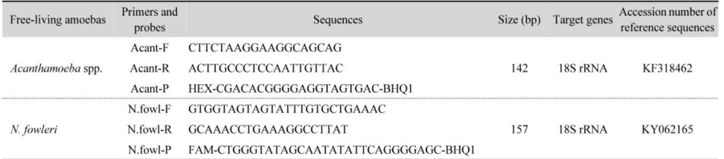
Table 1. Primers and probes used in real-time PCR for detecting Acanthamoeba spp. and Naegleria fowleri Free-living amoebas Primers and
probes Sequences Size (bp) Target genesAccession number of
reference sequences
Acanthamoeba spp.
Acant-F CTTCTAAGGAAGGCAGCAG
142 18S rRNA KF318462
Acant-R ACTTGCCCTCCAATTGTTAC
Acant-P HEX-CGACACGGGGAGGTAGTGAC-BHQ1
N. fowleri
N.fowl-F GTGGTAGTAGTATTTGTGCTGAAAC
157 18S rRNA KY062165
N.fowl-R GCAAACCTGAAAGGCCTTAT
N.fowl-P FAM-CTGGGTATAGCAATATATTCAGGGGAGC-BHQ1
5~7 days. The E. coli was inactivated by heating at 65°C for 30 min (Fig. 2).
Genomic DNA extraction and real-time PCR
Genomic DNA was extracted from the environmental samples and from the cultured reference strains as follows.
Cultured FLAs were raked from the incubated plates using a scraper and were each suspended in 200 ml of phosphate buffered saline solution. The Qiagen DNA mini kit (Qiagen) was then used to extract the genomic DNA from each sample, according to the manufacturer’s protocol. The extracted genomic DNA was then used as the template DNA for real-time PCR using our newly designed primers and probes (Table 1). The real-time PCR mixture (20 ml) was composed of 2X qPCR
premix (10 ml), 4X oligo mix (5 ml), extracted genomic DNA from the water sample culture (2 ml), and nuclease-free water (3 ml). The real-time PCR conditions were as follows: one 15 min denaturation step at 95°C, followed by 45 cycles of denaturation at 95°C for 15 sec and annealing/extension at 60°C for 45 sec. The real-time PCR was carried out using a Mic qPCR Cycler (Bio Molecular Systems).
Results and Discussion
Acanthamoeba spp. and N. fowleri are widespread in natural environments, including water. Since they can infect humans and cause fatal amoebic encephalitis, these FLAs are classified by the World Health Organization (WHO) as high health-impact
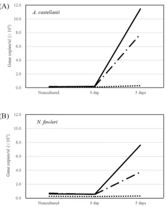
Fig. 3. Comparison of DNA amplification results for live, inactivated, and mixed amoebas after incubation. live; live + dead;
dead.
pathogenic protozoa that can spread through drinking water. In the United States, they are on the CCL and are managed by the EPA. The WHO (2017) recommends specific monitoring of these amoebae when considering their distribution in the environment. Therefore, it is important to develop a reliable method to detect these FLAs in aquatic environments. Although most probable number (MPN) methods have been used to quantitatively detect FLAs in water environments, it has been reported that MPN methods might lead to underestimations (Thomas and Ashbolt, 2011). For this reason, various molecular biological techniques, such as PCR, nested PCR, and others, have been used in several studies (Marciano-Carbal and Cabral, 2003; Qvarnstrom et al., 2006; Derda et al., 2014). Among these techniques, real-time PCR has the advantage of high detection sensitivity and relatively low technical difficulty (Valasek and Repa, 2005). However, since molecular biological methods are based on the detection of nucleic acids, they have the disadvantage of being unable to determine whether the detected organisms are alive or dead. This is the most important disadvantage of applying molecular biological methods to
organism is alive. This method involves the completion of a pretreatment process using either propidium monoazide or ethylene methyl acrylate before nucleic acid extraction and PCR (Nam et al., 2011). In our present study, the concentrated water sample was incubated on non-nutrient agar with heat- inactivated E. coli as a food source for any amoebas before extracting genomic DNA from the sample and performing the real-time PCR. This method can indirectly determine whether culturable amoebas exist in the water samples, by comparing the results from nonculture-based real-time PCR with those from culture-based real-time PCR (Fig. 2).
To test this method, we first conducted experiments with heat-inactivated and live reference strains of A. castellanii and N. fowleri. In the inactivated amoeba samples, no increases in the quantity of genomic DNA were seen during incubation (Fig. 3). In contrast, in case of living amoebas, the quantity of DNA steadily increased as the samples were incubated. We then tested an inactivated amoeba sample mixed with a living amoeba suspension in a 1:1 ratio. This mixture showed a moderate increase in genomic DNA (Fig. 3). These results indicate that real-time PCR after an incubation stage serves not only as a way to detect FLAs more sensitively, but also as a way to indirectly determine whether the detected FLAs are culturable. This represents an important practical advance over conducting real-time PCR immediately after extracting the genomic DNA from concentrated water samples, a procedure that can only detect the presence of FLA DNA, which does not necessarily reflect the presence of viable FLAs.
There have been several reports of pathogenic FLAs in the Korean water system, albeit from peripheral and small-scale surveys (Jung et al., 2008; NIER, 2009; KCDC, 2014). The present study represents a larger survey using culture-based real-time PCR to search for viable FLAs in raw water that will be used for the source water of major water treatment plants in Korea. As this raw water will eventually become tap water and FLAs are able to survive during the water treatment process, it is important to estimate the presence. A total of 52 raw water samples were selected in 2017 (Fig. 1) and investigated for the
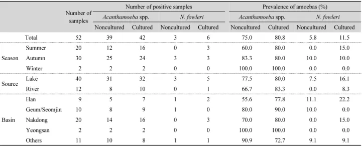
Table 2. Detection of Acanthamoeba spp. and Naegleria fowleri in water sources in Korea: season, source, and basin
Number of samples
Number of positive samples Prevalence of amoebas (%)
Acanthamoeba spp. N. fowleri Acanthamoeba spp. N. fowleri
Noncultured Cultured Noncultured Cultured Noncultured Cultured Noncultured Cultured
Total 52 39 42 3 6 75.0 80.8 5.8 11.5
Season
Summer 20 12 16 0 3 60.0 80.0 0.0 15.0
Autumn 30 25 24 3 3 83.3 80.0 10.0 10.0
Winter 2 2 2 0 0 100.0 100.0 0.0 0.0
Source Lake 40 31 32 3 5 77.5 80.0 7.5 16.1
River 12 8 10 0 1 66.7 83.3 0.0 8.3
Basin
Han 9 5 7 1 2 55.6 77.8 11.1 22.2
Geum/Seomjin 10 8 9 1 0 80.0 90.0 10.0 0.0
Nakdong 20 14 16 0 3 70.0 80.0 0.0 15.0
Yeongsan 2 2 2 0 0 100.0 100.0 0.0 0.0
Others 11 10 8 1 1 90.9 72.7 9.1 9.1
presence of infectious Acanthamoeba spp. and N. fowleri using the culture-based real-time PCR assay. As a result, Acanthamoeba spp. were detected in 42 samples with a detection rate of 80.8%, and N. fowleri was detected in six samples with a detection rate of 11.5% (Table 2 and Supplementary data Table S1). Regarding differences between the water sources, the Acanthamoeba spp.
showed no significant differences in detection rate between lake water (80.0%) and river water (83.3%), but the N. fowleri was detected slightly more often in lake water (16.1%) than river water (8.3%). Acanthamoeba spp. showed high detection rates (72.7~100%) in all water systems (Table 2). In contrast, N. fowleri was detected only in the Han River system (22.2%) and Nakdong River system (15.0%) with no detection in the Geum/Seomjin and Yeongsan River systems (Table 2).
Nonculture-based real-time PCR analysis detected Acantha- moeba spp. and N. fowleri in 39 (75.0%) and 3 (5.8%) samples, respectively (Table 2). These results indicated that culture-based real-time PCR is a more sensitive method for detecting FLAs from aquatic environmental samples than nonculture-based real-time PCR. Acanthamoeba, which has been very commonly detected in previous environmental studies (Marcino-Carbral and Carbral, 2003), was detected in 80.8% of the samples in the present study (Table 2).
N. fowleri is not as common as Acanthamoeba spp.
(Visvesvara, 2010). In the present study, the detection rate of N.
fowleri was also lower than that of Acanthamoeba spp. N.
fowleri is an amoeba that prefers warm water, and has been
detected at high rates during the hot summer season (Huizinga and McLaughlin, 1990; Hoffmann and Michel, 2001). We detected N. fowleri at high levels in the summer season, with 15.0% of the samples, and with high copy numbers for the detected DNA (Table 2 and Supplementary data Table S1).
N. fowleri was not detected in either of the two samples collected during winter (Table 2). In order to prevent infection by N. fowleri, it is necessary to intensively monitor and to properly control this amoeba during the summer season when water temperature rises. If this amoeba is detected as increased levels, it may be necessary for residents to refrain from some leisure activities such as swimming.
FLAs are strongly resistant to disinfectants such as chlorine (Thomas et al., 2004; Coulon et al., 2010; Goudot et al, 2014).
Infections through leisure activities in water such as swimming and tap water have also been reported (Shakoor et al., 2011;
Yoder et al., 2012; Cope et al., 2015). Therefore, it is necessary to completely remove the amoeba physically during the water treatment process by flocculation, sedimentation, and filtration.
The technique presented here can be used as a suitable method to discriminate whether amoebas detected in raw water or treated water are alive and monitor amoebas in treated water, tap water, and distribution systems, as well as raw water, in the future.
fowleri. Using this method, 52 samples from water sources were surveyed in 2017 for the presence of viable two FLAs. As a result, Acanthamoeba spp. were detected in 42 samples with a detection rate of 80.8%, and N. fowleri was detected in six samples with a detection rate of 11.5%. Nonculture-based real- time PCR analysis of these samples detected Acanthamoeba spp. and N. fowleri in 39 (75.0%) and 3 (5.8%) samples, respectively. These results indicate that real-time PCR after an incubation stage serves not only as a way to detect FLAs more sensitively, but also as a way to indirectly determine whether the detected FLAs are alive.
적 요
자유생활아메바인 가시아메바(Acanthamoeba spp.)와 파 울러자유아메바(Naegleria fowleri)는 아메바성 뇌염 등 치명 적인 질병을 일으키며, 물을 포함한 자연 환경에 널리 분포한 다. 가시아메바와 파울러자유아메바가 한국의 주요 상수원수 에 존재하는지 알아보기 위해 배양법에 기초한 실시간 중합효 소연쇄반응법을 이용하여 2017년 7월부터 12월 사이에 한국 의 52개 주요 상수원수를 조사하였다. 가시아메바와 파울러 자유아메바가 각각 42개 시료(80.8%)와 6개 시료(11.5%)에 서 검출되었다. 가시아메바의 경우 계절과 상관없이 고른 검 출율을 보였으나, 파울러자유아메바는 주로 여름과 가을에 검 출되었으며 겨울에는 검출되지 않았다. 이상의 결과는 이러한 자유생활아메바가 한국의 상수 원수에도 고루 존재한다는 것 을 의미한다.
Acknowledgements
This work was supported by a research grant from K-water (KWCI2018-080-032).
References
Cope JR, Ratard RC, Hill VR, Sokol T, Causey JJ, Yoder JS, Mirani G,
of Acanthamoeba cysts to disinfection treatments used in health care settings. J. Clin. Microbiol. 48, 2689–2697.
Cursons RT, Brown TJ, and Keys EA. 1980. Effect of disinfectants on pathogenic free-living amoebae: in axenic conditions. Appl.
Environ. Microbiol. 40, 62–66.
da Rocha-Azevedo B, Tanowitz HB, and Marciano-Cabral F. 2009.
Diagnosis of infections caused by pathogenic free-living amoebae.
Interdiscip. Perspect. Infect. Dis. 2009, 251406.
De Jonckheere JF. 2004. Molecular definition and the ubiquity of species in the genus Naegleria. Protist 155, 89–103.
De Jonckheere JF. 2012. The impact of man on the occurrence of the pathogenic free-living amoeboflagellate Naegleria fowleri. Future Microbiol. 7, 5–7.
De Jonckheere J and van de Voorde H. 1976. Differences in destruction of cysts of pathogenic and nonpathogenic Naegleria and Acanthamoeba by chlorine. Appl. Environ. Microbiol. 31, 294–297.
Derda M, Wojtkowiak-Giera A, and Hadas E. 2014. Comparative analyses of different genetic markers for the detection of Acan- thamoeba spp. isolates. Acta Parasitol. 59, 472–477.
Goudot S, Herbelin P, Mathieu L, Soreau S, Banas S, and Jorand FP.
2014. Biocidal efficacy of monochloramine against planktonic and biofilm-associated Naegleria fowleri cells. J. Appl. Microbiol.
116, 1055–1065.
Hoffmann R and Michel R. 2001. Distribution of free-living amoebae (FLA) during preparation and supply of drinking water. Int. J.
Hyg. Environ. Health 203, 215–219.
Huizinga HW and McLaughlin GL. 1990. Thermal ecology of Naegleria fowleri from a power plant cooling reservoir. Appl.
Environ. Microbiol. 56, 2200–2205.
Jeong HJ and Yu HS. 2005. The role of domestic tap water in Acanthamoeba contamination in contact lens storage cases in Korea. Korean J. Parasitol. 43, 47–50.
Jung EY, Jung ME, Park HG, Jung JM, Rho JS, and Ryu PJ. 2008.
Distribution of Acanthamoeba spp. in raw water and water treatment process. J. Environ. Sci. 17, 1121–1127.
Korea Centers for Disease Control and prevention (KCDC). 2014.
Settlement of diagnostic methods of Naegleria fowelri infection and detection methods for Naegleria fowleri form natural environments.
Madarova L, TrnKova K, Feikova S, Klement C, and Obernauerova M. 2010. A real-time PCR diagnostic method for detection of Naegleria fowleri. Exp. Parasitol. 126, 37–41.
Mahittikorn A, Mori H., Popruk S, Roobthaisong A, Sutthikornchai C, Marciano-Cabral F, MacLean R, Mensah A, and LaPat-Polasko L. 2003. Identification of Naegleria fowleri in domestic water sources by nested PCR. Appl. Environ. Microbiol. 69, 5864–5869.
Marciano-Cabral F and Cabral G. 2003. Acanthamoeba spp. as agents of disease in humans. Clin. Microbiol. Rev. 16, 273–307.
Martinez AJ and Visvesvara GS. 1997. Free-living, amphizoic and opportunistic amebas. Brain Pathol. 7, 583–598.
Mathers MD, Nelson SE, Lane JL, Wilson ME, Allen RC, and Folberg R. 2000. Confirmation of confocal microscopy diagnosis of Acanthamoeba keratitis using polymerase chain reaction analysis. Arch. Ophthalmol. 118, 178–183.
Moussa M, De Jonckheere JF, Guerlotte J, Richard V, Bastaraud A, Romana M, and Talarmin A. 2013. Survey of Naegleria fowleri in geothermal recreational waters of Guadeloupe (French West Indies). PLoS One 8, e54414.
Nam S, Kwon S, Kim MJ, Chae JC, Maeng PJ, Park JG, and Lee GC.
2011. Selective detection of viable Helicobacter pylori using ethidium monoazide or propidium monoazide in combination with real-time polymerase chain reaction. Microbiol. Immunol.
55, 841–846.
National Institute of Environmental Research (NIER). 2009. Settlement of the management and risk assessment system for environmental harmful microorganisms.
Ozcelik S, Coskun KA, Yunlu O, Alim A, and Malatyal E. 2012. The prevalence, isolation and morphotyping of potentially pathogenic free-living amoebae from tap water and environmental water sources in Sivas. Turkiye Parazitol. Derg. 36, 198–203.
Qvarnstrom Y, Visversvara GS, Sriram R, and da Silva AJ. 2006.
Multiplex real-time PCR assay for simultaneous detection of Acanthamoeba spp., Balamuthia mandrillaris, and Naegleria fowleri. J. Clin. Microbiol. 44, 3589–3595.
Sarkar P and Gerba CP. 2012. Inactivation of Naegleria fowleri by chlorine and ultraviolet light. Am. Water Works Assoc. 104, E173–E180.
Schroeder JM, Booton GC, Hay J, Niszl IA, Seal DV, Markus MB, Fuerst PA, and Byers TJ. 2001. Use of subgenic 18S ribosomal DNA PCR and sequencing for genus and genotype identification of Acanthamoeba from humans with keratitis and from sewage sludge. J. Clin. Microbiol. 39, 1903–1911.
Schuster FL and Visvesvara GS. 2004. Free-living amoebae as opportunistic and non-opportunistic pathogens of humans and animals. Int. J. Parasitol. 34, 1001–1027.
Shakoor S, Beg MA, Mahmood SF, Bandea R, Sriram R, Noman F, Ali F, Visvesvara GS, and Zafar A. 2011. Primary amebic meningoencephalitis caused by Naegleria fowleri, Karachi, Pakistan. Emerg. Infect. Dis. 17, 258–261.
Siddiqui R and Khan NA. 2012. Biology and pathogenesis of Acanthamoeba. Parasit. Vectors 5, 6.
Thomas JM and Ashbolt NJ. 2011. Do free-living amoebae in treated drinking water systems present an emerging health risk?
Environ. Sci. Technol. 45, 860–869.
Thomas V, Bouchez T, Nicolas V, Robert Sl, Loret JF, and Levi Y.
2004. Amoebae in domestic water systems: resistance to disinfection treatments and implication in Legionella persistence.
J. Appl. Microbiol. 97, 950–963.
Valasek MA and Repa JJ. 2005. The power of real-time PCR. Adv.
Physiol. Educ. 29, 151–159.
Visvesvara GS. 2010. Free-living amebae as opportunistic agents of human disease. J. Neroparasitology 1, 13.
Visvesvara GS, Moura H, and Schuster FL. 2007. Pathogenic and opportunistic free-living amoebae: Acanthamoeba spp., Balamuthia Mandrillaris, Naegleria fowleri and Sappinia diploidea. FEMS Immunol. Med. Microbiol. 50, 1–26.
World Health Organization (WHO). 2017. Guidelines for drinking water quality.
Yang HW, Lee YR, Inoue N, Jha BK, Danne DB, Kim HK, Lee J, Goo YK, Kong HH, Chung DI, et al. 2013. Loop-mediated isothermal amplification targeting 18S ribosomal DNA for rapid detection of Acanthamoeba. Korean J. Parasitol. 51, 269–277.
Yoder JS, Straif-Bourgeois S, Roy SL, Moore TA, Visvesvara GS, Ratard RC, Hill VR, Wilson JD, Linscott AJ, Crager R, et al.
2012. Primary amebic meningoencephalitis deaths associated with sinus irrigation using contaminated tap water. Clin. Infect.
Dis. 55, e79–85.