1
미세입자의 중력을 이용한 세포 자극기 개발에 관한 연구
김영훈 † · 김태진 *· 정효일**
Micro-bioreactor for Physical stimulation of endothelial cells using micro-bead impact by gravitational force
YOUNG HUN KIM†, TAE JIN KIM * and HYO IL JUNG **
Key Words : Endothelial Cell(상피세포), Physical Stimulation(물리적 자극), Micro Bead(마이크로 비드)
Abstract
Micro cell stimulation device is interested in many researchers because it has several advantages such as saving time and reagents. We introduce new micro-bioreactor using micro bead and conduct cell stimulation experiments to verify effective time because cell have operated by cell-cycle (G1, S, G2, and M phase). Micro-bioreactor was made by soft lithography and CAPE (calf pulmonary artery endothelial cell) was cultured in PDMS (polydimethylsiloxane) micro device for 12 hour and cell starvation process was performed for 24 hours. Micro glass beads were rolled only by slating device every hour during 15 hour because of minimizing other stimulation force like flow and pressure. The result represents that cells under exposed under micro bead stimulation show higher growth rate than normal condition and earlier and later stimulation time are more effective.
1. Introduction
현재 세포에 관한 관찰 및 자극을 위한 실험은 시간, 비용, 여러 종류의 시약 등이 다량으로 요구 된다. 이러한 단점을 극복하기 위하여 최근 많은 연구자들에 의해 마이크로 사이즈의 세포 배양기(bioreactor)에 관한 연구들이 진행 되고 있다. 마이크로 사이즈의 세포 배양기는 실험 시간, 공간을 줄여주고 세포 배양 및 관찰 등 수행하고자 하는 실험에 사용되는 시약의 양을 최소로 줄일 수 있다. 본 실험은 사람이나 동물에 있는 세포들은 물리적, 화학적, 전기적 자극에 노출 되어 있다는 점을 착안 하여 수행 되었다(1).
본 연구실에서 이 전에 수행된 실험은 마이크로 비드를 유동에 의해 움직여 자극을 수행하였다 (2). 특히, 세포는 약 16 시간에 걸친 세포 주기 (G1, S, G2, M 기)에 의해 분화가 진행 되므로 이 주기는 세포 자극의 가장 효과적인 시간에 영향을 준다 (3).
그러므로 이번 실험의 목적은 마이크로 비드의
충격에 의한 세포 자극을 최대화 하고 유동과 압력과 같은 다른 힘들은 최소화 하는 것과 가장 적절한 자극 시간을 찾는 것이다.
2. Experiment
2.1 Micro bioreactor
여기에 2.1 절의 내용을 입력한다. 16 시간 동안의 세포 주기 동안 실험을 수행하기 위하여 16 개의 device 가 PDMS (polydimethylsiloxane)을 이용한 soft lithography 과정을 통하여 제작 되었다. 각 device 는 매 시간 10 분 동안 경사를 주어 마이 크로 비드가 세포를 자극하도록 하였다. 각 device 는 10 개의 세포 배양 chamber (height = 100μm, width = 40μm, length = 80μm)을 가지고 있 다 (Figure 1 과 Figure 2). 이 device 에 주입된 세 포의 농도는 1.65x10⁶/ml, 마이크로 비드의 농도는 1.9x10⁵/ml 이다. 세포 관찰 및 비교는 4 시간에 한번씩 수행되었다.
2.1.1 Cell culture
제작 된 각 device 는 세포 배양 전 70% ethanol 과 UV 에 의한 살균 과정을 거친 후 RPMI 배지로 채널을 채운 후 세포를 주입 하여 12 시간 동안 배양하였다. 혈관 세포 CPAE 가 배양 유무를 확인 한 후 모든 세포의 주기를 G1 기로 고정하기 위하 여 5% serum 을 채널 내로 주입하여 24 시간 동안
* * 회원, 연세대학교, 기계공학과 E-mail : uridle7@yonsei.ac.kr
TEL : (02)2123-5814 FAX : (02)2123-2159
†, * 연세대학교, 기계공학과
1690 대한기계학회 2008년도 추계학술대회 논문집
2 cell starvation 과정을 진행 하였다.
3. Result
Figure 3 은 자극 시간 (stimulation time)에 따른 세 포의 증식 비율 (proliferation rate)을 나타낸 그래프 이다. 이 그래프에서 보면, 초기 자극 시간 (1 시 간)과 후기 자극 시간 (15 시간)이 다른 자극 시간 과 control 과 비교하여 CPAE 세포 성장 비율 (cell growth rate)이 더 크다는 것을 알 수 있다. 실험을 수행하기 전, 마이크로 세포 배양기 내 CPAE 세포 는 starvation 과정에 의해 G1 기에 고정 되었으므로 DNA 합성에 관여 된 G1 기나 S 기에서의 자극이 세포 증식에 더 도움이 된 다고 결론 내릴 수 있 다. 더욱이 다른 시간에서의 자극은 세포의 수가 감소한 것을 관찰 할 수 있었다.
4. conclusion
위 실험에서는 중력에 의하여 움직이는 마이크로 비드를 이용한 새로운 마이크로 사이즈의 세포 자 극 장치 (micro cell stimulation device)를 소개 하였 다. 이 PDMS device 는 세포 증식을 자극 하기 위 하여 이용되었다. CPAE 세포의 증식 비율은 1 시 간과 15 시간 에서의 자극이 다른 시간과 control 의 결과보다 더 효과적이라는 것을 알았다. 다른 시간에서의 자극은 control 과 비슷하거나 증식 비 율이 줄어드는 것이 관찰 되었다. 그러므로 본 연 구에서 고안된 마이크로 사이즈의 세포 배양기내 자극이 세포 증식에 도움이 된다는 것을 알 수 있 었고 자극 시간 (stimulation time)이 세포 분화에 큰 영향을 준다는 것을 확인 하였다. 세포는 4 종 류의 주기 (G1, S, G2, M 기)를 갖는데 이 중 DNA 합성에 관여하는 G1 기와 S 기에서의 세포 자극이 큰 효과를 가진다고 결론 내릴 수 있다.
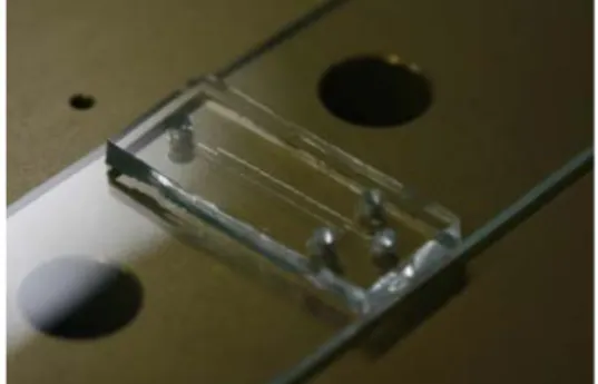
Fig 1. Photograph of device
Fig 2. Schematic diagram of device
Fig 3. Graph about stimulation time
참고문헌
(1) Matteucci, 2007, “Preliminary study of micromechanical stress delivery for cell biology studies”, Microelectron Eng, Vol.
84 ,pp.1729–1732.
(2) Tae-Jin Kim, Su-Jin Kim, Hyo-Il Jung, 2008,
“Physical stimulation of mammalian cells using micro-bead impact within a microfluidic environment to enhance growth rate”
Microfluid Nanofluid, on-line published
(3) Asako, 2008, “Visualizing spatiotemporal dynamics of multicellular cell-cycle progression”, Cell , Vol.132, pp.487-498
1691