Korean J Intern Med 2013;28:206-215 http://dx.doi.org/10.3904/kjim.2013.28.2.206
Is the frequency of metabolic syndrome higher in South Korean women with rheumatoid arthritis than in healthy subjects?
Seung-Geun Lee1, Ji-Min Kim1, Sun-Hee Lee1, Kye-Hyung Kim1, Ji-Hye Kim1, Ji-Won Yi1,
Woo-Jin Jung1, Young-Eun Park2, Seong-Hu Park3, Joung-Wook Lee4, Seung-Hoon Baek5, Jun-Hee Lee5, and Geun-Tae Kim6
1Department of Internal Medicine, Pusan National University School of Medicine, Busan; 2Department of Internal Medicine, Malgeunsem Hospital, Changwon; 3Department of Internal Medicine, Young Do Hospital, Busan; 4Department of Internal Medicine, Busan St.
Mary’s Medical Center, Busan;
5Department of Internal Medicine, Ilsin Christian Hospital, Busan;
6Division of Rheumatology, Department of Internal Medicine, Kosin University College of Medicine, Busan, Korea
Received: June 24, 2012 Revised : August 29, 2012 Accepted: August 31, 2012 Correspondence to Geun-Tae Kim, M.D.
Division of Rheumatology, Department of Internal Medi- cine, Kosin University College of Medicine, 262 Gamcheon-ro, Seo-gu, Busan 602-702, Korea Tel: +82-51-990-6722
Fax: +82-51-990-3010 E-mail: [email protected]
Background/Aims: To compare the frequency of metabolic syndrome (MetS) and magnitude of insulin resistance, measured by the homeostatic model assessment of insulin resistance (HOMA-IR), between South Korean women with rheuma- toid arthritis (RA) and healthy subjects, and to evaluate risk factors for MetS and increased HOMA-IR in patients with RA.
Methods: In a cross-sectional setting, 84 female patients with RA and 109 age- matched healthy female subjects were consecutively recruited at a university- affiliated rheumatology center in South Korea. MetS was defined according to the Third Report of the National Cholesterol Education Program’s Adult Treatment Panel (NCEP-ATP III) 2004 criteria.
Results: The frequency of MetS did not differ significantly between patients with RA (19%) and healthy subjects (15.6%, p = 0.566), although patients with RA had a higher HOMA-IR compared with healthy subjects (p < 0.001). Patients with RA met the NCEP-ATP III 2004 criteria for high blood pressure more often than healthy subjects (44% vs. 19.3%, p < 0.001), and low high density lipoprotein cho- lesterol was more prevalent in healthy subjects (33%) than in patients with RA (14.3%, p = 0.004). Although no obvious risk factors for the presence of MetS were identified in patients with RA, higher serum C-reactive protein and disease activ- ity score assessed using the 28-joint count for swelling and tenderness-erythro- cyte sedimentation rate significantly contributed to a higher HOMA-IR.
Conclusions: Despite their increased insulin resistance, South Korean women with RA did not have a significantly higher frequency of MetS compared with that in healthy subjects.
Keywords: Arthritis, rheumatoid; Metabolic syndrome X; Insulin resistance; Car- diovascular diseases
INTRODUCTION
Metabolic syndrome (MetS), also known as syndrome X or insulin resistance syndrome, comprises obesity, insulin resistance, impaired glucose tolerance or dia-
betes, hypertension, and dyslipidemia, all of which are known risk factors for atherosclerosis [1]. Among these factors, insulin resistance is recognized as the key pathophysiological factor for MetS. Moreover, insulin resistance per se increases the risk for cardiovascular
diseases (CVDs) and contributes to the association between MetS and coronary atherosclerosis [2,3]. Al- though the value of MetS as a predictor of cardiovascu- lar risk has been much debated, a recent meta-analysis showed that MetS is associated with a 2-fold increase in cardiovascular outcome and a 1.5-fold increase in all-cause mortality [4]. Hence, MetS has grown in importance in light of its contribution to the burden of cardiovascular morbidity and mortality in recent years.
Recent studies have demonstrated that in addition to insulin resistance, inflammation is closely associated with the pathogenesis of MetS [1,5,6]. A rise in acute- phase reactants such as C-reactive protein (CRP) and proinflammatory cytokines, including tumor necrosis factor-alpha (TNF-α) and interleukin-6 (IL-6), pro- mote insulin resistance [1,7,8]. Inflammatory biomark- ers are frequently elevated in subjects with MetS, and conversely, MetS is prevalent in patients with chronic inflammatory status such as rheumatic diseases [6].
Rheumatoid arthritis (RA) is a chronic systemic inf lammatory disease characterized by articular and extra-articular involvement. Patients with RA have an increased risk for CVDs due to accelerated athero- sclerosis as a result of both increased inf lammatory cytokines and a higher prevalence of traditional risk factors such as type 2 diabetes mellitus (DM) and hypertension [9,10]. MetS may provide an additional connection between accelerated atherosclerosis and inflammation in RA [7]. MetS is a common manifesta- tion in patients with RA, but previously reported fre- quencies of MetS among patients with RA vary widely, from 14% to 56% [11-24]. This diversity may be attribut- able to differences in the definition of MetS, ethnicity, geographic area, study design, and study population characteristics. Moreover, some studies have demon- strated a higher prevalence of MetS in patients with RA than in the general population [14,19,21], whereas others have not [12,13,22-24]. This discrepancy war- rants further exploration. In addition, the prevalence of MetS in South Korean women with RA has not been investigated to date.
The objectives of the present study were to compare the frequency of MetS between South Korean female patients with RA and healthy subjects and to evalu- ate factors associated with the presence of MetS in
patients with RA. Additionally, insulin resistance was measured by the homeostatic model assessment of insulin resistance (HOMA-IR) and compared between patients with RA and healthy subjects.
METHODS
Study design and subjects
We designed a cross-sectional study that included 84 female patients with RA and 109 age-matched female healthy subjects (± 2 years) (age range, 22 to 76). The entire study population was consecutively recruited at a university-aff iliated rheumatology center in South Korea from January 2008 to December 2009.
All patients with RA fulfilled the American College of Rheumatology 1987 revised classification criteria for RA [25]. Patients with rheumatic diseases other than RA or who refused to participate in this study were excluded. Healthy subjects were selected randomly from among applicants undergoing an annual health check in the same center and had no history of taking any medications such as glucocorticoids (GCs) or oral contraceptives that would affect insulin resistance and no current autoimmune or rheumatic diseases. Writ- ten informed consent was obtained from each subject based on the Declaration of Helsinki. This study was approved by the Research and Ethics Review Board of the Pusan National University Hospital, Busan, South Korea.
Assessments
All information was collected during an interview and by reviewing medical records. Anthropometric pa- rameters, including height, weight, body mass index (BMI), waist circumference, and blood pressure, were measured in all study subjects. BMI was calculated by dividing body weight by the square of height in meters (kg/m2), and waist circumference was measured using a tape at the midpoint between the lower part of the lowest rib and the highest point of the iliac crest on the mid-axillary line. Blood pressure was determined as the mean of two measurements taken at an interval of 5 minutes using a TM-2655P apparatus (A&D Com- pany Ltd., Tokyo, Japan). Hypertension was defined as blood pressure ≥ 140/90 mmHg or requiring antihy-
pertensive medication.
Study subjects also underwent biochemical assess- ments. Fasting blood samples of all participants were taken between 8:00 AM and 10:00 AM to determine total cholesterol (TC), triglycerides (TGs), low density lipoprotein cholesterol (LDL-C), high density lipopro- tein cholesterol (HDL-C), fasting glucose and insulin, and CRP. The concentrations of TC, TGs, and HDL-C were analyzed using an enzymatic colorimetric re- agent (Roche Diagnostics, Zurich, Switzerland) and a P800 Module (Roche Diagnostics). LDL-C was cal- culated using the Friedewald formula. CRP was mea- sured with a particle-enhanced immunoturbidimetric assay (Tina-quant C-reactive protein assay, Roche Diagnostics) using a P800 Module (Roche Diagnos- tics). Fasting glucose and insulin were assessed by the glucose oxidase method (Synchron LX-20, Beckman Coulter Inc., Fullerton, CA, USA) and radioimmunoas- say (Diagnostic Product Co., Los Angeles, CA, USA), respectively.
The following additional data were collected for pa- tients with RA: disease duration, medication records, erythrocyte sedimentation rate (ESR; mm/hr), CRP (mg/dL), immunoglobulin M-rheumatoid factor (RF;
IU/mL), and disease activity score assessed using the 28-joint count for swelling and tenderness (DAS28)- ESR. Medication records included the use of GCs;
disease modifying antirheumatic drugs (DMARDs), including methotrexate, hydroxychloroquine, sul- fasalazine, leflunomide, and tacrolimus; TNF inhibi- tors, and antihypertensive drugs. We calculated the cumulative GC dose by multiplying the current daily dose by the number of days the patient had been tak- ing GCs since they were first prescribed. RF was as- sessed by particle enhanced immunoturbidometric assay, and seropositivity was defined as > 14.0 IU/mL.
DAS28-ESR was calculated using the following for- mula [26]:
DAS28-ESR score = [0.56 × √ (tender joint count 28)]
+ [0.28 × √ (swollen joint count 28)] + (0.70 × in ESR) + (0.0014 × visual analogue scale)
MetS was defined according to the Third Report of the National Cholesterol Education Program’s Adult Treatment Panel (NCEP-ATP III) 2004 [27], using the Asian criteria for central obesity [28] when three or more of the following components were present: 1)
waist circumference ≥ 80 cm in women, 2) elevated blood pressure ≥ 130/85 mmHg or requiring drug therapy, 3) elevated serum TG level ≥ 150 mg/dL, 4) reduced serum HDL-C ≤ 50 mg/dL in women, and 5) elevated fasting glucose level ≥ 100 mg/dL or requir- ing drug therapy. Insulin resistance was evaluated by HOMA-IR, which was calculated with the formula de- fined by Matthews et al. [29] as follows:
HOMA-IR = [fasting serum insulin (µIU/mL) × fast- ing serum glucose (mg/dL) × 0.055 / 22.5]
Statistical analysis
Data are summarized as mean (standard deviation) or median (interquartile) for continuous variables and as number (percentage) for categorical variables.
The Kolmogorov-Smirnov test was used to assess the distributions of continuous variables. The two-tailed Student’s t test or Mann-Whitney U test was used to compare continuous variables between patients with RA and healthy subjects, and the chi-squared test or Fisher’s exact test was performed for categorical vari- ables. Univariate and multivariate logistic regression models were used to estimate unadjusted and adjusted odds ratios (ORs) for factors associated with the pres- ence of MetS. The multivariate logistic regression model included variables with p < 0.20 in the univari- ate logistic regression analysis. To approximate a nor- mal distribution, natural log-transformed HOMA- IR values were used in Pearson’s correlation analysis and stepwise multivariate linear regression analysis for estimating independent predictors of increased in- sulin resistance. Values of p < 0.05 were considered to indicate statistical significance. All statistical analyses were carried out using STATA version 11.1 for Win- dows (StataCorp LP, College Station, TX, USA).
RESULTS
Clinical characteristics of the study subjects
The demographics of the study subjects are summa- rized in Table 1. The mean ± SD age of the 84 female patients with RA was 50.6 ± 11.3 years, and the median (interquartile range) disease duration was 42.2 (24.6 to 76.5) months. Sixty-eight patients (81%) had RF sero- positivity, and the mean ± SD DAS28-ESR in patients
Table 1. Clinical characteristics of the study subjects
Characteristic RA
(n = 84)
Healthy controls
(n = 109) p value
Age, yr 50.6 ± 11.3 48.3 ± 11.3 0.157
Smoker 3 (3.6) 7 (6.4) 0.518
SBP, mmHg 122.7 ± 11.6 119.2 ± 16.4 0.083
DBP, mmHg 80.1 ± 8.2 73.0 ± 10.7 < 0.001
LDL-C, mg/dL 112.9 ± 31.3 118.1 ± 36.7 0.305
TG, mg/dL 90.0 (62.5–133.0) 78.0 (64.0–109.0) 0.101
HDL-C, mg/dL 66.8 ± 17.1 57.5 ± 12.9 < 0.001
TC, mg/dL 192.3 ± 39.3 194.7 ± 37.8 0.753
Weight, kg 56.9 ± 8.3 57.0 ± 8.0 0.911
Height, cm 157.1 ± 6.4 157.1 ± 5.8 0.956
BMI, kg/m2 23.1 ± 3.4 23.2 ± 3.5 0.912
Waist circumference, cm 76.2 ± 7.4 77.7 ± 8.4 0.189
Fasting glucose, mg/dL 87.0 (81.0–95.0) 87.0 (82.0–91.5) 0.753
Fasting insulin, µIU/mL 5.84 (4.03–7.78) 3.35 (2.30–5.84) < 0.001
Type 2 diabete mellitus 6 (7.1) 6 (5.5) 0.766
Serum CRP, mg/dL 0.20 (0.06–0.86) 0.04 (0.02–0.10) < 0.001
HOMA-IR 1.27 (0.81–1.65) 0.73 (0.48–0.99) < 0.001
Disease duration, mon 42.2 (24.6–76.5) - -
RF seropositivity 68 (81.0) - -
DAS28-ESR 3.49 ± 1.09 - -
Current medication
GC 80 (95.2) - -
Cumulative GC dose, g 2.60 (0.94–5.45) - -
Methotrexate 68 (81.0) - -
Hydroxycholorquine 28 (33.3) - -
Sulfasalazine 8 (9.5) - -
Lefluomide 35 (41.7) - -
Tacrolimus 6 (7.1) - -
TNF inhibitor 1 (1.2) - -
Antihypertensive 8 (9.5) - -
Values are presented as number (%), mean ± SD, or mean (interquartile range).
RA, rheumatoid arthritis; SBP, systolic blood pressure; DBP, diastolic blood pressure; LDL-C, low density lipoprotein cholesterol; TG, triglycerides; HDL-C, high density lipoprotein cholesterol; TC, total cholesterol; BMI, body mass index;
CRP, C-reactive protein; HOMA-IR, homeostatic model assessment of insulin resistance; RF, rheumatoid factor; DAS28-ESR, disease activity score 28-erythrocyte sedimentation rate; GC, glucocorticoid; TNF, tumor necrosis factor.
with RA was 3.49 ± 1.09. The majority of patients were taking GCs, and all but one of the patients with RA was treated with at least one DMARD. Compared with the healthy subjects, patients with RA had signifi- cantly higher diastolic blood pressure, HDL-C, fast- ing serum insulin, and serum CRP. No significant differences were observed according to age, propor- tion of smokers, systolic blood pressure, LDL-C, TG, BMI, waist circumference, fasting serum glucose, or percentage with type 2 DM between the two groups.
HOMA-IR was significantly higher in patients with RA than in healthy subjects (p < 0.001), suggesting that patients with RA were more insulin resistant than healthy subjects.
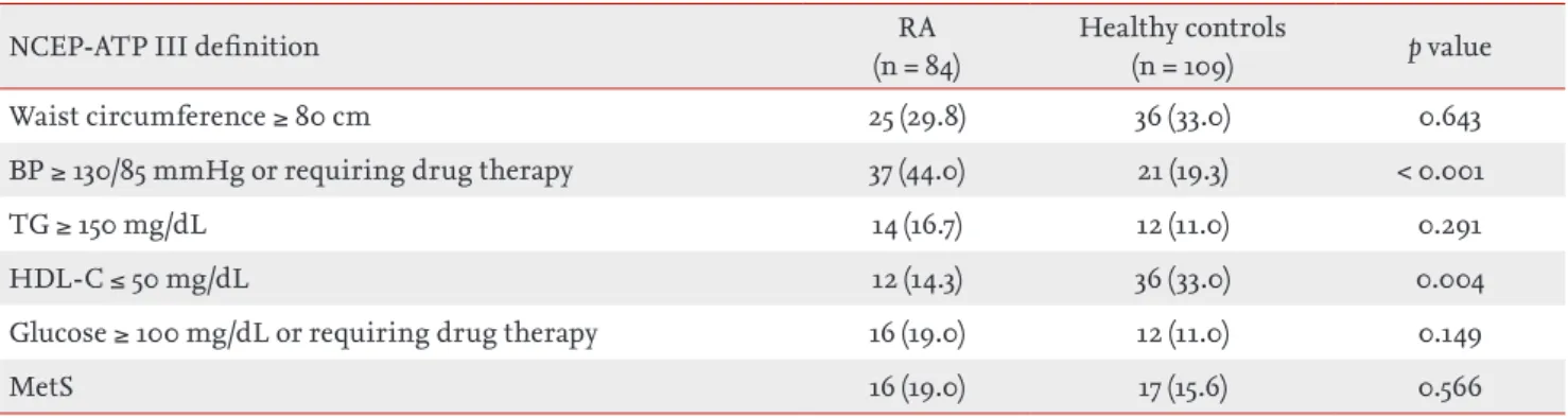
Frequency of MetS and MetS-related features Table 2 shows the frequency of the individual MetS criteria, according to the NCEP-ATP III 2004, in the study population. The frequency of MetS in patients with RA (19%) was not significantly higher than that in healthy subjects (15.6%, p = 0.566). Patients with RA met the NCEP-ATP III 2004 criteria for high blood pressure more often than healthy subjects (44% vs.
19.3%, p < 0.001), and low HDL-C was more prevalent in healthy controls than in patients with RA (33% vs.
14.3%, p = 0.004). No significant differences in waist circumstance, TG, or glucose criteria were seen be- tween the two groups.
Factors associated with MetS and increased insulin resistance
Table 3 shows the ORs of disease-related variables for the presence of MetS in 84 female patients with RA.
In the univariate analyses, higher age (OR, 1.08; 95%
confidence interval [CI], 1.02 to 1.14) and longer disease duration (OR, 1.01; 95% CI, 1.00 to 1.12) were related to an increased frequency of MetS, whereas serum CRP, RF seropositivity, DAS28-ESR, cumulative GC dose, and hydroxychloroquine and methotrexate use did not demonstrate a significant association. Longer disease duration tended to be associated with MetS in the multivariate logistic regression model, but did not remain statistically significant after adjusting for age (p = 0.084). To identify an independent relationship between disease duration and MetS, we entered dis- ease duration as a dichotomous variable, instead of a continuous variable, using the cutoff point defined by the median value (< 42, ≥ 42 months) in the multivari- ate model; age was still not statistically significant (data not shown).
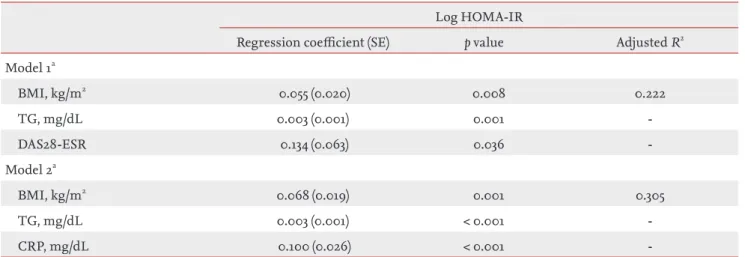
Because HOMA-IR was not normally distributed, we used natural log-transformed HOMA-IR values to assess the risk factors for insulin resistance. In the Pearson’s correlation analyses, age, TG, BMI, waist cir- cumference, serum CRP, and DAS28-ESR were posi- tively correlated with log HOMA-IR (data not shown).
Whether RA-specific variables such as serum CRP and DAS28-ESR are independent predictors for insulin resistance was of interest. However, the two variables were significantly correlated (p = 0.010). Two stepwise
Table 2. Frequency of metabolic syndrome criteria in study subjects
NCEP-ATP III definition RA
(n = 84)
Healthy controls
(n = 109) p value
Waist circumference ≥ 80 cm 25 (29.8) 36 (33.0) 0.643
BP ≥ 130/85 mmHg or requiring drug therapy 37 (44.0) 21 (19.3) < 0.001
TG ≥ 150 mg/dL 14 (16.7) 12 (11.0) 0.291
HDL-C ≤ 50 mg/dL 12 (14.3) 36 (33.0) 0.004
Glucose ≥ 100 mg/dL or requiring drug therapy 16 (19.0) 12 (11.0) 0.149
MetS 16 (19.0) 17 (15.6) 0.566
Values are presented as number (%)
NCEP-ATP III, Third Report of the National Cholesterol Education Program’s Adult Treatment Panel; RA, rheumatoid arthritis; BP, blood pressure; TG, triglycerides; HDL-C, high density lipoprotein cholesterol; MetS, metabolic syndrome.
multivariate linear regression models that included CRP and DAS28-ESR were constructed separately to avoid multicollinearity (Table 4). Both RA specific vari- ables remained significant, and the magnitude of the statistical association in the multivariate models was greater for CRP than for DAS28-ESR (p < 0.001 and p = 0.036, respectively). Higher TG and BMI were also in- dependent risk factors for log HOMA-IR, but age was not.
DISCUSSION
In the present study, no significant difference was observed in the frequency of MetS between female pa- tients with RA and healthy subjects, whereas the mag- nitude of insulin resistance in patients with RA was significantly higher than that in healthy subjects. De- spite the lack of obvious risk factors for the presence of MetS in patients with RA, RA inflammation (CRP), and disease activity (DAS28-ESR) significantly contrib-
Table 3. Odds ratio for the presence of metabolic syndrome in female patients with rheumatoid arthritis
Variable Crude OR (95% CI) Adjusted OR (95% CI)a
Age, yr 1.08 (1.02–1.14)b -
Disease duration, mon 1.01 (1.00–1.02)c 1.01 (1.00–1.02)d
Serum CRP, mg/dL 0.72 (0.37–1.39) -
RF seropositivity 0.64 (0.18–2.34) -
DAS28-ESR 1.10 (0.61–1.67) -
Cumulative GC dose, g 1.00 (0.98–1.01) -
Hydroxycholorquine use 0.61 (0.18–2.10) -
Methotrexate use 1.02 (0.25–4.13) -
OR, odds ratio; CI, confidence interval; CRP, C-reactive protein; RF, rheumatoid factor; DAS28-ESR, disease activity score 28-erythrocyte sedimentation rate; GC, glucocorticoid.
aAdjusted for age.
bp = 0.012.
cp = 0.049.
dp = 0.084.
Table 4. Stepwise multivariate linear regression models for insulin resistance in female patients with rheumatoid arthritis Log HOMA-IR
Regression coefficient (SE) p value Adjusted R2 Model 1a
BMI, kg/m2 0.055 (0.020) 0.008 0.222
TG, mg/dL 0.003 (0.001) 0.001 -
DAS28-ESR 0.134 (0.063) 0.036 -
Model 2a
BMI, kg/m2 0.068 (0.019) 0.001 0.305
TG, mg/dL 0.003 (0.001) < 0.001 -
CRP, mg/dL 0.100 (0.026) < 0.001 -
HOMA-IR, homeostatic model assessment of insulin resistance; SE, standard error; BMI, body mass index; TG, triglyceride;
DAS28-ESR, disease activity score 28-erythrocyte sedimentation rate; CRP, C-reactive protein.
aModels 1 and 2, adjusted for age.
uted to increased insulin resistance as measured by HOMA-IR.
A growing body of evidence indicates that both MetS and RA are closely related to increased cardiovascular morbidity and mortality, which has led to the need to evaluate whether MetS is more prevalent in patients with RA compared with healthy subjects in the last decade. Several studies have demonstrated a higher prevalence in patients with RA than in healthy con- trols [14,19,21], whereas others have not [12,13,22-24].
Our results yielded no difference in the frequency of MetS between patients with RA and healthy subjects.
Interestingly, Sahebari et al. [23] reported that the fre- quency of MetS in patients with RA was significantly lower than that in healthy controls. The epidemiologi- cal association between RA and MetS has not yet been fully determined, and various factors may be respon- sible for the differences in the prevalence of MetS in patients with RA. Among these factors, ethnicity and geographic area appeared to affect the difference in MetS between patients with RA and healthy controls.
Two of three previous studies in Asian subjects report- ed a similar prevalence of MetS in patients with RA than in controls [23,24]. Considering that our study in- cluded South Korean subjects only, it is presumed that MetS tends to be less prevalent among Asian patients with RA. This assumption was also suggested in a recent systematic review by Yiu et al. [30]. As the num- ber of studies is limited, further research is needed to confirm the relationship between RA and MetS and to evaluate the effect of ethnicity and geographic area on the frequency of MetS.
No significant risk factors for MetS were observed in female patients with RA. In some studies, methotrex- ate therapy was associated with reduced prevalence of MetS in patients with RA [16,18,19]. Toms et al. [18]
suggested that the anti-inflammatory effect of metho- trexate and concurrent folic acid supplementation may contribute to the decreased frequency of MetS in pa- tients with RA. However, these findings are not consis- tent with other studies [12,31]. Our study also showed no significant association between methotrexate use and MetS in patients with RA. A better understand- ing of the differences among these studies will require further investigation to elucidate a plausible protective effect of methotrexate against MetS in patients with
RA.
In addition, the use of GCs did not significantly con- tribute to the presence of MetS in patients with RA in previous reports [14,15,19]. As the majority of patients in our study were taking GCs, we assessed the OR of cumulative GC dose instead of GC use, and no signifi- cant relationship between GCs and MetS in patients with RA was observed, similar to previous reports. It has been recognized that GCs have a deleterious ef- fect on blood pressure, insulin resistance, and lipid metabolism [32] and that GCs lead to increase CVD in patients with RA [33]. GCs also have anti-inflammatory and immunosuppressive properties that could coun- teract undesirable side effects in patients with RA [34].
Hence, verifying the contribution of GCs to MetS in patients with RA is complicated by the intricacy of GC actions.
Among various direct and indirect methods for measuring insulin resistance, HOMA-IR, derived from fasting blood insulin and glucose concentra- tions, is a simple and useful clinical index, particu- larly in epidemiological studies [8,35]. In our study, both serum CRP and DAS28-ESR were independent risk factors for increased HOMA-IR in female patients with RA. These findings largely agreed with some pre- vious studies evaluating predictors for HOMA-IR in patients with RA [36-38]. Over the last decade, increas- ing evidence has suggested a relationship between chronic inf lammation and insulin resistance in RA [39]. Proinf lammatory cytokines such as TNF-α and IL-6 are key players in the pathogenesis of RA and are closely related to insulin resistance [6,7,39]. Further- more, the role of various adipokines in both RA and insulin resistance has been highlighted recently [6].
Taken together, our results suggest a connection be- tween inflammation and insulin resistance in patients with RA.
In the present study, HOMA-IR was significantly higher in female patients with RA than in healthy sub- jects, which agreed with previous studies [14,24,36,38].
Taken together, these findings suggest that patients with RA had significantly increased insulin resistance compared with healthy controls. However, consider- ing that insulin resistance is the major catalyst in MetS, the lack of a significant difference in the fre- quency of MetS between patients with RA and healthy
controls in our study was unforeseen and interesting.
Similarly, Karimi et al. [24] also demonstrated higher HOMA-IR in women patients with RA compared with controls, yet there was no difference in the prevalence of MetS between the two groups. We assumed that the complexity in MetS characteristics could explain these findings. MetS is a constellation of different, but correlated, metabolic abnormalities rather than a par- ticular disease entity, and insulin resistance may be necessary, but not sufficient, for MetS [40,41]. Hence, increased HOMA-IR may not always result in the pres- ence of MetS.
Whether patients with RA have significantly higher probabilities of traditional cardiovascular risk factors such as high blood pressure, dyslipidemia, diabetes, and smoking compared with those in the general pop- ulation is somewhat controversial [30]. In our study, diastolic blood pressure and the frequency of the NCEP-ATP III 2004 criteria for high blood pressure were significantly higher in patients with RA than in healthy controls (Tables 1 and 2). Numerous factors, in- cluding obesity, inflammation, physical inactivity, and medications, may increase blood pressure in patients with RA [42]. Therefore, our findings are unlikely to be generalizable. In addition, HDL-C was significantly higher in patients with RA than in healthy subjects (Table 1). High disease activity in patients with RA is associated with low HDL-C levels, and antirheumatic treatment, including GCs, can reverse this dyslipid- emia [43]. As mentioned above, most patients with RA in our study were taking antirheumatic medications that can alleviate dyslipidemia.
The findings in the present study must be consid- ered in light of major limitations. First, this was an observational study with a cross-sectional design.
The majority of our patients with RA were receiving GCs and DMARDs. Thus, we could not fully adjust for the effect of various medications on MetS and in- sulin resistance. Second, subjects in our study were recruited from only a single center located in a harbor city. Hence, most of our study subjects resided in a seacoast region, which was assumed to affect the over- all frequency of MetS in our study (RA, 19.6%; healthy controls, 15.6%). Similarly, the prevalence of MetS in 2,519 healthy female subjects in the same center, re- ported by Kang et al. [44] in 2008, was 15.6% according
to criteria used in our study. However, using the same criteria, the prevalence of MetS in a nationwide survey in South Korea in 2005 was 38.7% [45], which appears to be higher than that in our center. Taken together, we speculate that the geographic characteristics of the area could have acted as a confounding factor that affected the difference in MetS frequency between patients with RA and controls. Therefore, our results should be carefully interpreted. Last, the number of subjects in the present study might not have been suf- ficient to investigate all potential associated factors.
In conclusion, the frequency of MetS in South Kore- an women with RA was comparable to that in healthy subjects, although HOMA-IR was significantly higher in patients with RA than in healthy subjects. As many epidemiological factors, particularly ethnicity, may affect the frequency of MetS in patients with RA, it is still not clear whether patients with RA have a higher prevalence of MetS. Further studies are needed to con- firm the relationship between RA and MetS.
Conflict of interest
No potential conflict of interest relevant to this article is reported.
Acknowledgments
We thank the late professor Sung-Il Kim who devoted himself to patient care, research, and education at the Division of Rheumatology, Department of Internal Medicine, Pusan National University School of Medi- cine.
KEY MESSAGE
1. The frequency of metabolic syndrome in South Korean women with rheumatoid arthritis (RA) was similar to than in that in healthy subjects.
2. RA patients had a higher homeostatic model assessment of insulin resistance (HOMA-IR), a measure of insulin resistance, than healthy sub- jects.
3. In RA patients, higher C-reactive protein and disease activity were associated with increased HOMA-IR.
REFERENCES
1. Dandona P, Aljada A, Chaudhuri A, Mohanty P, Garg R. Metabolic syndrome: a comprehensive perspective based on interactions between obesity, diabetes, and in- flammation. Circulation 2005;111:1448-1454.
2. Hanley AJ, Williams K, Stern MP, Haffner SM. Homeo- stasis model assessment of insulin resistance in rela- tion to the incidence of cardiovascular disease: the San Antonio Heart Study. Diabetes Care 2002;25:1177-1184.
3. Reilly MP, Wolfe ML, Rhodes T, Girman C, Mehta N, Rader DJ. Measures of insulin resistance add incre- mental value to the clinical diagnosis of metabolic syndrome in association with coronary atherosclerosis.
Circulation 2004;110:803-809.
4. Mottillo S, Filion KB, Genest J, et al. The metabolic syndrome and cardiovascular risk a systematic review and meta-analysis. J Am Coll Cardiol 2010;56:1113-1132.
5. Pereira RM, de Carvalho JF, Bonfa E. Metabolic syn- drome in rheumatological diseases. Autoimmun Rev 2009;8:415-419.
6. Santos MJ, Fonseca JE. Metabolic syndrome, inflamma- tion and atherosclerosis: the role of adipokines in health and in systemic inflammatory rheumatic diseases. Acta Reumatol Port 2009;34:590-598.
7. Sidiropoulos PI, Karvounaris SA, Boumpas DT. Meta- bolic syndrome in rheumatic diseases: epidemiology, pathophysiology, and clinical implications. Arthritis Res Ther 2008;10:207.
8. Nakanishi N, Shiraishi T, Wada M. Association between C-reactive protein and insulin resistance in a Japanese population: the Minoh Study. Intern Med 2005;44:542- 547.
9. Ozbalkan Z, Efe C, Cesur M, et al. An update on the relationships between rheumatoid arthritis and athero- sclerosis. Atherosclerosis 2010;212:377-382.
10. Gremese E, Ferraccioli G. The metabolic syndrome: the crossroads between rheumatoid arthritis and cardio- vascular risk. Autoimmun Rev 2011;10:582-589.
11. Dessein PH, Tobias M, Veller MG. Metabolic syndrome and subclinical atherosclerosis in rheumatoid arthritis.
J Rheumatol 2006;33:2425-2432.
12. Karvounaris SA, Sidiropoulos PI, Papadakis JA, et al.
Metabolic syndrome is common among middle-to- older aged Mediterranean patients with rheumatoid ar- thritis and correlates with disease activity: a retrospec-
tive, cross-sectional, controlled, study. Ann Rheum Dis 2007;66:28-33.
13. La Montagna G, Cacciapuoti F, Buono R, et al. Insulin resistance is an independent risk factor for athero- sclerosis in rheumatoid arthritis. Diab Vasc Dis Res 2007;4:130-135.
14. Chung CP, Oeser A, Solus JF, et al. Prevalence of the metabolic syndrome is increased in rheumatoid arthri- tis and is associated with coronary atherosclerosis. Ath- erosclerosis 2008;196:756-763.
15. Toms TE, Panoulas VF, Douglas KM, Griffiths HR, Kitas GD. Lack of association between glucocorticoid use and presence of the metabolic syndrome in patients with rheumatoid arthritis: a cross-sectional study. Ar- thritis Res Ther 2008;10:R145.
16. Zonana-Nacach A, Santana-Sahagun E, Jimenez-Bal- deras FJ, Camargo-Coronel A. Prevalence and factors associated with metabolic syndrome in patients with rheumatoid arthritis and systemic lupus erythemato- sus. J Clin Rheumatol 2008;14:74-77.
17. Elkan AC, Hakansson N, Frostegard J, Cederholm T, Hafstrom I. Rheumatoid cachexia is associated with dyslipidemia and low levels of atheroprotective natural antibodies against phosphorylcholine but not with di- etary fat in patients with rheumatoid arthritis: a cross- sectional study. Arthritis Res Ther 2009;11:R37.
18. Toms TE, Panoulas VF, John H, Douglas KM, Kitas GD.
Methotrexate therapy associates with reduced preva- lence of the metabolic syndrome in rheumatoid arthri- tis patients over the age of 60- more than just an anti- inflammatory effect? A cross sectional study. Arthritis Res Ther 2009;11:R110.
19. Dao HH, Do QT, Sakamoto J. Increased frequency of metabolic syndrome among Vietnamese women with early rheumatoid arthritis: a cross-sectional study. Ar- thritis Res Ther 2010;12:R218.
20. Raterman HG, van Eijk IC, Voskuyl AE, et al. The meta- bolic syndrome is amplified in hypothyroid rheumatoid arthritis patients: a cross-sectional study. Ann Rheum Dis 2010;69:39-42.
21. Crowson CS, Myasoedova E, Davis JM 3rd, et al. In- creased prevalence of metabolic syndrome associated with rheumatoid arthritis in patients without clinical cardiovascular disease. J Rheumatol 2011;38:29-35.
22. Mok CC, Ko GT, Ho LY, Yu KL, Chan PT, To CH. Preva- lence of atherosclerotic risk factors and the metabolic
syndrome in patients with chronic inf lammatory ar- thritis. Arthritis Care Res (Hoboken) 2011;63:195-202.
23. Sahebari M, Goshayeshi L, Mirfeizi Z, et al. Investigation of the association between metabolic syndrome and disease activity in rheumatoid arthritis. Scientif icWorldJournal 2011;11:1195-1205.
24. Karimi M, Mazloomzadeh S, Kafan S, Amirmoghadami H. The frequency of metabolic syndrome in women with rheumatoid arthritis and in controls. Int J Rheum Dis 2011;14:248-254.
25. Arnett FC, Edworthy SM, Bloch DA, et al. The American Rheumatism Association 1987 revised criteria for the classification of rheumatoid arthritis. Arthritis Rheum 1988;31:315-324.
26. Prevoo ML, van 't Hof MA, Kuper HH, van Leeuwen MA, van de Putte LB, van Riel PL. Modified disease ac- tivity scores that include twenty-eight-joint counts: de- velopment and validation in a prospective longitudinal study of patients with rheumatoid arthritis. Arthritis Rheum 1995;38:44-48.
27. Grundy SM, Cleeman JI, Daniels SR, et al. Diagno- sis and management of the metabolic syndrome: an American Heart Association/National Heart, Lung, and Blood Institute Scientific Statement. Circulation 2005;112:2735-2752.
28. Heng D, Ma S, Lee JJ, et al. Modification of the NCEP ATP III definitions of the metabolic syndrome for use in Asians identifies individuals at risk of ischemic heart disease. Atherosclerosis 2006;186:367-373.
29. Matthews DR, Hosker JP, Rudenski AS, Naylor BA, Treacher DF, Turner RC. Homeostasis model assess- ment: insulin resistance and beta-cell function from fasting plasma glucose and insulin concentrations in man. Diabetologia 1985;28:412-419.
30. Yiu KH, Tse HF, Mok MY, Lau CS. Ethnic differences in cardiovascular risk in rheumatic disease: focus on Asians. Nat Rev Rheumatol 2011;7:609-618.
31. Raterman HG, Voskuyl AE, Dijkmans BA, Nurmo- hamed MT. Use of methotrexate therapy is not associ- ated with decreased prevalence of metabolic syndrome.
Arthritis Res Ther 2009;11:413.
32. Maxwell SR, Moots RJ, Kendall MJ. Corticosteroids: do they damage the cardiovascular system? Postgrad Med J 1994;70:863-870.
33. Gonzalez-Gay MA. Glucocorticoid-related cardiovascu- lar and cerebrovascular events in rheumatic diseases:
myth or reality? Arthritis Rheum 2007;57:191-192.
34. Hoes JN, Jacobs JW, Buttgereit F, Bijlsma JW. Current view of glucocorticoid co-therapy with DMARDs in rheumatoid arthritis. Nat Rev Rheumatol 2010;6:693- 702.
35. Singh B, Saxena A. Surrogate markers of insulin resis- tance: a review. World J Diabetes 2010;1:36-47.
36. Dessein PH, Joffe BI. Insulin resistance and impaired beta cell function in rheumatoid arthritis. Arthritis Rheum 2006;54:2765-2775.
37. Chung CP, Oeser A, Solus JF, et al. Inflammation-asso- ciated insulin resistance: differential effects in rheuma- toid arthritis and systemic lupus erythematosus define potential mechanisms. Arthritis Rheum 2008;58:2105- 2112.
38. Shahin D, Eltoraby E, Mesbah A, Houssen M. Insulin resistance in early untreated rheumatoid arthritis pa- tients. Clin Biochem 2010;43:661-665.
39. Wasko MC, Kay J, Hsia EC, Rahman MU. Diabetes mel- litus and insulin resistance in patients with rheumatoid arthritis: risk reduction in a chronic inflammatory dis- ease. Arthritis Care Res (Hoboken) 2011;63:512-521.
40. Kendall DM, Harmel AP. The metabolic syndrome, type 2 diabetes, and cardiovascular disease: understand- ing the role of insulin resistance. Am J Manag Care 2002;8(20 Suppl):S635-S653.
41. Balkau B, Charles MA. Comment on the provisional report from the WHO consultation: European Group for the Study of Insulin Resistance (EGIR). Diabet Med 1999;16:442-443.
42. Panoulas VF, Metsios GS, Pace AV, et al. Hyperten- sion in rheumatoid arthritis. Rheumatology (Oxford) 2008;47:1286-1298.
43. Boers M, Nurmohamed MT, Doelman CJ, et al. Influ- ence of glucocorticoids and disease activity on total and high density lipoprotein cholesterol in patients with rheumatoid arthritis. Ann Rheum Dis 2003;62:842-845.
44. Kang YH, Min HG, Kim IJ, Kim YK, Son SM. Compari- son of alanine aminotransferase, white blood cell count, and uric acid in their association with metabolic syn- drome: a study of Korean adults. Endocr J 2008;55:1093- 1102.
45. Choi SH, Ahn CW, Cha BS, et al. The prevalence of the metabolic syndrome in Korean adults: comparison of WHO and NCEP criteria. Yonsei Med J 2005;46:198-205.